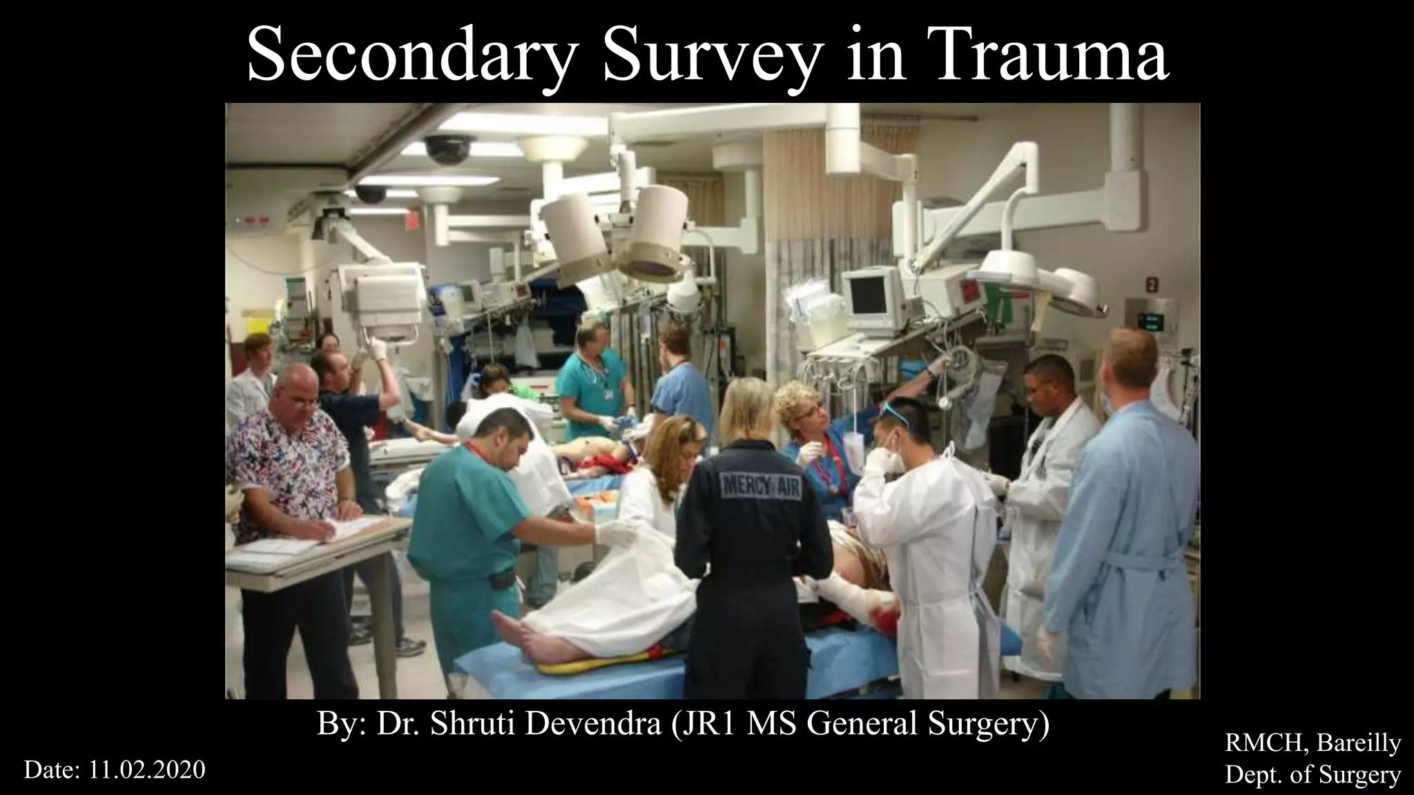
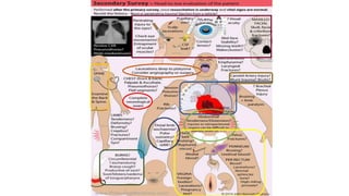
Secondary Survey in Trauma
By: Dr. Shruti Devendra (JR1 MS General Surgery)
Date: 11.02.2020
RMCH, Bareilly
Dept. of Surgery
Secondary Survey
The secondary survey is a head-to-toe evaluation of
the trauma patient, including a complete history, physical examination,
and the reassessment of all vital signs.
Why? To find what we missed
What to see? Look everything, Palpate everything
Secondary survey should not be performed until:
The primary survey has been completed
Resuscitation has been initiated
Normalization of vital signs has begun.
History taking
Patient (if conscious)
Relatives, survivors
Ambulance team
Environment
Pregnancy
SAMPLE
Examination
Scalp: hematoma, laceration, fracture.
Eye exam: Pupil size and reactivity, visual acuity, haemorrhage of globe,
ocular entrapment (lateral canthotomy to release peri-orbital pressure.
Tympanic membrane: hemotympanum, otorrhea, or rupture, which may
signal an underlying head injury.
Basilar skull fracture
Anterior facial structures: This entails palpating for bony step-off of the
facial bones and instability of the midface (by grasping the upper palate
and seeing if this moves separately from the patient’s head).
Mandible, maxillary fracture, nasal fracture
(No NG tube inserted in case of facial and basilar skull fracture)
Cervical spine injury: Due to the devastating consequences of
quadriplegia, a diligent evaluation for occult cervical spine injuries is
mandatory. In the awake patient, the presence of posterior midline pain
or tenderness should provoke a thorough radiologic evaluation.
Additionally, intubated patients, patients with distracting injuries, or
another identified spine fracture should undergo CT imaging
Neck: Carefully palpated for any tenderness or bruise which may indicate
underlying C-spine injury.
Symptomatic zone I and III should be further evaluated If the patient is
stable.
Symptoms like dysphagia, hoarseness, hematoma, venous
bleeding, minor haemoptysis, and subcutaneous
emphysema should undergo CTA.
<15% of neck injury patients undergo neck exploration
Surgery. Asymptomatic patients are typically observed
for 6 to12 hours.
Chest: Palpate the entire chest wall for crepitus (subcutaneous emphysema) and
tenderness.
The area over the sternum and clavicles requires special attention as fractures
involving these bones may suggest significant force and need further evaluation
for other intrathoracic injuries.
Assess any respiratory effort and work at breathing. Evaluate whether breath
sounds are symmetrical and heart sounds are normal and not muffled.
Any patient who undergoes an intervention in the ED—endotracheal
intubation, central line placement, tube thoracostomy—needs a repeat
chest radiograph to document the adequacy of the procedure.
Patients with hemothorax must have a chest radiograph documenting
complete evacuation of the chest; a persistent hemothorax that is not
drained by two chest tubes is termed a caked hemothorax.
Abdomen: distension, bowel sounds, bruising or tenderness, seatbelt
sign should prompt further evaluation.
absence of abdominal tenderness does not eliminate the possibility of
abdominal injury. The abdominal examination may not be reliable in the
following cases:
• Elderly population
• Presence of distracting injuries
• Altered mental state
• Pregnant patient, especially late pregnancy
Perineum: inspected for any evidence of injury. digital rectal
examination should be performed when there is a suspicion of urethral
injury or penetrating rectal injury.
Look for the following:
• Gross blood in the rectal vault, which may indicate bowel injury
• Displaced or high-riding prostate, which may suggest urethral injury
• Abnormal sphincter tone, which may be due to a spinal cord injury
In case of blood at the meatus retrograde urethrography should be
performed before a Foley catheter is inserted.
Consider vaginal injury in patients with lower abdominal pain, pelvic
fracture or perineal laceration. In such situations, a vaginal examination
should be performed.
Extremities: assessed for fractures by carefully palpating each extremity
over its entire length for tenderness and decreased the range of motion.
Assess the integrity of uninjured joints by both active and passive
movements.
Injured joints should also be immobilized, and radiographs should be
obtained.
Check pulses, the capillary refill time and evaluate each compartment.
The presence of significant pain or tense compartments. Pain with
passive movement may indicate a development of the compartment
syndrome.
Pelvis: The pubis and anterior iliac spines should be evaluated for any
signs of pelvic instability. The presence of ecchymosis over the iliac
wings, pubis, labia, or scrotum and tenderness along the pelvic ring
also, requires diagnostic evaluation.
Neurologic exam: the sensory and motor functions should be assessed,
and the Glasgow Coma Scale score should be repeated. This is
important, since a patient's condition may change rapidly over time.
Skin: lacerations, abrasions, ecchymosis, hematoma, marks or bruises.
Pay attention to the following areas:
• Scalp
• Axillary abdominal and gluteal folds
• Perineum
• Back should be evaluated by log-rolling the patient, and the spine
should be palpated for step-offs or focal tenderness.
Blunt Trauma
High energy transfer
Motor-vehicle accidents
>20mph speed
Motorcycle collisions
Fall from >20ft height
low energy transfer
struck with club
Falling from bicycle
May not be widely distributed
Can cause substantial injury to any
particular location
Unrestrained driver sustains a frontal impact, the head strikes the
windshield, the chest and upper abdomen hit the steering column, and
the legs or knees contact the dashboard.
The resultant injuries can include facial fractures, cervical spine
fractures, laceration of the thoracic aorta, myocardial contusion, injury
to the spleen and liver, and fractures of the pelvis and lower extremities.
Collisions with side impact also carry the risk of cervical spine and
thoracic trauma, diaphragm rupture, and crush injuries of the pelvic
ring, solid organ injury usually is limited to either the liver or spleen
based on the direction of Impact.
The "seat belt sign" was first described by Garrett and Braunstein in
1962 as an ecchymosis across the abdominal wall in the location of a lap
belt. The "seat belt syndrome" referred to the musculoskeletal and
visceral injuries associated with the force transmitted by the seat belt.
Skin abrasions and bruising occur on the surface, while common
internal injuries are bowel and mesenteric injuries, along with fractures
of the lumbar spine. Solid organ injuries such as liver and spleen
laceration can occur in these patients. Likewise, injuries to the pancreas,
kidneys, and any other visceral organ can occur.
Penetrating trauma
Stab wound, gunshot wound, or shotgun wound
High
velocity
Low
velocity
Bullet speed >2000 ft/s
Infrequent in civilian setting
Close range: (<20 feet): entire energy focused
to small area with devastating results
Long range: In contrast, long-range shotgun
blasts result in a diffuse pellet pattern in which
many pellets miss the victim. Low energy if
strike.
Thank You

Secondary survey in trauma

  • 1.
    Secondary Survey inTrauma By: Dr. Shruti Devendra (JR1 MS General Surgery) Date: 11.02.2020 RMCH, Bareilly Dept. of Surgery
  • 2.
    Secondary Survey The secondarysurvey is a head-to-toe evaluation of the trauma patient, including a complete history, physical examination, and the reassessment of all vital signs. Why? To find what we missed What to see? Look everything, Palpate everything
  • 3.
    Secondary survey shouldnot be performed until: The primary survey has been completed Resuscitation has been initiated Normalization of vital signs has begun.
  • 4.
    History taking Patient (ifconscious) Relatives, survivors Ambulance team
  • 5.
  • 8.
    Examination Scalp: hematoma, laceration,fracture. Eye exam: Pupil size and reactivity, visual acuity, haemorrhage of globe, ocular entrapment (lateral canthotomy to release peri-orbital pressure. Tympanic membrane: hemotympanum, otorrhea, or rupture, which may signal an underlying head injury. Basilar skull fracture
  • 9.
    Anterior facial structures:This entails palpating for bony step-off of the facial bones and instability of the midface (by grasping the upper palate and seeing if this moves separately from the patient’s head). Mandible, maxillary fracture, nasal fracture (No NG tube inserted in case of facial and basilar skull fracture) Cervical spine injury: Due to the devastating consequences of quadriplegia, a diligent evaluation for occult cervical spine injuries is mandatory. In the awake patient, the presence of posterior midline pain or tenderness should provoke a thorough radiologic evaluation. Additionally, intubated patients, patients with distracting injuries, or another identified spine fracture should undergo CT imaging
  • 10.
    Neck: Carefully palpatedfor any tenderness or bruise which may indicate underlying C-spine injury. Symptomatic zone I and III should be further evaluated If the patient is stable. Symptoms like dysphagia, hoarseness, hematoma, venous bleeding, minor haemoptysis, and subcutaneous emphysema should undergo CTA. <15% of neck injury patients undergo neck exploration Surgery. Asymptomatic patients are typically observed for 6 to12 hours.
  • 11.
    Chest: Palpate theentire chest wall for crepitus (subcutaneous emphysema) and tenderness. The area over the sternum and clavicles requires special attention as fractures involving these bones may suggest significant force and need further evaluation for other intrathoracic injuries. Assess any respiratory effort and work at breathing. Evaluate whether breath sounds are symmetrical and heart sounds are normal and not muffled.
  • 12.
    Any patient whoundergoes an intervention in the ED—endotracheal intubation, central line placement, tube thoracostomy—needs a repeat chest radiograph to document the adequacy of the procedure. Patients with hemothorax must have a chest radiograph documenting complete evacuation of the chest; a persistent hemothorax that is not drained by two chest tubes is termed a caked hemothorax.
  • 13.
    Abdomen: distension, bowelsounds, bruising or tenderness, seatbelt sign should prompt further evaluation. absence of abdominal tenderness does not eliminate the possibility of abdominal injury. The abdominal examination may not be reliable in the following cases: • Elderly population • Presence of distracting injuries • Altered mental state • Pregnant patient, especially late pregnancy
  • 14.
    Perineum: inspected forany evidence of injury. digital rectal examination should be performed when there is a suspicion of urethral injury or penetrating rectal injury. Look for the following: • Gross blood in the rectal vault, which may indicate bowel injury • Displaced or high-riding prostate, which may suggest urethral injury • Abnormal sphincter tone, which may be due to a spinal cord injury In case of blood at the meatus retrograde urethrography should be performed before a Foley catheter is inserted.
  • 15.
    Consider vaginal injuryin patients with lower abdominal pain, pelvic fracture or perineal laceration. In such situations, a vaginal examination should be performed. Extremities: assessed for fractures by carefully palpating each extremity over its entire length for tenderness and decreased the range of motion. Assess the integrity of uninjured joints by both active and passive movements. Injured joints should also be immobilized, and radiographs should be obtained.
  • 16.
    Check pulses, thecapillary refill time and evaluate each compartment. The presence of significant pain or tense compartments. Pain with passive movement may indicate a development of the compartment syndrome. Pelvis: The pubis and anterior iliac spines should be evaluated for any signs of pelvic instability. The presence of ecchymosis over the iliac wings, pubis, labia, or scrotum and tenderness along the pelvic ring also, requires diagnostic evaluation.
  • 17.
    Neurologic exam: thesensory and motor functions should be assessed, and the Glasgow Coma Scale score should be repeated. This is important, since a patient's condition may change rapidly over time. Skin: lacerations, abrasions, ecchymosis, hematoma, marks or bruises. Pay attention to the following areas: • Scalp • Axillary abdominal and gluteal folds • Perineum • Back should be evaluated by log-rolling the patient, and the spine should be palpated for step-offs or focal tenderness.
  • 18.
    Blunt Trauma High energytransfer Motor-vehicle accidents >20mph speed Motorcycle collisions Fall from >20ft height low energy transfer struck with club Falling from bicycle May not be widely distributed Can cause substantial injury to any particular location
  • 19.
    Unrestrained driver sustainsa frontal impact, the head strikes the windshield, the chest and upper abdomen hit the steering column, and the legs or knees contact the dashboard. The resultant injuries can include facial fractures, cervical spine fractures, laceration of the thoracic aorta, myocardial contusion, injury to the spleen and liver, and fractures of the pelvis and lower extremities.
  • 20.
    Collisions with sideimpact also carry the risk of cervical spine and thoracic trauma, diaphragm rupture, and crush injuries of the pelvic ring, solid organ injury usually is limited to either the liver or spleen based on the direction of Impact.
  • 21.
    The "seat beltsign" was first described by Garrett and Braunstein in 1962 as an ecchymosis across the abdominal wall in the location of a lap belt. The "seat belt syndrome" referred to the musculoskeletal and visceral injuries associated with the force transmitted by the seat belt. Skin abrasions and bruising occur on the surface, while common internal injuries are bowel and mesenteric injuries, along with fractures of the lumbar spine. Solid organ injuries such as liver and spleen laceration can occur in these patients. Likewise, injuries to the pancreas, kidneys, and any other visceral organ can occur.
  • 23.
    Penetrating trauma Stab wound,gunshot wound, or shotgun wound High velocity Low velocity Bullet speed >2000 ft/s Infrequent in civilian setting Close range: (<20 feet): entire energy focused to small area with devastating results Long range: In contrast, long-range shotgun blasts result in a diffuse pellet pattern in which many pellets miss the victim. Low energy if strike.
  • 24.