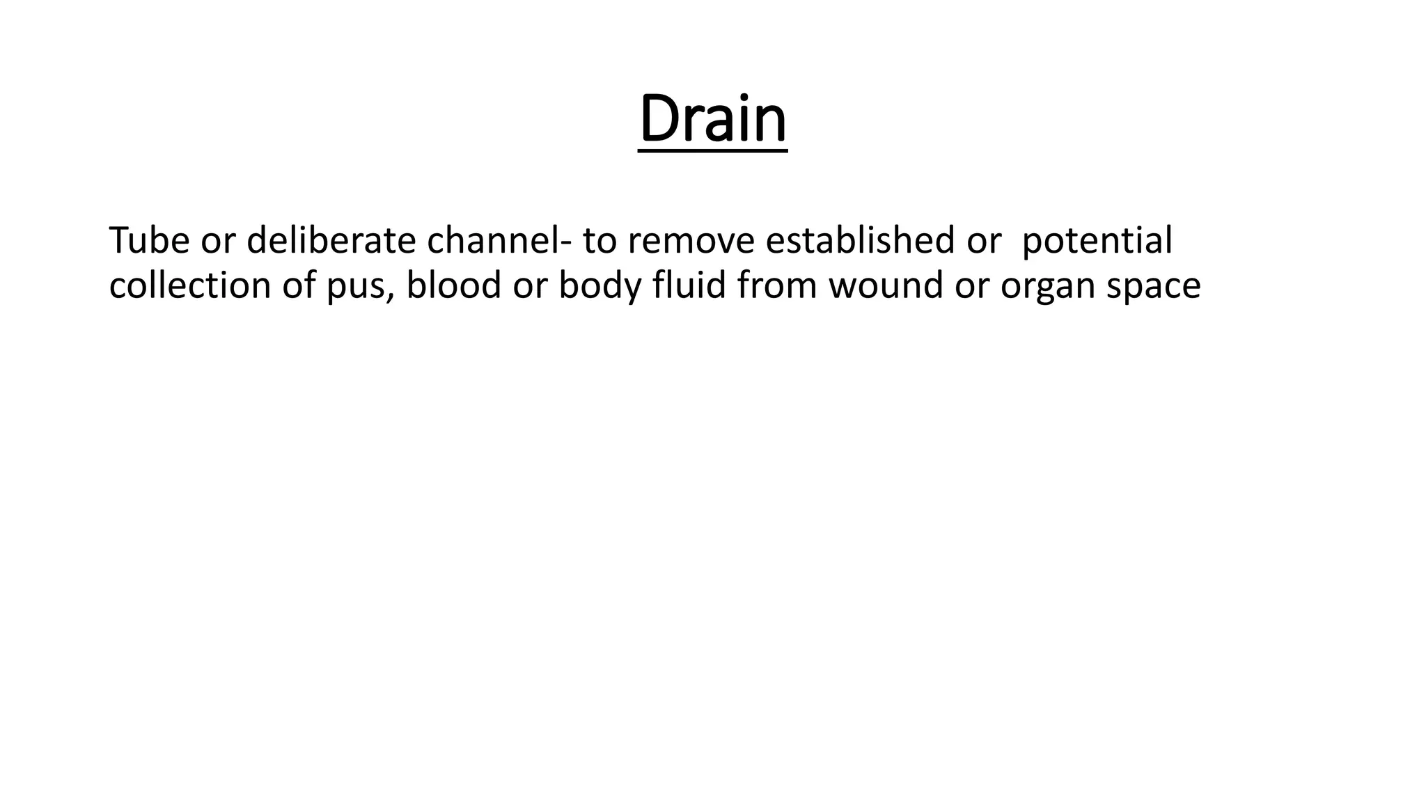
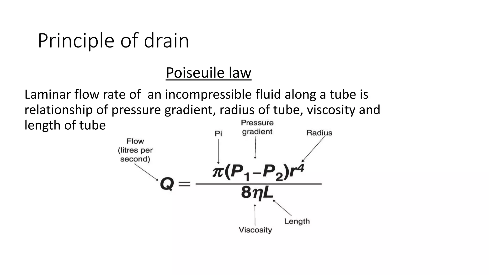
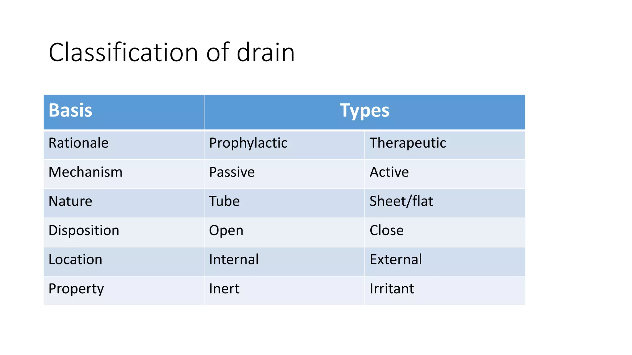
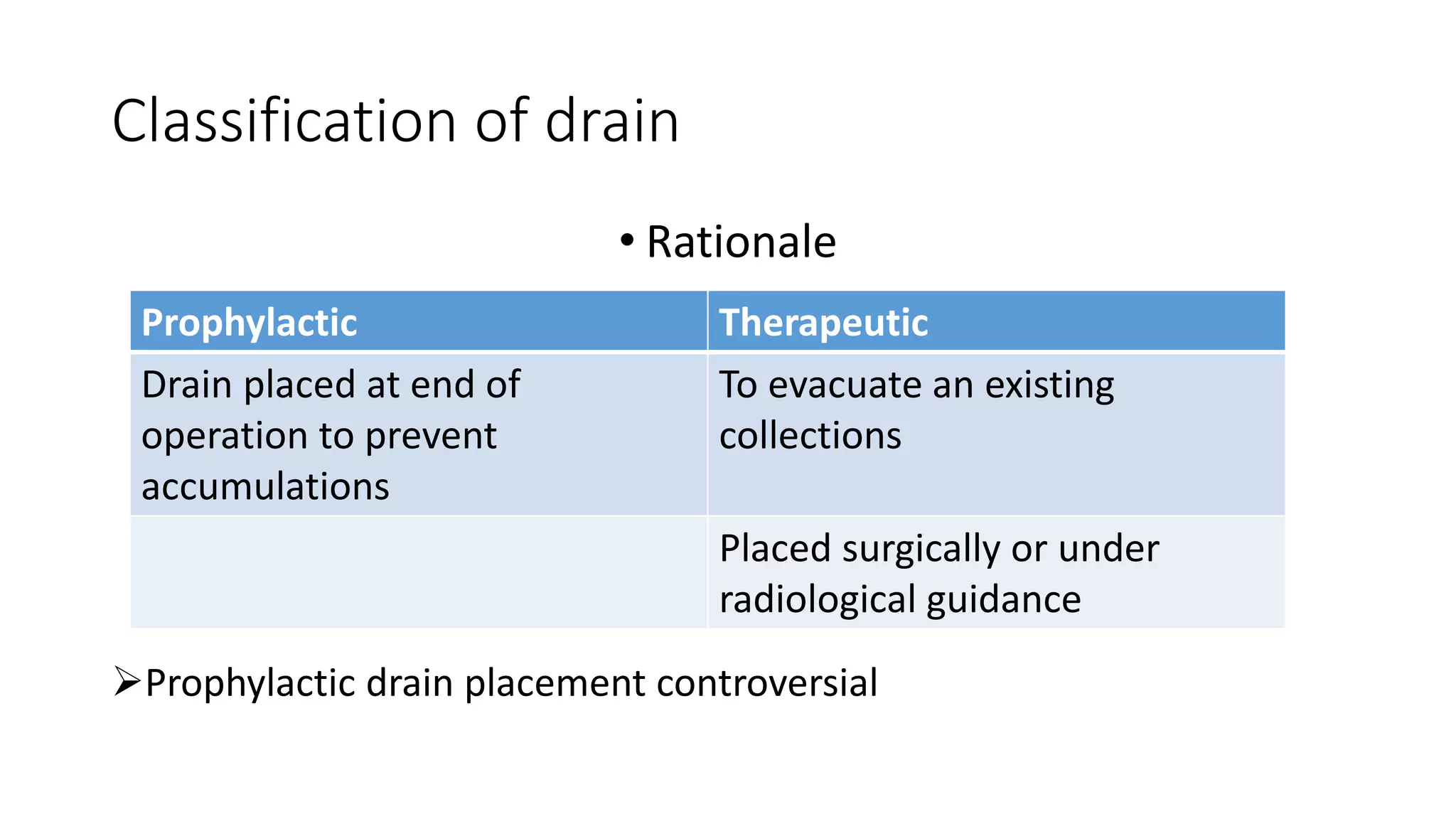
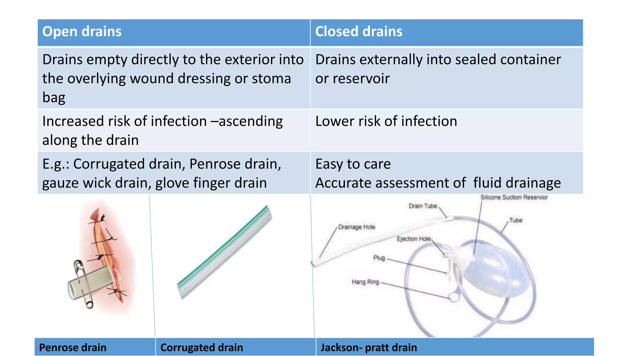
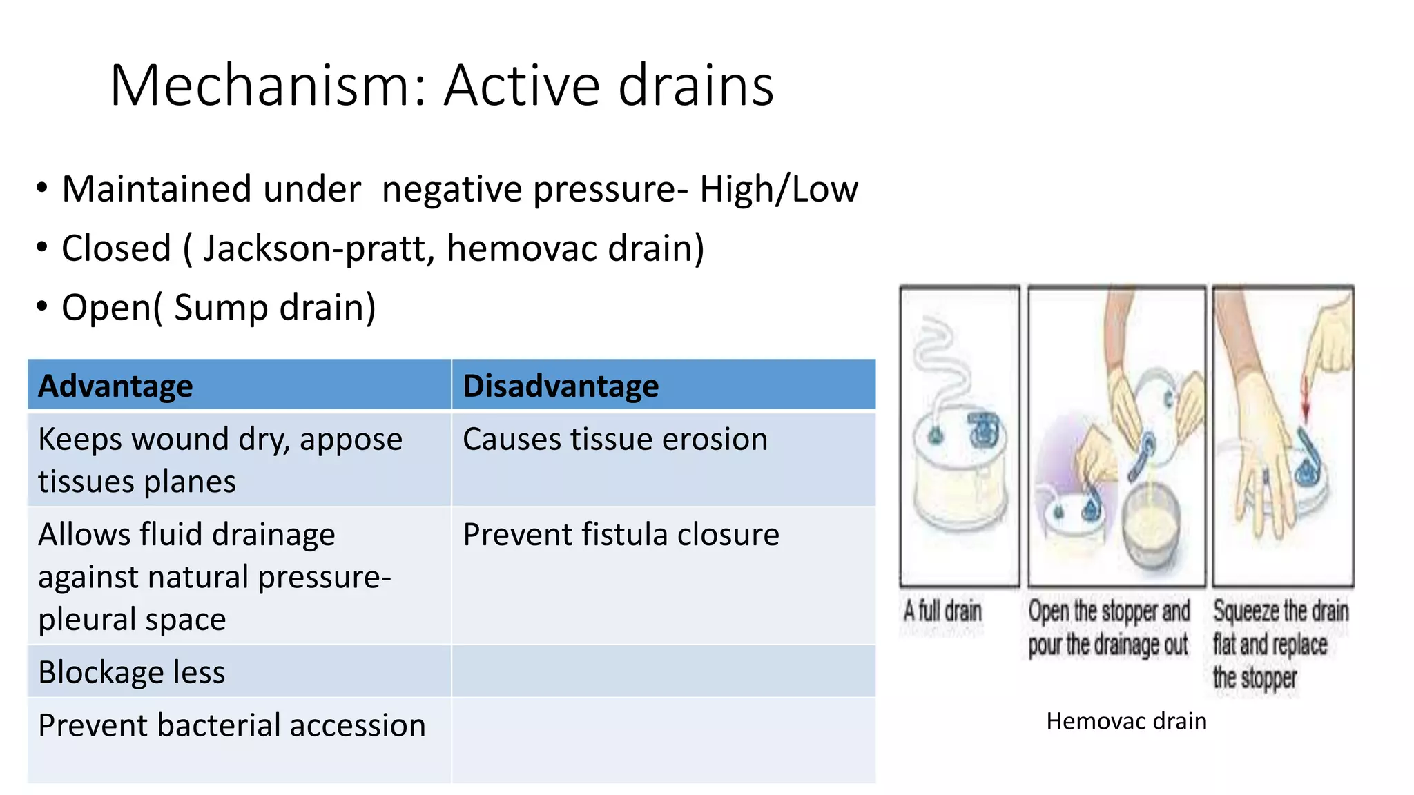
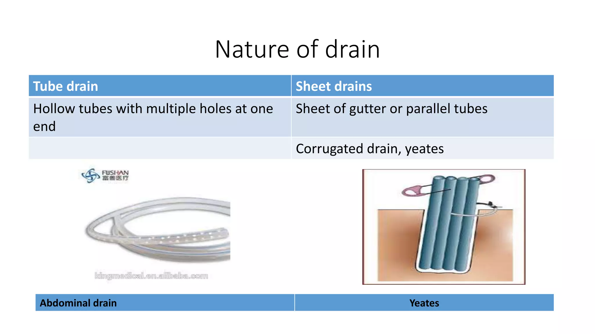
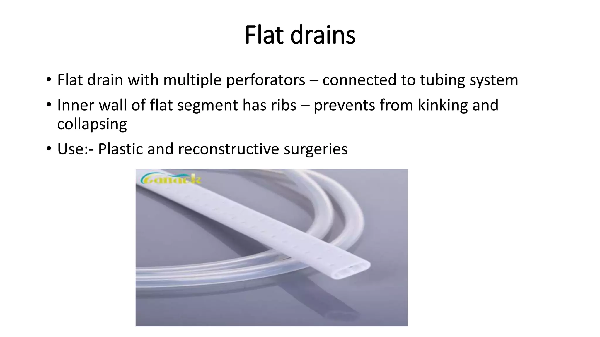
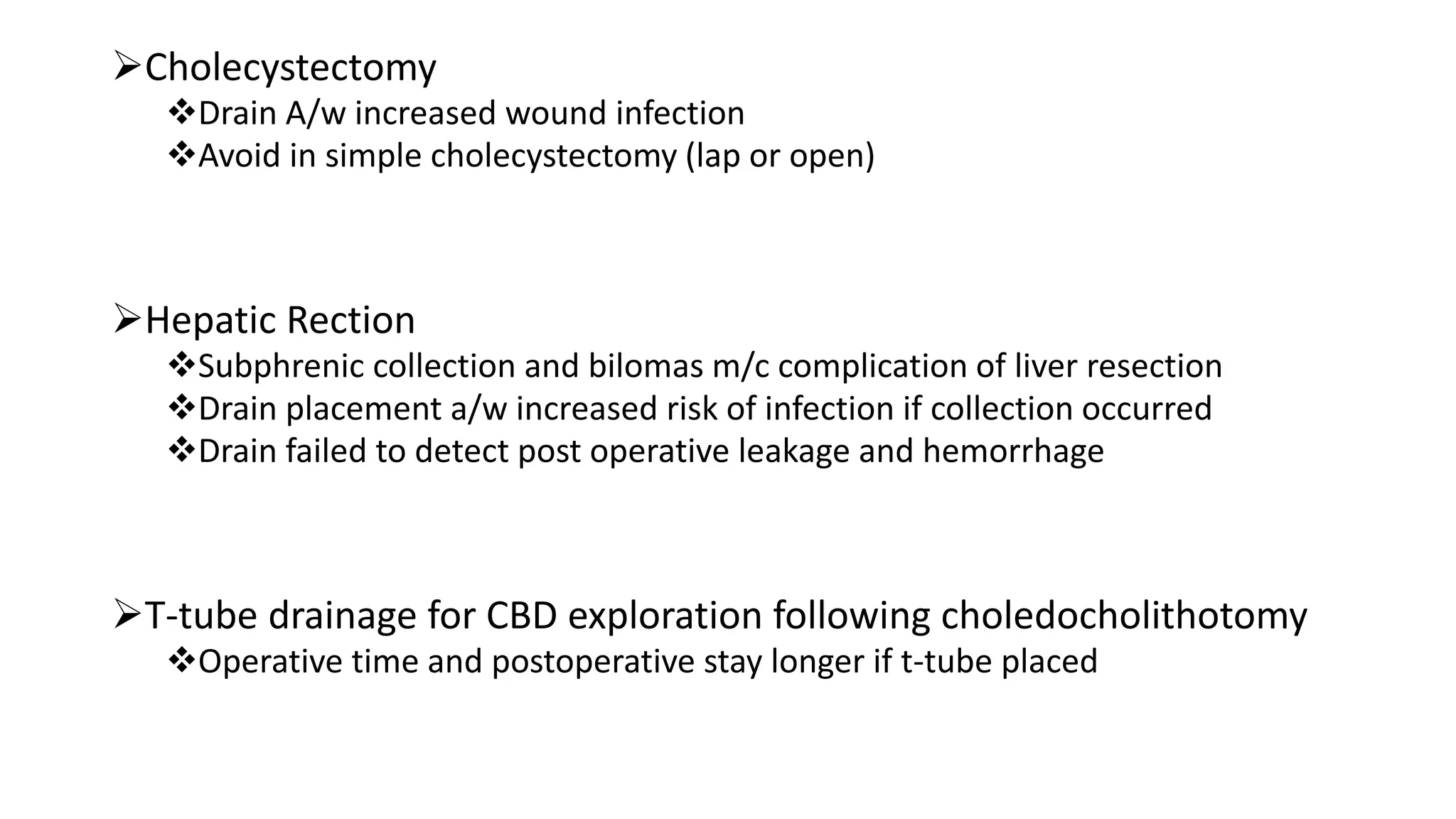
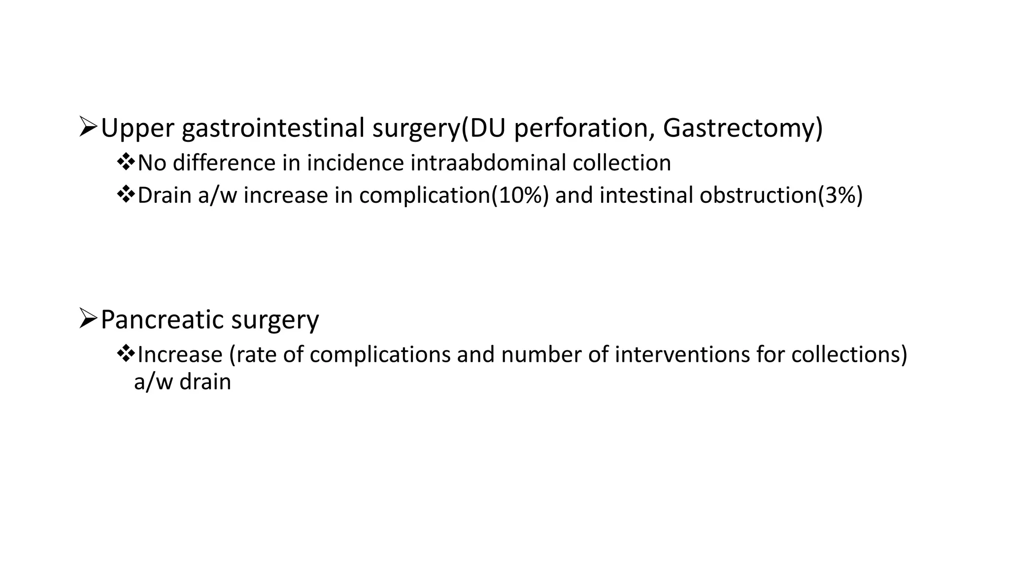
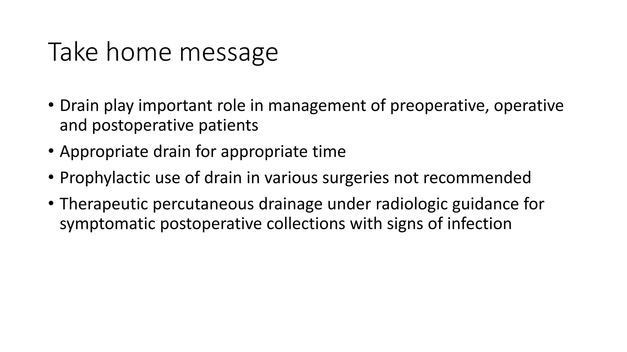
Tubes and Drains
Presenter: Dr. Annush Tha
Moderators: Dr. David Shrestha/ Dr. Dilip Baral/ Dr. Gani Alam
Department of Surgery
Pokhara Academy of Health Sciences
2077/05/01
Objectives
 Principles of drain usage
Drain and its types
Indication/ purpose of drains
Insertion, care and Removal of drain
Complications of drains
Recommendations in surgical practice based on evidence
Drain
Tube or deliberate channel- to remove established or potential
collection of pus, blood or body fluid from wound or organ space
Principle of drain
Poiseuile law
Laminar flow rate of an incompressible fluid along a tube is
relationship of pressure gradient, radius of tube, viscosity and
length of tube
Classification of drain
Basis Types
Rationale Prophylactic Therapeutic
Mechanism Passive Active
Nature Tube Sheet/flat
Disposition Open Close
Location Internal External
Property Inert Irritant
Classification of drain
• Rationale
Prophylactic drain placement controversial
Prophylactic Therapeutic
Drain placed at end of
operation to prevent
accumulations
To evacuate an existing
collections
Placed surgically or under
radiological guidance
Open drains Closed drains
Drains empty directly to the exterior into
the overlying wound dressing or stoma
bag
Drains externally into sealed container
or reservoir
Increased risk of infection –ascending
along the drain
Lower risk of infection
E.g.: Corrugated drain, Penrose drain,
gauze wick drain, glove finger drain
Easy to care
Accurate assessment of fluid drainage
Penrose drain Corrugated drain Jackson- pratt drain
Mechanism: Active drains
• Maintained under negative pressure- High/Low
• Closed ( Jackson-pratt, hemovac drain)
• Open( Sump drain)
Advantage Disadvantage
Keeps wound dry, appose
tissues planes
Causes tissue erosion
Allows fluid drainage
against natural pressure-
pleural space
Prevent fistula closure
Blockage less
Prevent bacterial accession Hemovac drain
Mechanism: Passive Drain
• Drains by capillary action, pressure gradient or gravity
• No suction
• Closed ( NG tube, Foley’s catheter, T-tube, Under water seal drain)
• Open( Penrose, corrugated drain, gauge wick drain)
Nature of drain
Tube drain Sheet drains
Hollow tubes with multiple holes at one
end
Sheet of gutter or parallel tubes
Corrugated drain, yeates
Abdominal drain Yeates
Flat drains
• Flat drain with multiple perforators – connected to tubing system
• Inner wall of flat segment has ribs – prevents from kinking and
collapsing
• Use:- Plastic and reconstructive surgeries
Sump drain
• Double lumen, radio opaque
• 1st lumen- suction of gastric
contents
• 2nd lumen- blue extension open to
room air to maintain continuous
flow of atmospheric air into
stomach
• Controls the amount of suction
pressure on tissue/organ and
prevents blockage
• Reduces amount of tissue damage
Pigtail drain
• Removes unwanted fluid from organ, duct or abscess
• Inserted under radiological guidance
Disposition
External Internal
Open externally outside body wall Placed internally within luminal organs to create a
route or to connect two luminal organs
Fluid channeled from deepest part of cavity to
exterior
Diverts retained fluid from primary drainage site
/area distal to body passage or cavity to bypass
obstruction
E.g.: ventriculo-jugular shunt, ventriculo-atrial shunt,
Dj stents
Nature/Drain Materials
Irritant Inert
Rubber or latex Polyurethrane, silicone,
polytetrafluoroethylene
Induces fibrous reaction, allergic rxn Non irritative, no fibrous rxn
Indication/ Purpose Of Drains
Therapeutic – remove fluid/pus/gas
• Abscess cavity
• Seroma
• Pleural fluid
• Acute urinary Retention
Palliative- to allow diversion of body fluids/bypass luminal
obstruction
• Advanced Ca oesophagus
• Hydrocephalus
Access route- to body space or cavity
• Percutaneous nephrolithotomy,
Diagnostic- injection of dye/contrast about underlying cavity or
fistula to reach diagnosis
• Biliary fistula
• T-tube cholangiogram for retained gall stones in common bile duct
Prophylactic-
 prevents post operative complications arising from fluid collection in wound
cavity
 Promote tissue appostion
Post thyroidectomy
Thoracotomy
Monitoring- of fluid volume and quality
 Gastrointestinal bleeding
 Urinary Catheterisation
Drain insertion
Exteriorized via shortest , safest route and through a stab wound not
through the surgical incision
Reach deepest and most dependent part of the cavity or wound
Placed lower than the incision site
Appropriate size and length of drain
Secured at exit to prevent migration of drain-
Silk, safety pin, drain clip, adhesives
Tubing free of kinks, debris, clots—free drainage
Securing the drain
A Modified Technique for Securing Drains to the Skin.
Mura S1 , Guarneri GF1 , Parodi PC1 1. Department of Plastic and Reconstructive
Surgery, University of Udine, Udine, Italy.
Different ways of securing drain
Drain Care
• Is the patient well?
• Drain secure?
• Signs of infection, excoriation, peritubal leakage at skin site?
• Tube kinked/ clogged/ damaged?
• Drain connected properly?
• Quality and quantity of drainage fluid?
• Change in nature or volume of effluent?
• Vacuum suction working or not?
Drain Removal
• Drain has lived up to its function
• Remove drain with steady gentle traction (avoid sudden jerks)
• Release suction prior to removal
Complications and prevention
• Tissue reaction and necrosis
• Hemorrhage
• Source of infection- increase with prolonged drain
placement
• Bowel herniation- if complicated by infection
• Drain entrapment- due to fibrosis
• Fluid and electrolyte loss-high output
• Migration of drain- not secured properly
• Erosion of viscera- in peritoneal cavity without well defined
abscess cavity
Evidence based findings
Routine NG drainage after abdominal surgery not recommended
Delay return of gastrointestinal function a/w drain( time to passage of flatus)
NG use –Acute gastric dilation, GOO, Small bowel obstruction, NG feeding
Colorectal surgery-
Drain only if anastomotic leak
Avoid Prophylactic drainage
Appendiectomy
Despite of severity ( gangrenous or perforated or uncomplicated appendicitis)
drain placement a/w increased wound infection and faecal fistula
Cholecystectomy
Drain A/w increased wound infection
Avoid in simple cholecystectomy (lap or open)
Hepatic Rection
Subphrenic collection and bilomas m/c complication of liver resection
Drain placement a/w increased risk of infection if collection occurred
Drain failed to detect post operative leakage and hemorrhage
T-tube drainage for CBD exploration following choledocholithotomy
Operative time and postoperative stay longer if t-tube placed
Upper gastrointestinal surgery(DU perforation, Gastrectomy)
No difference in incidence intraabdominal collection
Drain a/w increase in complication(10%) and intestinal obstruction(3%)
Pancreatic surgery
Increase (rate of complications and number of interventions for collections)
a/w drain
Therapeutic Drainage
Percutaneous drainage under image guidance ( USG or CT) for
symptomatic post operative collection with signs of infection is
recommended for treatment.
Take home message
• Drain play important role in management of preoperative, operative
and postoperative patients
• Appropriate drain for appropriate time
• Prophylactic use of drain in various surgeries not recommended
• Therapeutic percutaneous drainage under radiologic guidance for
symptomatic postoperative collections with signs of infection
References
• James A. caton ,Dileep N. Lobo.The use of drains in Gastrointestinal
surgery, Recent Advances of surgery.31 Edition
• JG M, EA A. Surgical drains: what the resident needs to know. Nigerian
journal of Medicine. 2008 Jul;17(3).
• Durai R, Mownah A, Philip CN. Use of drains in surgery: a review.
Journal of perioperative practice. 2009 Jun;19(6):180-6.
• SRB’s manual of surgery
Thank you

Tubes and drains

  • 1.
    Tubes and Drains Presenter:Dr. Annush Tha Moderators: Dr. David Shrestha/ Dr. Dilip Baral/ Dr. Gani Alam Department of Surgery Pokhara Academy of Health Sciences 2077/05/01
  • 2.
    Objectives  Principles ofdrain usage Drain and its types Indication/ purpose of drains Insertion, care and Removal of drain Complications of drains Recommendations in surgical practice based on evidence
  • 3.
    Drain Tube or deliberatechannel- to remove established or potential collection of pus, blood or body fluid from wound or organ space
  • 4.
    Principle of drain Poiseuilelaw Laminar flow rate of an incompressible fluid along a tube is relationship of pressure gradient, radius of tube, viscosity and length of tube
  • 5.
    Classification of drain BasisTypes Rationale Prophylactic Therapeutic Mechanism Passive Active Nature Tube Sheet/flat Disposition Open Close Location Internal External Property Inert Irritant
  • 6.
    Classification of drain •Rationale Prophylactic drain placement controversial Prophylactic Therapeutic Drain placed at end of operation to prevent accumulations To evacuate an existing collections Placed surgically or under radiological guidance
  • 7.
    Open drains Closeddrains Drains empty directly to the exterior into the overlying wound dressing or stoma bag Drains externally into sealed container or reservoir Increased risk of infection –ascending along the drain Lower risk of infection E.g.: Corrugated drain, Penrose drain, gauze wick drain, glove finger drain Easy to care Accurate assessment of fluid drainage Penrose drain Corrugated drain Jackson- pratt drain
  • 8.
    Mechanism: Active drains •Maintained under negative pressure- High/Low • Closed ( Jackson-pratt, hemovac drain) • Open( Sump drain) Advantage Disadvantage Keeps wound dry, appose tissues planes Causes tissue erosion Allows fluid drainage against natural pressure- pleural space Prevent fistula closure Blockage less Prevent bacterial accession Hemovac drain
  • 9.
    Mechanism: Passive Drain •Drains by capillary action, pressure gradient or gravity • No suction • Closed ( NG tube, Foley’s catheter, T-tube, Under water seal drain) • Open( Penrose, corrugated drain, gauge wick drain)
  • 10.
    Nature of drain Tubedrain Sheet drains Hollow tubes with multiple holes at one end Sheet of gutter or parallel tubes Corrugated drain, yeates Abdominal drain Yeates
  • 11.
    Flat drains • Flatdrain with multiple perforators – connected to tubing system • Inner wall of flat segment has ribs – prevents from kinking and collapsing • Use:- Plastic and reconstructive surgeries
  • 12.
    Sump drain • Doublelumen, radio opaque • 1st lumen- suction of gastric contents • 2nd lumen- blue extension open to room air to maintain continuous flow of atmospheric air into stomach • Controls the amount of suction pressure on tissue/organ and prevents blockage • Reduces amount of tissue damage
  • 13.
    Pigtail drain • Removesunwanted fluid from organ, duct or abscess • Inserted under radiological guidance
  • 14.
    Disposition External Internal Open externallyoutside body wall Placed internally within luminal organs to create a route or to connect two luminal organs Fluid channeled from deepest part of cavity to exterior Diverts retained fluid from primary drainage site /area distal to body passage or cavity to bypass obstruction E.g.: ventriculo-jugular shunt, ventriculo-atrial shunt, Dj stents
  • 15.
    Nature/Drain Materials Irritant Inert Rubberor latex Polyurethrane, silicone, polytetrafluoroethylene Induces fibrous reaction, allergic rxn Non irritative, no fibrous rxn
  • 16.
    Indication/ Purpose OfDrains Therapeutic – remove fluid/pus/gas • Abscess cavity • Seroma • Pleural fluid • Acute urinary Retention Palliative- to allow diversion of body fluids/bypass luminal obstruction • Advanced Ca oesophagus • Hydrocephalus Access route- to body space or cavity • Percutaneous nephrolithotomy,
  • 17.
    Diagnostic- injection ofdye/contrast about underlying cavity or fistula to reach diagnosis • Biliary fistula • T-tube cholangiogram for retained gall stones in common bile duct Prophylactic-  prevents post operative complications arising from fluid collection in wound cavity  Promote tissue appostion Post thyroidectomy Thoracotomy Monitoring- of fluid volume and quality  Gastrointestinal bleeding  Urinary Catheterisation
  • 18.
    Drain insertion Exteriorized viashortest , safest route and through a stab wound not through the surgical incision Reach deepest and most dependent part of the cavity or wound Placed lower than the incision site Appropriate size and length of drain Secured at exit to prevent migration of drain- Silk, safety pin, drain clip, adhesives Tubing free of kinks, debris, clots—free drainage
  • 19.
    Securing the drain AModified Technique for Securing Drains to the Skin. Mura S1 , Guarneri GF1 , Parodi PC1 1. Department of Plastic and Reconstructive Surgery, University of Udine, Udine, Italy. Different ways of securing drain
  • 20.
    Drain Care • Isthe patient well? • Drain secure? • Signs of infection, excoriation, peritubal leakage at skin site? • Tube kinked/ clogged/ damaged? • Drain connected properly? • Quality and quantity of drainage fluid? • Change in nature or volume of effluent? • Vacuum suction working or not?
  • 21.
    Drain Removal • Drainhas lived up to its function • Remove drain with steady gentle traction (avoid sudden jerks) • Release suction prior to removal
  • 22.
    Complications and prevention •Tissue reaction and necrosis • Hemorrhage • Source of infection- increase with prolonged drain placement • Bowel herniation- if complicated by infection • Drain entrapment- due to fibrosis • Fluid and electrolyte loss-high output • Migration of drain- not secured properly • Erosion of viscera- in peritoneal cavity without well defined abscess cavity
  • 23.
    Evidence based findings RoutineNG drainage after abdominal surgery not recommended Delay return of gastrointestinal function a/w drain( time to passage of flatus) NG use –Acute gastric dilation, GOO, Small bowel obstruction, NG feeding Colorectal surgery- Drain only if anastomotic leak Avoid Prophylactic drainage Appendiectomy Despite of severity ( gangrenous or perforated or uncomplicated appendicitis) drain placement a/w increased wound infection and faecal fistula
  • 24.
    Cholecystectomy Drain A/w increasedwound infection Avoid in simple cholecystectomy (lap or open) Hepatic Rection Subphrenic collection and bilomas m/c complication of liver resection Drain placement a/w increased risk of infection if collection occurred Drain failed to detect post operative leakage and hemorrhage T-tube drainage for CBD exploration following choledocholithotomy Operative time and postoperative stay longer if t-tube placed
  • 25.
    Upper gastrointestinal surgery(DUperforation, Gastrectomy) No difference in incidence intraabdominal collection Drain a/w increase in complication(10%) and intestinal obstruction(3%) Pancreatic surgery Increase (rate of complications and number of interventions for collections) a/w drain
  • 26.
    Therapeutic Drainage Percutaneous drainageunder image guidance ( USG or CT) for symptomatic post operative collection with signs of infection is recommended for treatment.
  • 27.
    Take home message •Drain play important role in management of preoperative, operative and postoperative patients • Appropriate drain for appropriate time • Prophylactic use of drain in various surgeries not recommended • Therapeutic percutaneous drainage under radiologic guidance for symptomatic postoperative collections with signs of infection
  • 28.
    References • James A.caton ,Dileep N. Lobo.The use of drains in Gastrointestinal surgery, Recent Advances of surgery.31 Edition • JG M, EA A. Surgical drains: what the resident needs to know. Nigerian journal of Medicine. 2008 Jul;17(3). • Durai R, Mownah A, Philip CN. Use of drains in surgery: a review. Journal of perioperative practice. 2009 Jun;19(6):180-6. • SRB’s manual of surgery
  • 29.