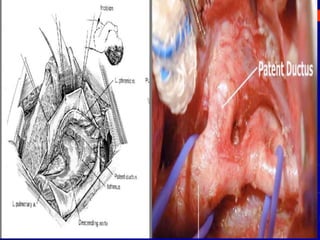
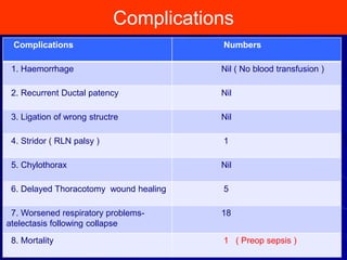
Patent Ductus Arteriosus
Surgical Closure in Premature Infants
1
PDA Ligation in Preterms
Surgical ligation is performed for PDA closure when medical
treatment fails or is contraindicated.
The success rate of indomethacin therapy for PDA closure is 79% if
birth weight is < 1750 g.
The failure rate is up to 40–50% if birth weight is < 800g.
Fragility of the tissues and also duct is a challenge on a low birth
weight infant compared to a normal weight one
2
Protocol for Surgical Referral
3
Premature neonates < 29/40 all get an Echo within 4-24 hours after birth
PDA > 1.5mm are treated with a course of indomethacin/ibuprofen.
Ductus still significantly open on Re-Echo after the 1st course of NSAIDs,
then a 2nd course is given.
If the 2nd course of NSAIDs fails to close the ductus and it remains large
or problematic --- Causing high ventilation requirements/failure to wean
off ventilation --- decision may be to refer that baby to cardiology for
consideration of Surgical closure
Guidelines for Surgical Management
4
Decision for PDA ligation should be a Consensus among: the Neonatologist on
service, Cardiologist performing 2D ECHO . and the Paediatric cardiothoracic
surgeon performing the procedure.
All PDA ligation in our institute need a JCC discussion .
A PDA may be considered for ligation if it is large both size and shunt volume,
with clinical indicators of pulmonary over-circulation (Intubated with escalating
respiratory support) with or without clinical indicators of systemic hypo-perfusion.
The side of arch , PDA dimensions and any other cardiac anomaly is clearly
Documented.
The reason for surgical ligation of the Ductus arteriosus should be clearly
indicated and documented by the neonatologist on service.
Guidelines for Surgical Management
5
Echocardiogram should be performed by paediatric cardiologist at least once
before PDA ligation to exclude duct-dependent congenital heart disease .
Rule out spontaneous duct closure prior to surgery- Clinical / Echo
On the day before PDA ligation surgery:
-Chest x-ray: As baseline prior to surgery.
- Complete blood count.( Platelet count )
- Coagulation profile
- Group & Hold – 1 PRBC/ 1 Platelet
- Rule out MRSA/MSSA
NEONATAL PDAs – 5 year review
6
8
12
10
8 8
0
2
4
6
8
10
12
14
2018 2017 2016 2015 2014
GESTATIONAL
AGE
Infants (n)
≤ 25 26
26–30 5
31-34 15
Birth Weight Gestational Age
BIRTH WEIGHT Infants (n)
≤ 750 gm 24
751–1000 gm 8
1000–1500 gm 14
Procedure Performed
8
22
8
10
6
0
5
10
15
20
25
Clipped Transfixation Clipped & Transfixed Clipped & Ligated
Procedures
9
10
Complications
11
Complications Numbers
1. Haemorrhage Nil ( No blood transfusion )
2. Recurrent Ductal patency Nil
3. Ligation of wrong structre Nil
4. Stridor ( RLN palsy ) 1
5. Chylothorax Nil
6. Delayed Thoracotomy wound healing 5
7. Worsened respiratory problems-
atelectasis following collapse
18
8. Mortality 1 ( Preop sepsis )
Case Discussion
12
Kid was born with Extreme prematurity (24 weeks)
Extremely low birth weight (639 g)
Respiratory Distress – Consistent with Hyaline membrane disease- Intubated
Gastric Perforation – Day 12 of life – Laprotomy done.
MRSA Bacteraemia – positive blood culture – treated with 2 week
Vancomycin
MRSA Conjunctivitis – treated with 5 days of chloramphenicol
Patent ductus arteriosus -Dilated LV , Increasing size pf PDA – 1.6 TO 1.8
mm – Referred for PDA ligation.
Case Discussion
13
PDA ligation – deferred – Falling platelet levels / Dearranged Coagulation
Clipping of PDA done. Procedure uneventful.Postop 2D Echo – Nil concenrs
Postop Echo
14
POSTNATAL CMV INFECTION
15
Initial Urine CMV on was negative (result sent as part of work-up for
causes of gastric perforation).
Repeat urine CMV was sent on day 31 of life to investigate
unexplained thrombocytopenia (nadir 60) and mildly abnormal liver
enzymes (ALT 70).
There was also an associated fever to 38 on day 30 of life (thought
to be transfusion related).
The repeat urine CMV was later found to be positive.
Severe respiratory deterioration in his last few hours of life was
likely related to a CMV infection or pneumonitis
Post Ligation Cardiac Syndrome
16
PLCS is characterised clinically by a fall in systolic blood
pressure (usually < 3rd centile for age) requiring one or more
cardiotropic agents, and increasing ventilator requirements,
necessitating an increase in mean air way pressure and FiO2
by at least 20%.
Do a Chest x-ray within one hour of surgical intervention to
exclude air leaks or hyperinflation.
2d Echo postoperatively – showcasing unobstructed arch and
closed ductus.
Post Ligation Cardiac Syndrome
17
Pay close attention to post-operative respiratory management (consider frequent
blood gases) due to expected improvement of lung compliance after resolution of
pulmonary over circulation.
Consider an indwelling arterial line for enhanced blood pressure monitoring and 6-
8 hrly blood gas/lactate analysis.
Mean arterial pressure (MAP) may be less reliable in the setting of PDA ligation
due to trends seen in systolic (SAP) and diastolic (DAP) arterial pressure. The
decline in SAP secondary to LV dysfunction may be masked when MAP only is
considered.
Thresholds for intervention should include a SAP or DAP < 3rd percentile.
In the setting of low SAP, low fractional shortening, inodilator therapy is
preferable. Common agents used in this setting may include dobutamine or
milrinone
PDA ligation and adrenal insufficiency
18
Hydrocortisone therapy is occasionally indicated
for refractory hypotension.
These patients are typified by an early fall in
both SAP and DAP.
Measurement of pre- and postoperative cortisol,
or a preoperative ACTH stimulation test may
guide any decisions related to steroid treatment.
Summary
19
PDA ligation may lead to resolution of the primary clinical
problem, but potentially exposes the infant to significant
cardiorespiratory instability.
The physiologic changes accompanying PDA ligation and
multifactorial nature of the circulatory compromise require
focused neonatal cardiac care.
Standardized guidelines are likely to be beneficial, given
the predictable nature of the clinical deterioration.
Thank You

Surgical closure Neonatal PDA

  • 1.
    Patent Ductus Arteriosus SurgicalClosure in Premature Infants 1
  • 2.
    PDA Ligation inPreterms Surgical ligation is performed for PDA closure when medical treatment fails or is contraindicated. The success rate of indomethacin therapy for PDA closure is 79% if birth weight is < 1750 g. The failure rate is up to 40–50% if birth weight is < 800g. Fragility of the tissues and also duct is a challenge on a low birth weight infant compared to a normal weight one 2
  • 3.
    Protocol for SurgicalReferral 3 Premature neonates < 29/40 all get an Echo within 4-24 hours after birth PDA > 1.5mm are treated with a course of indomethacin/ibuprofen. Ductus still significantly open on Re-Echo after the 1st course of NSAIDs, then a 2nd course is given. If the 2nd course of NSAIDs fails to close the ductus and it remains large or problematic --- Causing high ventilation requirements/failure to wean off ventilation --- decision may be to refer that baby to cardiology for consideration of Surgical closure
  • 4.
    Guidelines for SurgicalManagement 4 Decision for PDA ligation should be a Consensus among: the Neonatologist on service, Cardiologist performing 2D ECHO . and the Paediatric cardiothoracic surgeon performing the procedure. All PDA ligation in our institute need a JCC discussion . A PDA may be considered for ligation if it is large both size and shunt volume, with clinical indicators of pulmonary over-circulation (Intubated with escalating respiratory support) with or without clinical indicators of systemic hypo-perfusion. The side of arch , PDA dimensions and any other cardiac anomaly is clearly Documented. The reason for surgical ligation of the Ductus arteriosus should be clearly indicated and documented by the neonatologist on service.
  • 5.
    Guidelines for SurgicalManagement 5 Echocardiogram should be performed by paediatric cardiologist at least once before PDA ligation to exclude duct-dependent congenital heart disease . Rule out spontaneous duct closure prior to surgery- Clinical / Echo On the day before PDA ligation surgery: -Chest x-ray: As baseline prior to surgery. - Complete blood count.( Platelet count ) - Coagulation profile - Group & Hold – 1 PRBC/ 1 Platelet - Rule out MRSA/MSSA
  • 6.
    NEONATAL PDAs –5 year review 6 8 12 10 8 8 0 2 4 6 8 10 12 14 2018 2017 2016 2015 2014
  • 7.
    GESTATIONAL AGE Infants (n) ≤ 2526 26–30 5 31-34 15 Birth Weight Gestational Age BIRTH WEIGHT Infants (n) ≤ 750 gm 24 751–1000 gm 8 1000–1500 gm 14
  • 8.
    Procedure Performed 8 22 8 10 6 0 5 10 15 20 25 Clipped TransfixationClipped & Transfixed Clipped & Ligated Procedures
  • 9.
  • 10.
  • 11.
    Complications 11 Complications Numbers 1. HaemorrhageNil ( No blood transfusion ) 2. Recurrent Ductal patency Nil 3. Ligation of wrong structre Nil 4. Stridor ( RLN palsy ) 1 5. Chylothorax Nil 6. Delayed Thoracotomy wound healing 5 7. Worsened respiratory problems- atelectasis following collapse 18 8. Mortality 1 ( Preop sepsis )
  • 12.
    Case Discussion 12 Kid wasborn with Extreme prematurity (24 weeks) Extremely low birth weight (639 g) Respiratory Distress – Consistent with Hyaline membrane disease- Intubated Gastric Perforation – Day 12 of life – Laprotomy done. MRSA Bacteraemia – positive blood culture – treated with 2 week Vancomycin MRSA Conjunctivitis – treated with 5 days of chloramphenicol Patent ductus arteriosus -Dilated LV , Increasing size pf PDA – 1.6 TO 1.8 mm – Referred for PDA ligation.
  • 13.
    Case Discussion 13 PDA ligation– deferred – Falling platelet levels / Dearranged Coagulation Clipping of PDA done. Procedure uneventful.Postop 2D Echo – Nil concenrs
  • 14.
  • 15.
    POSTNATAL CMV INFECTION 15 InitialUrine CMV on was negative (result sent as part of work-up for causes of gastric perforation). Repeat urine CMV was sent on day 31 of life to investigate unexplained thrombocytopenia (nadir 60) and mildly abnormal liver enzymes (ALT 70). There was also an associated fever to 38 on day 30 of life (thought to be transfusion related). The repeat urine CMV was later found to be positive. Severe respiratory deterioration in his last few hours of life was likely related to a CMV infection or pneumonitis
  • 16.
    Post Ligation CardiacSyndrome 16 PLCS is characterised clinically by a fall in systolic blood pressure (usually < 3rd centile for age) requiring one or more cardiotropic agents, and increasing ventilator requirements, necessitating an increase in mean air way pressure and FiO2 by at least 20%. Do a Chest x-ray within one hour of surgical intervention to exclude air leaks or hyperinflation. 2d Echo postoperatively – showcasing unobstructed arch and closed ductus.
  • 17.
    Post Ligation CardiacSyndrome 17 Pay close attention to post-operative respiratory management (consider frequent blood gases) due to expected improvement of lung compliance after resolution of pulmonary over circulation. Consider an indwelling arterial line for enhanced blood pressure monitoring and 6- 8 hrly blood gas/lactate analysis. Mean arterial pressure (MAP) may be less reliable in the setting of PDA ligation due to trends seen in systolic (SAP) and diastolic (DAP) arterial pressure. The decline in SAP secondary to LV dysfunction may be masked when MAP only is considered. Thresholds for intervention should include a SAP or DAP < 3rd percentile. In the setting of low SAP, low fractional shortening, inodilator therapy is preferable. Common agents used in this setting may include dobutamine or milrinone
  • 18.
    PDA ligation andadrenal insufficiency 18 Hydrocortisone therapy is occasionally indicated for refractory hypotension. These patients are typified by an early fall in both SAP and DAP. Measurement of pre- and postoperative cortisol, or a preoperative ACTH stimulation test may guide any decisions related to steroid treatment.
  • 19.
    Summary 19 PDA ligation maylead to resolution of the primary clinical problem, but potentially exposes the infant to significant cardiorespiratory instability. The physiologic changes accompanying PDA ligation and multifactorial nature of the circulatory compromise require focused neonatal cardiac care. Standardized guidelines are likely to be beneficial, given the predictable nature of the clinical deterioration.
  • 20.