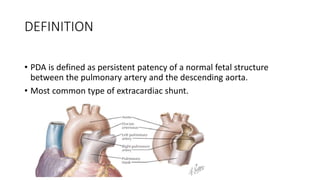
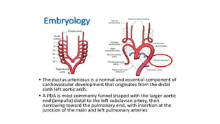
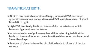
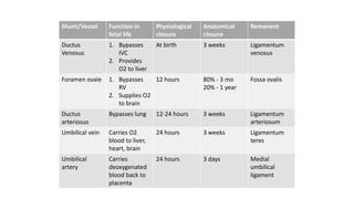
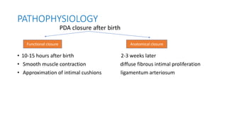
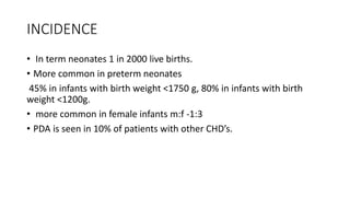
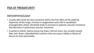
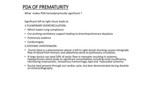
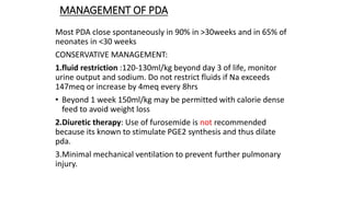
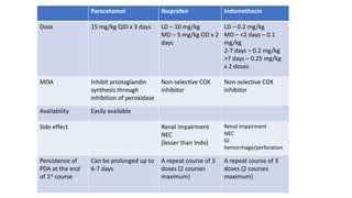
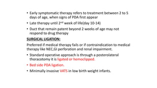
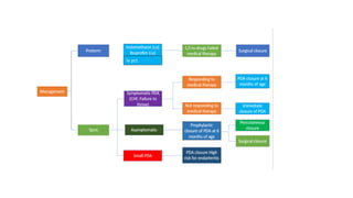
2. DEFINITION
• PDA is defined as persistent patency of a normal fetal structure
between the pulmonary artery and the descending aorta.
• Most common type of extracardiac shunt.
3.
4. umbilicus venous blood
Hepatic circulation 50% joins IVC via ductus venosus superior venacava
mixes with venous blood from
Mixes with venous blood
from lower part of body
RA foramen ovale LA
LV
Ascending aorta
(predominantly supplies
fetal upper body and brain)
RA tricuspid valve RV
Pulmonary artery
Due to vasoconstriction 5%
of RV volume enters lungs
Rest enters aorta via ductus
arteriosus and supplies lower
part of body
Only 10% of fetal cardiac output
flows around aortic arch to
descending aorta. Rest flows to
placenta via umbilical artery
5. TRANSITION AT BIRTH
At birth mechanical expansion of lungs, increased PO2, increased
systemic vascular resistance, decreased PVR leads to reversal of shunt
from left to right.
High PO2 eventually leads to closure of ductus arteriosus which
becomes ligamentum arteriosum.
Increased volume of pulmonary blood flow returning to left atrium
leads to closure of foramen ovale, functional closure occurs by around
3 months of age.
Removal of placenta from the circulation leads to closure of ductus
venosus
6. Shunt/Vessel Function in
fetal life
Physiological
closure
Anatomical
closure
Remanent
Ductus
Venosus
1. Bypasses
IVC
2. Provides
O2 to liver
At birth 3 weeks Ligamentum
venosus
Foramen ovale 1. Bypasses
RV
2. Supplies O2
to brain
12 hours 80% - 3 mo
20% - 1 year
Fossa ovalis
Ductus
arteriosus
Bypasses lung 12-24 hours 3 weeks Ligamentum
arteriosum
Umbilical vein Carries O2
blood to liver,
heart, brain
24 hours 3 weeks Ligamentum
teres
Umbilical
artery
Carries
deoxygenated
blood back to
placenta
24 hours 3 days Medial
umbilical
ligament
7. PATHOPHYSIOLOGY
PDA closure after birth
• 10-15 hours after birth 2-3 weeks later
• Smooth muscle contraction diffuse fibrous intimal proliferation
• Approximation of intimal cushions ligamentum arteriosum
Functional closure Anatomical closure
8. INCIDENCE
• In term neonates 1 in 2000 live births.
• More common in preterm neonates
45% in infants with birth weight <1750 g, 80% in infants with birth
weight <1200g.
• more common in female infants m:f -1:3
• PDA is seen in 10% of patients with other CHD’s.
11. CLASSIFICATION BASED ON ETIOLOGY
• 1.PDA OF PREMATURIY
• 2.PDA OF DUCTAL DEPENDANT LEISON
• 3.PDA OF PPHN
• 4.PDA OF ACYANOTIC HEART DISEASE
12. PDA OF PREMATURITY
PATHOPHYSIOLOGY
• Usually after birth the duct constricts within the first 24hrs of life aided by
expansion of the lungs, increase in oxygenation and a fall in vasodilator
prostaglandins which ultimately leads to increase in systemic vascular resistance
and decrease in pulmonary vascular resistance
• In preterm infants, ductus tissue has lower intrinsic tone, less smooth muscle
fiber and fewer subendothelial cushions that can cause a delay or failure of
ductus to close postnatally.
13. PDA OF PREMATURITY
What makes PDA hemodynamically significant ?
Significant left to right shunt leads to
1.PULMONARY OVERCIRCULATION:
• Which lowers lung compliance
• Can prolong ventilatory support leading to bronchopulmonary dysplasia
• Pulmonary oedema
• Cardiomegaly
2.SYSTEMIC HYPOTENSION:
• Ductal Steal is a phenomenon where a left to right ductal shunting causes retrograde
flow of blood from thoracic and abdominal aorta to pulmonary circulation.
• A large ductus can steal 50% of aortic flow in neonates resulting in systemic
hypoperfusion which leads to significant comorbidities including renal insufficiency,
necrotizing enterocolitis, intravenous hemorrhage, bpd and myocardial ischemia
• Ductal steal present through out cardiac cycle, but best demonstrated during diastole
on echocardiography
14. HOW WIL YOU CLINICALLY SUSPECT PDA???
• Inability to wean the infant from ventilator or a need to increase ventilator settings or
oxygen requirements in a 4-7 day old premature infant.
• Persistent tachycardia
• Episodes of apnea or bradycardia
• Wide pulse pressure.
• Bounding pulse
• Hyper active precordium(visible pulsations in >2 rib spaces)
• Systolic murmur(ejection systolic ),continuous machinery murmur, some times mid
diastolic murmur across mitral valve is seen.
• ROLE OF 2D ECHO
Gold standard for diagnosis
Helps in assessing severity of shunting
Indicated in all preterm neonates for screening and used to follow up for closure of PDA
15.
16.
17. CXR:
• prominent pulmonary artery, increased
pulmonary vascular markings
Ascending aortic dilatation
• In case of significant PDA leading to
congestive heart failure features like
cardiomegaly, pulmonary plethora are
seen.
ROLE OF BIOMARKERS:
• ANP ,BNP ,cardiac troponin T
• BNP most widely studied biomarker
which has good correlation with
echocardiographic markers of PDA
(specificity 92% and sensitivity 88%)
18. MANAGEMENT OF PDA
Most PDA close spontaneously in 90% in >30weeks and in 65% of
neonates in <30 weeks
CONSERVATIVE MANAGEMENT:
1.fluid restriction :120-130ml/kg beyond day 3 of life, monitor
urine output and sodium. Do not restrict fluids if Na exceeds
147meq or increase by 4meq every 8hrs
• Beyond 1 week 150ml/kg may be permitted with calorie dense
feed to avoid weight loss
2.Diuretic therapy: Use of furosemide is not recommended
because its known to stimulate PGE2 synthesis and thus dilate
pda.
3.Minimal mechanical ventilation to prevent further pulmonary
injury.
19. Paracetamol Ibuprofen Indomethacin
Dose 15 mg/kg QID x 3 days LD – 10 mg/kg
MD – 5 mg/kg OD x 2
days
LD – 0.2 mg/kg
MD – <2 days – 0.1
mg/kg
2-7 days – 0.2 mg/kg
>7 days – 0.25 mg/kg
x 2 doses
MOA Inhibit prostaglandin
synthesis through
inhibition of peroxidase
Non-selective COX
inhibitor
Non-selective COX
inhibitor
Availability Easily available
Side effect Renal impairment
NEC
(lesser than Indo)
Renal impairment
NEC
GI
hemorrhage/perforation
Persistence of
PDA at the end
of 1st course
Can be prolonged up to
6-7 days
A repeat course of 3
doses (2 courses
maximum)
A repeat course of 3
doses (2 courses
maximum)
20. • Early symptomatic therapy refers to treatment between 2 to 5
days of age, when signs of PDA first appear
• Late therapy until 2nd week of life(day 10-14)
• Duct that remain patent beyond 2 weeks of age may not
respond to drug therapy
SURGICAL LIGATION:
Preferred if medical therapy fails or if contraindication to medical
therapy like NEC,GI perforation and renal impairment.
• Standard operative approach is through a posterolateral
thoracotomy it is ligated or hemoclipped.
• Bed side PDA ligation.
• Minimally invasive VATS in low birth weight infants.
22. • Complications
1. Immediate
a. IVH
b. NEC
c. Respiratory distress
d. Increased FiO2 requirement
2. Intermediate
a. BPD
3. Late
a. Cerebral palsy
b. Cognitive impairment
POST LIGATION SYNDROME
Seen in one third of extremely low birth weight babies
manifested in post op period as profound hypotension
refractory to inotropes.
Increase in ventilator requirement.
CAUSE :change in hemodynamics, that include decreased
preload due to less blood return from pulmonary vein and
increase in afterload due to increase in peripheral vascular
resistance
23. SUMMARY
• PDA of prematurity is the most common type
• Incidence is 65% in <28 weeks gestation
• Pathophysiology is delayed closure due to decreased response to o2,low
intrinsic tone of ductus tissue, less smooth muscle fiber and few sub-
endothelial tissue
• Hemodynamic consequences are due to pulmonary over circulation and
systemic hypotension
• Clinical signs include bounding pulse, wide pulse pressure, hyperactive
precordium, systolic murmur, persistent tachycardia, tachypnea
• 2D ECHO is the gold standard for diagnosis and in case of lab parameters
BNP has significant correlation
• management:
Conservative
• Pharmacological closure with indomethacin, ibuprofen or Paracetamol.
• If medical management fails or if there are contraindications to medical
management, then surgical ligation is preferred