The study analyzed 201 preterm neonates treated medically or surgically for a patent ductus arteriosus (PDA). The mean PDA diameter was significantly larger in patients where medical treatment failed compared to those where it succeeded. An index of PDA diameter squared divided by birth weight below 9 mm2/kg correctly predicted successful medical closure in 87.5% of patients, while an index above 9 mm2/kg correctly predicted medication failure in 41.5% of patients. Respiration time was significantly longer before PDA closure in patients where medication failed compared to those where it succeeded, but similar after closure between groups. Larger PDA size and lower birth weight make medical treatment more likely to fail.
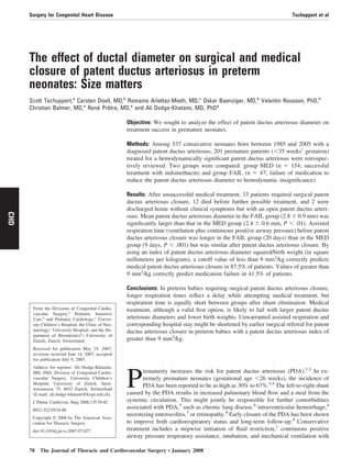
![In our sample 87.5% of patients in the MED group had
a value of less than 9 mm2
/kg, at which medical treatment
appropriately achieved PDA closure. By using this cutoff
value, only 12.5% of the MED group had a value of greater
than 9 mm2
/kg and would have inappropriately been sent
directly to surgical intervention without a prior medical trial
(false-positive result). On the other hand, 41.5% of the
patients in the FAIL group had an index of greater than 9
mm2
/kg and could have benefited from direct surgical re-
ferral, without wasting time for indomethacin failure, if this
index is validated. The cutoff value at 9 mm2
/kg might
represent a useful value for predicting in which patients the
medical treatment will succeed and which patients should
probably undergo surgical intervention rapidly after an ini-
tial medical trial (Figure 1).
As described in Table 1, the preoperative left atrium/aortic
root ratio was significantly smaller in the MED group (1.57 Ϯ
0.26) compared with that in the FAIL group (1.72 Ϯ 0.39, P ϭ
.01). The comorbidities associated with PDA (chronic lung
disease, intraventricular hemorrhage, necrotizing enteroco-
litis, or retinopathy) were comparable in both groups. When
specifically considering the 33 surgically treated infants of
the FAIL group compared with those of the MED group,
there is a significant difference with regard to chronic lung
disease: 16 (48%) of 33 versus 44 (29%) of 154 in the MED
group (P Ͻ .05). Importantly, there was a significant dif-
ference in the amount of infants receiving preoperative
inotropes, with 29% in the MED group compared with 64%
in the FAIL group (P Ͻ .001). This illustrates the number of
patients in an unstable hemodynamic condition who either
make it to surgical intervention or die before surgical clo-
sure is possible (12/47 [25.5%]; Table 1).
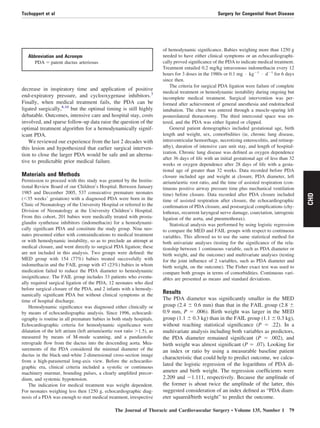
Comparison between the assisted respiration and hospi-
talization times of the 2 groups is depicted in Table 2.
Assisted respiration time before PDA closure was signifi-
cantly (P Ͻ .001) shorter in the MED group (9 Ϯ 7.2 days)
than in the FAIL group (20 Ϯ 14 days). There was no
significant difference in assisted respiration time after PDA
closure between the groups (MED group, 12 Ϯ 13.2 days;
FAIL group, 11 Ϯ 14.9 days). The time spent in the inten-
sive care unit differed significantly between the 2 groups
(MED group: 21 Ϯ 15.1 days vs FAIL group: 34 Ϯ 30.8
days, P ϭ .001). The MED group had a significantly (P Ͻ .01)
shorter hospitalization time (75 Ϯ 33.8 days) than the FAIL
group (100 Ϯ 35.1 days). When counting only the time of
hospitalization after the PDA was closed, there was no signif-
Figure 1. Scatter plot of all patients from both groups depicting
the relationship between PDA diameter squared and weight at
birth. PDA, Persistent ductus arteriosus.
TABLE 1. Characteristics of the 2 groups
Successful medical closure,
MED group (n ؍ 154)
Failed medical closure,
FAIL group (n ؍ 47) P value
PDA diameter 2.4 (0.6); n ϭ 88 2.8 (0.9); n ϭ 41 Ͻ.01
Birth weight (kg) 1.1 (0.3) 1.1 (0.3) NS (.069)
LA/Ao ratio 1.57 (0.26); n ϭ 118 1.72 (0.39); n ϭ 36 .01
Inotropes 30 (64%) 44 (29%) Ͻ.001
Chronic lung disease 44 (29%) 16 (34%) NS
Bradycardia-apnea syndrome 118 (77%) 29 (62%) NS
Intraventricular hemorrhage 43 (28%) 17 (36%) NS
Necrotizing enterocolitis 13 (8%) 9 (19%) NS
Retinopathy 33 (21%) 10 (21%) NS
Means (standard deviation) are shown for continuous variables, and counts (percentage) are shown for binary variables. PDA, Patent ductus arteriosus;
NS, not significant; LA/Ao, left atrium/aortic root.
Surgery for Congenital Heart Disease Tschuppert et al
80 The Journal of Thoracic and Cardiovascular Surgery ● January 2008
CHD](https://image.slidesharecdn.com/bf96ea1c-6b8d-42db-90c2-5a609f8dcbbf-161218180022/85/PDA-size-matters-3-320.jpg)
![icant difference between the 2 groups (MED group: 64 Ϯ 33.1
days vs FAIL group: 69 Ϯ 37.3 days, Table 2).
The overall mortality was 14%. There was a significant
difference in mortality between the 2 groups (8% [13/154]
in the MED group and 34% [16/47] in the FAIL group, P Ͻ
.001). Comparing only the surgically treated infants from
the FAIL group (4/33 [12%]) with the MED group, there
was no significant difference in mortality. The mortality of
surgically treated neonates who underwent surgical inter-
vention without a prior medical trial because of contraindi-
cations or hemodynamic instability was 11% (1/9). Among
all surgical patients, there was 1 postoperative chylothorax
and 1 recurrent nerve paresis.
Discussion
In premature neonates, a PDA is a common and potentially
life-threatening condition.1,10-14
and early closure of a PDA
improves cardiorespiratory status.9
Medical and technologic
advances have improved the survival of preterm infants,
thus extending the limits of viability.12
Common treatment
of a PDA is a stepwise escalation and begins with cycloox-
ygenase inhibitors, although protocols in centers vary
strongly with regard to patient selection and when and how
to treat them.13
The indication for surgical intervention is
usually given when medical treatment fails,15-18
when there
are contraindications to medication, or in the face of hemo-
dynamic instability despite inotropic support.7
In the ab-
sence of these settings, no study has demonstrated clear
indications for earlier “elective” surgical referral, predicting
a favorable outcome. In their series of 197 premature infants
undergoing PDA ligation, Raval and colleagues15
reported
greater gestational age, older age, larger size, and lesser
ventilatory support at the time of surgical intervention to be
associated with better outcomes, although they found no
absolute cutoff values.
Early surgical closure, compared with ligation performed
at a later stage, is associated with improved short-term
ventilatory parameters and pulmonary function, more rapid
achievement of full oral feeding, and improved body
growth.11,14
In our series there was a higher tendency to-
ward comorbidities in the FAIL group compared with the
MED group. More specifically, when considering only the
surgical group, the incidence of preoperative chronic lung
disease was significantly more increased compared with that
in the MED group. It is only speculative to think that earlier
PDA closure with direct surgical referral of neonates having an
index of greater than 9 mm2
/kg would reduce the incidence of
chronic lung disease through reduced assisted respiration times
and intensive care stay. Indeed, other groups have demon-
strated that delaying surgical ligation might increase the like-
lihood of morbidity, mortality, or both.17,18
We found relatively rapid improvements in pulmonary
function after surgical PDA closure, with postclosure as-
sisted respiration times similar to those after medical treat-
ment, despite the longer preoperative and total assisted
respiration times, higher inotrope requirements, and longer
intensive care unit stays with potential iatrogenic complica-
tions in the group of patients who underwent surgical inter-
vention. This would argue in favor of early shunt elimina-
tion, with its beneficial effect on lung function. Contrary to
our findings, Raval and colleagues15
did not observe rapid
improvements in cardiorespiratory status, with a significant
portion of patients going on to have chronic lung disease,
even after shunt elimination. Nonetheless, they do not ad-
vocate delaying surgical closure and find no contraindica-
tions to attempt PDA ligation, regardless of how small,
premature, or unstable the patient’s status preoperatively.
The significant difference in mortality between the 2
groups might be misleading: the FAIL group includes pa-
tients who died before they were able to be transferred to the
surgical department. When taking into account only the
surgically treated neonates, there is no significant difference
in mortality compared with the MED group, with only 4
postoperative deaths, the last in 1991. Therefore mortality
does not seem to be related to the surgical procedure itself
but rather to the underlying disease and complications of
prematurity.
A larger PDA has been a significant predictor of high
failure of indomethacin therapy.16
Our study supports these
results in that prostaglandin synthetase inhibitors failed to
close the PDA with larger diameters, indexed to the respec-
tive birth weight of the patient. Based on our findings, we
propose an index, dividing the squared PDA diameter (in
square millimeters) by the birth weight (in kilograms,
Figure 1). Assuming a cutoff value of greater than 9
mm2
/kg, it is possible to correctly predict 41.5% of
TABLE 2. Respiration and hospitalization times in the 2 groups
Successful medical closure,
MED group (n ؍ 141)
Failed medical closure,
FAIL group (n ؍ 31) P value
Respiration time before PDA closure (d) 9 (7.2) 20 (14) Ͻ.001
Respiration time after PDA closure (d) 12 (13.2) 11 (14.8) NS
Total hospitalization time (d) 75 (33.8) 100 (35.1) Ͻ.01
Hospitalization time after PDA closure (d) 64 (33.1) 69 (37.3) NS
Data are presented as means (standard deviation). PDA, Patent ductus arteriosus; NS, not significant.
Tschuppert et al Surgery for Congenital Heart Disease
The Journal of Thoracic and Cardiovascular Surgery ● Volume 135, Number 1 81
CHD](https://image.slidesharecdn.com/bf96ea1c-6b8d-42db-90c2-5a609f8dcbbf-161218180022/85/PDA-size-matters-4-320.jpg)


![Patent ductus arteriosus [PDA], shane stanley.pptx](https://cdn.slidesharecdn.com/ss_thumbnails/patentductusarteriosuspdashanestanley-251016112835-ae3342d7-thumbnail.jpg?width=640&height=640&fit=bounds)


