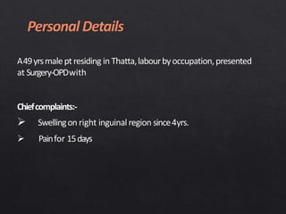
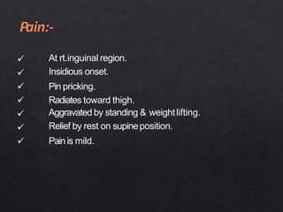
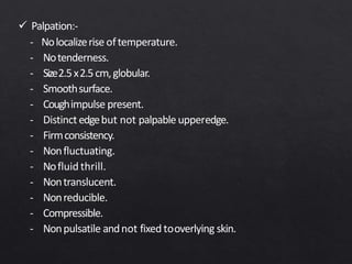
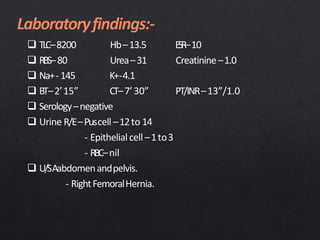
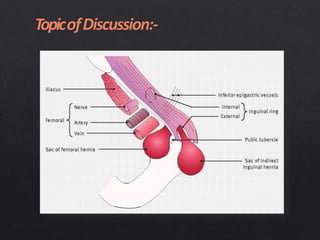
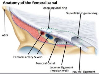
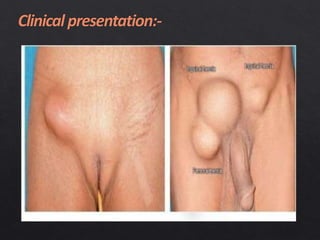
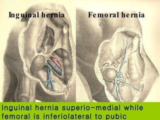
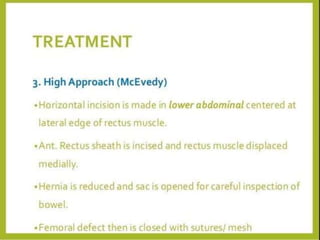
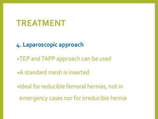
A49yrsmale pt residing in Thatta,labourbyoccupation,presented
at Surgery-OPDwith
Chiefcomplaints:-
 Swellingon right inguinal region since4yrs.
 Painfor 15days
 Swelling on right inguinal region:-
 Since4 yrs.
 Develops spontaneously.
 Associated with pain.
 Grows slowly.
 Not a/w fever, ulceration, loss ofbody weight and
swelling is single.







At rt.inguinal region.
Insidious onset.
Pin pricking.
Radiates toward thigh.
Aggravated by standing & weightlifting.
Relief by rest on supineposition.
Painis mild.
Pasthistory:-
 Nosignificantpastmedical( like:-chroniccough,constipation and
retention of urine )andsurgicalhistory in past.
Personalhistory:-
 Patient isnon-alcoholic,non-smoker,non-vegetariandiet.
Familyhistory:-
 Nosignificantfamilyhistory.
 Eightmember family with five kids.
 Generalcondition –fair
 Jaundice
 Anemia
 Cyanosis
 Clubbing
 Lymphadenopathy
 Edema
 Wellhydrated
Vitalsigns:-
B.P.- 110/70 mm ofHg.




Pulse–76beats/minute –regular.
Temperature–98.F
RR–18breathe / minute
Spo2–98%
Not found
Chest:-normal vesicularbreathe soundpresent.
CVS:- S1+S2+0
CNS:- grosslyintact.
Perabdomen:-
 Inspection:-
 Scaphoidshape.
 Umbilicusinverted andcentrallylocated
 All quadrants movesequally withrespiration.
 Visiblepulsation andperistalsisareabsent.
 Protrusion from right sidedsaphenousopening.
 Onpalpation:-
 Nolocalizeriseof
temperature.
 Notenderness.
 Noorganomegaly
detected.
 Onpercussion:-
 Tympanicsoundfound.
 OnAuscultation:-
 Bowelsoundpresent –3/ minute.
 Onstandingposition:-
 Inspection:- singleswelling
- at upper –medialthigh.
- coughimpulsepresent.
- similar to skincolor.
- globular.
- 2.5x2.5cm
- well definededge.
- no pulsation &peristalsis.
- no movewithrespiration.
- skinover swellingnormal.
 Onsupineposition:-
 Thereisno alterationof
shapeandsizeof the
swelling i.e. similar to
standingposition.
 Palpation:-
- Nolocalizerise oftemperature.
- Notenderness.
- Size2.5x2.5cm,globular.
- Smoothsurface.
- Coughimpulse present.
- Distinct edgebut not palpable upperedge.
- Firmconsistency.
- Nonfluctuating.
- Nofluid thrill.
- Nontranslucent.
- Nonreducible.
- Compressible.
- Nonpulsatile andnot fixed tooverlying skin.
 onpercussion:-
- Dull onpercussion.
 OnAuscultation:-
- Nobruits.
- Nogurglingandbowelsound.
 TLC–8200 Hb–13.5 ESR–10
 RBS–80 Urea–31 Creatinine–1.0
 Na+- 145 K+-4.1
 BT–2’15” CT–7’30” PT/INR–13”/1.0
 Serology–negative
 Urine R/E–Puscell–12to 14
- Epithelialcell–1to3
- RBC–nil
 U/SAabdomenandpelvis.
- RightFemoralHernia.
- Irreducible RightFemoralHernia( Non–obstructed).
Differentialdiagnosis:-
 Rt.inguinalhernia.
 Rt.inguinallymphnode.
 Lipoma.
 Psoasabscess.
 FemoralArteryAneurysm.
 Opensurgery–LOCKWOODapproachunder Spinal
Anesthesia.
 Intra operative findings are:- Rt.femoral hernia withperitoneal
fluids approx.amount 20ml
Boundaryof femoralcanal:-
 Supero-anteriorly:-
Inguinalligament.
 Infero-posteriorly:-
Iliopectinealligament
(cooper)
 Medially:- Lacunarligament
( Gimbernatsligament).
 Laterally:-femoralvein
 Usuallypresentswith painor discomfortin groin.
 Groinlump.
 Usuallynot reducible.
 Mild pain exacerbatedbybendingorlifting.
 Mild tenderness.
In caseof obstruction:-
 Colickyabdominalpain.
 Vomiting,constipation
 Abdominaldistension.
 Lumpirreducible andtender.
 Lumpisverytender.
 Skinover the lump red andhot.
 Featuresof shock.
 HistoryandClinicalexamination.
 Ultrasonography–abdomenandpelvis.
 Inguinalhernia.
 Saphenavarix.
 Lipoma.
 Femoralarteryaneurysm.
 Psoasabscess.
 Ectopictestis.
Femoral Hernia: A case presentation

Femoral Hernia: A case presentation

Editor's Notes