62 year old male presented to Dr. Pashman after a previous fusion at C4/5. The patient had severe degeneration from C2/3-C5/6. Dr. Pashman treated the patient with an Anterior Cervical fusion followed by a Posterior Cervical Fusion.
Case Review #8: 62 year old female with cervical spinal stenosisRobert Pashman
62 year old female with neck pain and left arm weakness. On MRI, the patient was found to have spinal stenosis. Dr. Pashman treated the patient with an Anterior Cervical Discecomy and fusion C4-/7.
Case Review #39: 55 year old male with Progressive ScoliosisRobert Pashman
A 55 year old male presented with Progressive Adult Idiopathic Scoliosis. While he was preparing for surgery, he lifted a heavy item, and had neck pain and pain going down his arm. The patient was found to have myeloradiculopathy and spinal cord effacement and required an Anterior Cervical Fusion prior to scoliosis surgery. The following year he had a posterior spinal fusion for Scoliosis.
Case Review #3: 53 year old male with severe spinal cord compressionRobert Pashman
A 53-year-old male presented with severe cervical kyphosis centered at C4-5, C5-6, and C6-7, causing spinal cord compression and myeloradiculopathy. Pre-op x-rays showed 78 degrees of kyphosis. The surgical strategy involved segmental spinal instrumentation from C4 to C7, radical diskectomies and deformity correction through osteotomies and vertebrectomies at C4-5, C5-6, and C6-7. Post-op x-rays showed successful reconstruction of natural cervical alignment and removal of 78 degrees of kyphosis.
Case Review #6: 57 year old female with severe spinal cord compressionRobert Pashman
A 57 year old female presented to Dr. Pashman with severe spinal cord compression cervical kyphosis, and degenerative disc disease. Dr. Pashman treated the patient with an anterior cervical discectomy and fusion C4-C7.
Case Review #6: 62 year old male presented with C5/6 Disc HerniationRobert Pashman
A 62-year-old male presented with a herniated disc at C5-C6 causing right arm weakness and numbness. He underwent a radical discectomy and bilateral neural foraminotomy to remove the herniated disc and decompress the spinal stenosis. An artificial disc was implanted at C5-C6. Post-operatively, the patient's symptoms resolved and he had regained strength and mobility.
Case Review #4: 51 year old female with adjacent segment degenerationRobert Pashman
A 51 year old female, presented after a previous 3 level cervical spine fusion. The patient developed a cervical disc herniation adjacent to the previous fusion. Dr. Pashman treated her by extending her cervical fusion.
Case Review #9: 40 year old female treated with a Bryan Cervical DiscRobert Pashman
A 40 year old female presented with a C5/6 disc herniation. Dr. Pashman treated the patient with an anterior cervical discectomy and a Bryan Cervical Disc.
Case Review #5: 46 year old female treated with a Prestige DiscRobert Pashman
This document summarizes a case review of a 46-year old female patient who underwent a Prestige Artificial Disc replacement at the C5-C6 level. She presented with neck pain and numbness in both hands due to a C5-C6 disc herniation causing cord compression. Surgery included a discectomy, vertebrectomy, and replacement of the disc with a 7x16mm Prestige disc. Post-operatively, the patient's cervical lordosis was restored and she was doing well.
Case Review #2: 46 year old female with Prestige Disc ReplacementRobert Pashman
A 46-year-old female with C5/6 disc herniation and neck and arm pain underwent a C5-6 artificial disc replacement after conservative therapy failed. Pre-op imaging showed degeneration, osteophytes, and spinal cord compression at C5-6. The surgery involved a discectomy, vertebrectomy to remove osteophytes causing stenosis, and implantation of a Prestige artificial disc. Post-op, the patient's neck and arm pain were resolved.
Case Review #4: Cervical Spine Surgery with Prestige DiscRobert Pashman
A 57 year old male presented after a C6/7 fusion. He had a 2nd level supra adjacent disc herniation. (two levels above the fusion). Dr. Pashman treated him with a Prestige Artificial Disc.
classification and managenement of paediatric craniocervical junction injuriesDieu Merci KABULO
The document discusses injuries to the pediatric spine. It notes that the pediatric spine is more mobile than the adult spine, particularly in the occipitoatlantoaxial region, making it more prone to injury. Common injuries include atlantooccipital dislocation, Jefferson fractures, and atlantoaxial subluxations. Imaging like CT and MRI are important for diagnosis but should be used judiciously due to radiation exposure risks. External immobilization methods like halo rings or Minerva braces are often used initially to treat injuries before considering surgery.
Case Review #3: 44 year old male with a subadjacent disc herniationRobert Pashman
A 44 year old male presented after a C5/6 fusion. On MRI he was found to have a subadjacent disc herniation. Dr. Pashman treated the patient with a Prestige Artificial Disc.
The cervical spine anatomy is specialized to support the cranium while allowing a large range of motion. C1 (atlas) has no vertebral body and unique articular pillars. C2 (axis) has a dens that is embryologically derived from C1's body. The ligaments of the cervical spine, including the tectorial membrane and transverse ligament, allow for wide range of motion while maintaining stability. Common cervical spine injuries include flexion teardrop fractures from hyperflexion, wedge fractures from compression, hangman's fractures from hyperextension, and Jefferson fractures from axial loading. Odontoid fractures also occur from hyperextension or hyperflexion forces on the neck. Radiographic evaluation of
Evolution of surgical strategies instrumentation in scoliosis- dr.shashidhar...Dr. Shashidhar B K
SCOLIOISIS SURGEON BANGALORE
SCOLIOSIS SURGEON INDIA
Website: http://spinesurgeonbangalore.com/
My goal is to provide spine care with a patient centeric-holistic approach in Bangalore, encompassing all aspects of non-operative and operative management of spinal disorders with special interest in the management of spinal deformities (scoliosis and kyphosis).
Bangalore Spine Specilaist Clinic. For Appointment contact : Call: 08025442552( 9 am to 9 pm). Whatsapp: +919448311068. Email: drshashidharbk@gmail.com.
The document discusses spinal surgeries performed at Shonan Fujisawa Tokushukai Hospital in Japan using the Artis zeego robotic imaging system. [1] The hospital has a hybrid operating room dedicated to spine and scoliosis surgery equipped with Artis zeego, an operating table, and navigation system. [2] Surgeries discussed include cervical, thoracic, lumbar spine procedures as well as scoliosis corrections using dorsal and lateral approaches. [3] Intraoperative CT-like imaging from Artis zeego and navigation guidance enable precise screw placement and correction of misplaced screws.
Case Review #46: 56 year old female with Adult Idiopathic Scoliosis and Burst...Robert Pashman
56 year old female with Idiopathic Scoliosis, status post burst fracture, presented with junctional kyphosis. Dr. Pashman treated the patient with a posterior spinal fusion from T2-pelvis. Spinal curvature was a KIM/SRP Classification 3.
This document discusses congenital high scapula, defined as the failure of the scapula to descend from its embryonic position in the neck during intrauterine development. Radiological criteria are provided to diagnose the condition. Differential diagnoses include elevated scapula due to old paralysis or nerve injuries. Treatment involves scapular osteotomy.
Cervical spine injuries are common and can be difficult to diagnose, especially in polytrauma patients. A thorough understanding of cervical spine anatomy is important for accurate diagnosis and treatment planning. Goals of management are prompt recognition and prevention of secondary neurological damage. While plain radiographs are usually the initial imaging method, other imaging such as CT, MRI, and flexion-extension views may be needed to fully evaluate the injury. Dynamic imaging should only be performed once the patient is pain free and able to fully move the cervical spine. SCIWORA describes spinal cord injuries identified by MRI where plain radiographs show no abnormality, and can occur in both children and adults.
Case Review #44: 64 male presented with Denovo Scoliosis and InfectionRobert Pashman
A 64 year old male presented with collapsing denova scoliosis, after several failed back surgeries. The patient was found to have an infection and required salvage surgery.
This document discusses imaging of injuries to the cranio-cervical junction, including dens fractures, hangman's fractures, and atlanto-occipital dissociation. Dens fractures are the most common cervical spine fracture in those over 65 and can be subtle on radiographs but seen as a step off on CT. Hangman's fractures involve the pars interarticularis and result from hyperextension and axial loading. Atlanto-occipital dissociation is an uncommon injury where the skull is displaced from the atlas in one of three directions. It can be assessed using measurements like the basion-dental interval and basion-axial interval on radiographs and CT.
Case Review #9: Adult Idiopathic Scoliosis with a Double CurvatureRobert Pashman
A 54 year old female presented with Adult Idiopathic Scoliosis. In addition to lower back pain, she noticed that her height was decreasing. Her spine was significantly rotated and she required a spinal fusion.
This document provides an overview of Sprengel's deformity, a rare congenital anomaly where the scapula is elevated and rotated medially. Key points:
- It results from interrupted migration of the scapula during embryonic development.
- Clinical features include an elevated shoulder, restricted shoulder movement, and sometimes scoliosis. Severity is graded based on elevation level.
- Diagnosis is made through x-rays showing elevated scapula position. CT scans can show associated bone anomalies.
- Surgical treatment aims to lower the scapula through muscle releases and reattachments, with the goal of improving function and asymmetry. Complications can include brachial plexus injury or sc
This document discusses adjacent level disease in the cervical spine. It provides background on a 62-year-old woman who presented with neck and occipital pain and had a history of anterior cervical discectomy and fusion surgeries at C5-C6 and C6-C7 levels in 1990 and 2007. It then reviews literature on adjacent segment degeneration rates of about 3% per year and 25% within 10 years, with risk factors including pre-existing degeneration and fusions at lower cervical levels.
This document presents a case report of a 21-year-old male diagnosed with Type II Klippel-Feil syndrome and chronic kidney disease. He presented with shortness of breath, fatigue, and reduced urine output. Imaging revealed cervical vertebral fusion, left Sprengel's deformity, right renal agenesis, and a solitary pelvic left kidney. He was found to have anemia and end-stage renal disease, and was advised to start hemodialysis.
Sprengle shoulder (congenital elevation of scapula)Gaurav Singh
Sprengel deformity is a congenital condition where the scapula is elevated higher than normal due to failed descent during development. It can range from mild where motion is slightly limited, to severe where the scapula touches the head. Surgery is aimed at lowering the scapula, and involves releasing muscles and reattaching them in a lower position. Complications include brachial plexus injury, so preventative clavicle resection may be used. Sprengel deformity is classified based on the degree of elevation and angle of the scapula.
Case Review #2: 40 year old female with a subadjacent cervical disc herniationRobert Pashman
A 40 year old female presented with a cervical disc herniation subjacent to previous cervical fusion. Dr. Pashman performed a revision surgery. The surgical procedure was an anterior cervical discectomy and fusion.
Case Review #50: 29 year old woman presents with dislodged instrumentation fo...Robert Pashman
A 29-year old woman presented with dislodged hardware from three previous surgeries for adolescent idiopathic scoliosis. She had retained loose instrumentation from her past operations that posed a medical risk. The surgical strategy was to remove the retained hardware through osteotomies, perform spinal fusions from T4 to L3, and place new segmental instrumentation from L2 to L3.
Case Review #7: 51 year old female with severe flatback after multiple surge...Robert Pashman
A 51 year old female status post multiple spine surgeries presented to Dr. Pashman with severe Flatback Syndrome and psueoarthrosis. Dr. Pashman treated the patient with a Posterior Spinal Fusion T4-Pelvis.
The document discusses cervical spine injuries and cervical spondylotic myelopathy. It presents early experience with anterior decompression, fusion and plating for cervical spine injuries in Abuja, Nigeria. It describes surgical techniques used including anterior cervical discectomy and fusion. It highlights the importance of early referral for surgery and discusses challenges including lack of intensive care and rehabilitation facilities.
Case Review #5: 67 year old woman with flatback syndrome following 5 spinal s...Robert Pashman
A 67 year old female presented to Dr. Pashman with severe Flat back Syndrome after 5 previous spine surgeries for Adult Idiopathic Scoliosis. Dr. Pashman treated her with a Posterior Spinal Fusion from T8 to S1.
This document provides an overview of radiographic anatomy of the spine and discusses spine trauma. It begins with defining normal anatomical features visible on spine radiographs, including vertebral bodies, discs, facet joints, and transverse/spinous processes. Common patterns of cervical spine injuries are then described, such as flexion injuries causing compression fractures and facet dislocations. The roles of imaging in spine trauma are outlined as diagnosing fractures, assessing stability, and evaluating neurological involvement. Specific cervical spine views and radiographic signs of trauma are also detailed.
techniques, methods, indications and complications of various fusion techniques for subaxial cervical spine. comparison of anterior versus posterior techniques, their indications and complication.
Case Review #2: 66 year old female with severe Flatback SyndromeRobert Pashman
A 66 year old female presented with severe Flat back Syndrome, Kyphosis, and critical stenosis. Dr. Pashman treated the patient with a posterior spinal fusion T2 to the pelvis. The patient was able to stand up straight following surgery.
A 71-year-old woman was in a car accident and presented with an incomplete spinal cord injury at C6-7. Imaging showed a distractive injury at C6-7. She underwent surgical treatment with lateral mass screws at C5-C6 and pedicle screws at C7-T1, which restored alignment. After 6 months, she had some neurologic improvement. Subaxial cervical fractures are common from C3-C7 and can occur from compression, burst, flexion, or extension mechanisms. Classification systems include the Subaxial Injury Classification Score and Cervical Spine Injury Severity Score to determine treatment. Nonoperative and operative treatment options were discussed.
This document discusses the classification, causes, symptoms, and treatment of kyphosis, which is an excessive curvature of the spine. It is classified into 15 major groups including postural disorders, Scheuermann's kyphosis, congenital disorders, paralytic disorders, and others. Treatment involves exercise, bracing, medication management, and surgery to correct the deformity and relieve pain or neurological symptoms. Surgical techniques range from posterior fusion to osteotomies to combined anterior-posterior procedures depending on the severity and rigidity of the deformity. The goals of surgery are to restore spinal alignment and remove any neural compression.
Case Review #3: 65 year old woman with 55 degree Thoracolumbar ScoliosisRobert Pashman
A 65 year old female with a 55° thoracolumbar curve, spondylolisthesis, and flatback syndrome. Treated with an Anterior/Posterior Spinal Fusion. KIM/SRP Classification 3.
Case Review 4: A 58 year old female presented with Scheurmann's KyphosisRobert Pashman
A 58-year-old female with severe rigid thoracolumbar kyphosis due to Scheuermann's disease and degeneration underwent a posterior spinal fusion from T2 to the pelvis. She had a 70 degree curvature that was rigid. The surgery involved segmental spinal instrumentation, multiple osteotomies to correct the curvature, decompression of neural elements, and an 18-level posterior fusion. Post-operatively, the patient's curvature was fully corrected and she gained 2 inches in height.
Case Review #3: 17 year old male with Scheurmann's KyphosisRobert Pashman
A 17 year old male presented with progressive Schuermann's Kyphosis. The patient had a 75° spinal curvature. Dr. Pashman treated the patient with a posterior fusion from T3 to L1.
Birmingham mid-head resection arthroplasty of hip for avascular necrosis of f...Apollo Hospitals
To study the outcome of Birmingham mid-head resection (BMHR) arthroplasty of the hip in young and active patients with avascular necrosis of femoral head with gross defects.
Case Review #7: 51 year old female with Prestige Cervical Disc ReplacementRobert Pashman
51 year old female with a C5/6 disc herniation. traveled to Los Angeles from Vietnam for treatment. Dr. Pashman performed an anterior cervical discectomy and placed a Prestige Artificial Disc.
Case Review #7: 42 year old male with KyphoscoliosisRobert Pashman
A 42-year-old male with congenital kyphoscoliosis and neck pain underwent a 17-level posterior spinal fusion from T2 to the pelvis. The surgery included vertebrectomies at T9, T11 and T10, with a vertebral column resection at T10 to correct his 90 degree kyphosis. Post-operatively, his spine was realigned to 35 degrees with resolution of his neurological symptoms and pain managed conservatively.
Case Review #29: 57 year old female with Adult Thorcolumber ScoliosisRobert Pashman
57 year old female with Adult Idiopathic Scoliosis. She presented with a 62 degree thoracolumbar curve. Dr. Pashman treated the patient with a posterior spinal fusion from T10-Pelvis. Dr. Pashman took great care with incision closure to preserve the patient's tattoo. KIM/SRP Classification 2.
Old unreduced dislocations of the hip and shoulder can be caused by trauma, neglect, or comorbid injuries that distract from the dislocation. Treatment depends on factors like time since injury, bone integrity, and viability of the femoral head. Options include closed or open reduction, osteotomy, arthroplasty, or no treatment. Open reduction may be necessary to address bone defects, fibrosis, and instability.
Similar to Case Review #5: 62 year old male with degenerative disc disease C2-C6 (20)
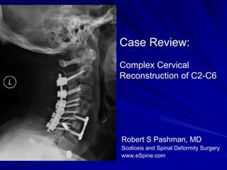
Case Review #22: 21 year old with Progressive Adolescent Idiopathic ScoliosisRobert Pashman
A 21 year old female presented to Dr. Pashman with Progressive Adolescent Idiopathic Scoliosis. The patient had been followed for scoliosis, and was compliant wearing her brace. Her spinal curvature progressed despite physical therapy and bracing. Dr. Pashman treated her with a Posterior Spinal Fusion T3-L1.
Case Review #11: Progressive Adolescent Idiopathic ScoliosisRobert Pashman
A 17 year old female with Progressive Adolescent Idiopathic Scoliosis presented to Dr. Pashman for a surgical opinion. The patient presented with a 36° thoracic curve which progressed to 48°. Dr. Pashman treated the patient with a posterior fusion T3-L1.
Case Review #42: 39 year old female with Adult Congenital ScoliosisRobert Pashman
A 39 year old female with Congenital Scoliosis presented to Dr. Pashman. The patient had a fusion at age six, and her spinal curve continued to progress. Dr. Pashman treated the patient with a posterior spinal fusion from T3-Pelvis. KIM/SRP Classification 3.
Case Review #6: 45 year old woman with flatback syndrome after several surger...Robert Pashman
A 45-year old female with a flat back due to multiple spine surgeries for scoliosis underwent surgery to correct her sagittal imbalance. The surgical strategy included a T10 to sacrum fusion with pedicle subtraction osteotomy at L3 to induce lordosis. Post-operatively, sagittal balance was restored but a slight coronal imbalance remained due to prior fusion in a shifted position. Good functional outcomes are expected with less than 4cm of coronal imbalance.
Case Review #34: 44 Year Old Woman with Adult Idiopathic ScoliosisRobert Pashman
A 44 year old woman presented with Adult Idiopathic Scoliosis. She wore a brace as a child, and the curvature progressed to the point she required surgery.
Case Review #35: 43 year old female with Adult Scoliosis and a Transitional V...Robert Pashman
1. This case review summarizes the treatment of a 43-year-old female patient with adult scoliosis and a lumbar sacral transitional vertebra.
2. She had a 40 degree lumbar curve and a fused L5-S1 region on the right side.
3. Her treatment plan involved an anterior interbody fusion at L4-5 and L5-S1 followed by posterior segmental spinal instrumentation, spinal osteotomies, and posterior spinal fusion to correct her deformities and relieve her pain.
Case Review #B: Spondylolisthesis Surgery Robert Pashman
A 16-year-old female presented with lower back and left leg pain following a motor vehicle accident. Imaging showed spondylolisthesis at L5 from fractures of the pars interarticularis. She underwent microscopic bilateral L5 laminotomy, foraminotomy, and repair of the L5 pars fractures with bone grafting and instrumentation. Post-operatively, her symptoms resolved and she was able to return to normal activities.
Case Review #2: Isthmic Spondylolisthesis Grade IVRobert Pashman
Case review: A 58 year old female with Isthmic Spondylolisthesis status post surgery. She continued to experience back pain and leg pain, and was treated with spinal surgery.
Case Review #A: Major League Baseball Player has a Spinal FusionRobert Pashman
25 year old Major League Baseball Player presented with low back pain. Upon review of CT scans, he was discovered to have a pars fracture and require surgery,
Case Review #12: 14 Year Old Female with Adolescent Idiopathic ScoliosisRobert Pashman
A 14 year old female presented with Adolescent Idiopathic Scoliosis. The patient was non-compliant with bracing. The Scoliosis curvature and Kyphosis curvature progressed, and she required surgery.
Case Review #27: 59 Year Old Female with Progressive Adult ScoliosisRobert Pashman
59 year old female presented with Progressive Adult Idiopathic Scoliosis, Spondylolisthesis, Flatback Deformity, and Stenosis. The patient was treated with a spinal fusion,
Case Review #31: 60 Year Old Female with Adult Idiopathic ScoliosisRobert Pashman
60 year old female presented with Adult Idiopathic Scoliosis and Grade 2 Isthmic Spondylolisthesis. She was treated with an anterior and posterior spinal fusion.
Case Review #7: Progressive Adult Idiopathic Scoliosis with a 75 degree curva...Robert Pashman
The patient, a 19-year-old female, presented with a progressive 75° thoracic scoliosis that had increased 21° over four years and was causing her significant pain. Pre-operative images showed the curvature was rigid. The surgical plan was to perform segmental spinal instrumentation from T3 to L1, posterior spinal fusion from T3 to L2, and spinal osteotomies from T4 to T9 to correct the rigid curvature. Post-operatively, a 43° correction was achieved, reducing the curvature to 27°, and the patient was able to resume her normal activities with no restrictions after one year.
Case Review #C: Adolescent Idiopathic ScoliosisRobert Pashman
A 14 year old female presented with Adolescent Idiopathic Scoliosis with a Thoracolumbar Curve. She was treated with an anterior throaco-abdominal approach.
Case Review #19: 40 year old Male with Adult Idiopathic Scoliosis with Flatba...Robert Pashman
A 40 year old male presented after scoliosis surgery at age 14. He presented with Flatback Syndrome and increasing low back pain and required revision surgery.
Case Review #13: 13 year old female softball player with Adolescent Idiopathi...Robert Pashman
A 13 year old female softball player presented with Adolescent Idiopathic Scoliosis. The degree of her scoliosis curve progressed to 48 degrees and she required a spinal fusion.
Case Review #4: Adolescent Idiopathic Scoliosis with 61 degree curvatureRobert Pashman
A 22 year old female presented with Adolescent Idiopathic Scoliosis. She was braced as a child, and the curve continued to progress until she required surgical intervention.
Basavarajeeyam is a Sreshta Sangraha grantha (Compiled book ), written by Neelkanta kotturu Basavaraja Virachita. It contains 25 Prakaranas, First 24 Chapters related to Rogas& 25th to Rasadravyas.
Adhd Medication Shortage Uk - trinexpharmacy.comreignlana06
The UK is currently facing a Adhd Medication Shortage Uk, which has left many patients and their families grappling with uncertainty and frustration. ADHD, or Attention Deficit Hyperactivity Disorder, is a chronic condition that requires consistent medication to manage effectively. This shortage has highlighted the critical role these medications play in the daily lives of those affected by ADHD. Contact : +1 (747) 209 – 3649 E-mail : sales@trinexpharmacy.com
Muktapishti is a traditional Ayurvedic preparation made from Shoditha Mukta (Purified Pearl), is believed to help regulate thyroid function and reduce symptoms of hyperthyroidism due to its cooling and balancing properties. Clinical evidence on its efficacy remains limited, necessitating further research to validate its therapeutic benefits.
Title: Sense of Smell
Presenter: Dr. Faiza, Assistant Professor of Physiology
Qualifications:
MBBS (Best Graduate, AIMC Lahore)
FCPS Physiology
ICMT, CHPE, DHPE (STMU)
MPH (GC University, Faisalabad)
MBA (Virtual University of Pakistan)
Learning Objectives:
Describe the primary categories of smells and the concept of odor blindness.
Explain the structure and location of the olfactory membrane and mucosa, including the types and roles of cells involved in olfaction.
Describe the pathway and mechanisms of olfactory signal transmission from the olfactory receptors to the brain.
Illustrate the biochemical cascade triggered by odorant binding to olfactory receptors, including the role of G-proteins and second messengers in generating an action potential.
Identify different types of olfactory disorders such as anosmia, hyposmia, hyperosmia, and dysosmia, including their potential causes.
Key Topics:
Olfactory Genes:
3% of the human genome accounts for olfactory genes.
400 genes for odorant receptors.
Olfactory Membrane:
Located in the superior part of the nasal cavity.
Medially: Folds downward along the superior septum.
Laterally: Folds over the superior turbinate and upper surface of the middle turbinate.
Total surface area: 5-10 square centimeters.
Olfactory Mucosa:
Olfactory Cells: Bipolar nerve cells derived from the CNS (100 million), with 4-25 olfactory cilia per cell.
Sustentacular Cells: Produce mucus and maintain ionic and molecular environment.
Basal Cells: Replace worn-out olfactory cells with an average lifespan of 1-2 months.
Bowman’s Gland: Secretes mucus.
Stimulation of Olfactory Cells:
Odorant dissolves in mucus and attaches to receptors on olfactory cilia.
Involves a cascade effect through G-proteins and second messengers, leading to depolarization and action potential generation in the olfactory nerve.
Quality of a Good Odorant:
Small (3-20 Carbon atoms), volatile, water-soluble, and lipid-soluble.
Facilitated by odorant-binding proteins in mucus.
Membrane Potential and Action Potential:
Resting membrane potential: -55mV.
Action potential frequency in the olfactory nerve increases with odorant strength.
Adaptation Towards the Sense of Smell:
Rapid adaptation within the first second, with further slow adaptation.
Psychological adaptation greater than receptor adaptation, involving feedback inhibition from the central nervous system.
Primary Sensations of Smell:
Camphoraceous, Musky, Floral, Pepperminty, Ethereal, Pungent, Putrid.
Odor Detection Threshold:
Examples: Hydrogen sulfide (0.0005 ppm), Methyl-mercaptan (0.002 ppm).
Some toxic substances are odorless at lethal concentrations.
Characteristics of Smell:
Odor blindness for single substances due to lack of appropriate receptor protein.
Behavioral and emotional influences of smell.
Transmission of Olfactory Signals:
From olfactory cells to glomeruli in the olfactory bulb, involving lateral inhibition.
Primitive, less old, and new olfactory systems with different path
ABDOMINAL TRAUMA in pediatrics part one.drhasanrajab
Abdominal trauma in pediatrics refers to injuries or damage to the abdominal organs in children. It can occur due to various causes such as falls, motor vehicle accidents, sports-related injuries, and physical abuse. Children are more vulnerable to abdominal trauma due to their unique anatomical and physiological characteristics. Signs and symptoms include abdominal pain, tenderness, distension, vomiting, and signs of shock. Diagnosis involves physical examination, imaging studies, and laboratory tests. Management depends on the severity and may involve conservative treatment or surgical intervention. Prevention is crucial in reducing the incidence of abdominal trauma in children.
- Video recording of this lecture in English language: https://youtu.be/kqbnxVAZs-0
- Video recording of this lecture in Arabic language: https://youtu.be/SINlygW1Mpc
- Link to download the book free: https://nephrotube.blogspot.com/p/nephrotube-nephrology-books.html
- Link to NephroTube website: www.NephroTube.com
- Link to NephroTube social media accounts: https://nephrotube.blogspot.com/p/join-nephrotube-on-social-media.html
These lecture slides, by Dr Sidra Arshad, offer a quick overview of the physiological basis of a normal electrocardiogram.
Learning objectives:
1. Define an electrocardiogram (ECG) and electrocardiography
2. Describe how dipoles generated by the heart produce the waveforms of the ECG
3. Describe the components of a normal electrocardiogram of a typical bipolar lead (limb II)
4. Differentiate between intervals and segments
5. Enlist some common indications for obtaining an ECG
6. Describe the flow of current around the heart during the cardiac cycle
7. Discuss the placement and polarity of the leads of electrocardiograph
8. Describe the normal electrocardiograms recorded from the limb leads and explain the physiological basis of the different records that are obtained
9. Define mean electrical vector (axis) of the heart and give the normal range
10. Define the mean QRS vector
11. Describe the axes of leads (hexagonal reference system)
12. Comprehend the vectorial analysis of the normal ECG
13. Determine the mean electrical axis of the ventricular QRS and appreciate the mean axis deviation
14. Explain the concepts of current of injury, J point, and their significance
Study Resources:
1. Chapter 11, Guyton and Hall Textbook of Medical Physiology, 14th edition
2. Chapter 9, Human Physiology - From Cells to Systems, Lauralee Sherwood, 9th edition
3. Chapter 29, Ganong’s Review of Medical Physiology, 26th edition
4. Electrocardiogram, StatPearls - https://www.ncbi.nlm.nih.gov/books/NBK549803/
5. ECG in Medical Practice by ABM Abdullah, 4th edition
6. Chapter 3, Cardiology Explained, https://www.ncbi.nlm.nih.gov/books/NBK2214/
7. ECG Basics, http://www.nataliescasebook.com/tag/e-c-g-basics
TEST BANK For Community Health Nursing A Canadian Perspective, 5th Edition by...Donc Test
TEST BANK For Community Health Nursing A Canadian Perspective, 5th Edition by Stamler, Verified Chapters 1 - 33, Complete Newest Version Community Health Nursing A Canadian Perspective, 5th Edition by Stamler, Verified Chapters 1 - 33, Complete Newest Version Community Health Nursing A Canadian Perspective, 5th Edition by Stamler Community Health Nursing A Canadian Perspective, 5th Edition TEST BANK by Stamler Test Bank For Community Health Nursing A Canadian Perspective, 5th Edition Pdf Chapters Download Test Bank For Community Health Nursing A Canadian Perspective, 5th Edition Pdf Download Stuvia Test Bank For Community Health Nursing A Canadian Perspective, 5th Edition Study Guide Test Bank For Community Health Nursing A Canadian Perspective, 5th Edition Ebook Download Stuvia Test Bank For Community Health Nursing A Canadian Perspective, 5th Edition Questions and Answers Quizlet Test Bank For Community Health Nursing A Canadian Perspective, 5th Edition Studocu Test Bank For Community Health Nursing A Canadian Perspective, 5th Edition Quizlet Test Bank For Community Health Nursing A Canadian Perspective, 5th Edition Stuvia Community Health Nursing A Canadian Perspective, 5th Edition Pdf Chapters Download Community Health Nursing A Canadian Perspective, 5th Edition Pdf Download Course Hero Community Health Nursing A Canadian Perspective, 5th Edition Answers Quizlet Community Health Nursing A Canadian Perspective, 5th Edition Ebook Download Course hero Community Health Nursing A Canadian Perspective, 5th Edition Questions and Answers Community Health Nursing A Canadian Perspective, 5th Edition Studocu Community Health Nursing A Canadian Perspective, 5th Edition Quizlet Community Health Nursing A Canadian Perspective, 5th Edition Stuvia Community Health Nursing A Canadian Perspective, 5th Edition Test Bank Pdf Chapters Download Community Health Nursing A Canadian Perspective, 5th Edition Test Bank Pdf Download Stuvia Community Health Nursing A Canadian Perspective, 5th Edition Test Bank Study Guide Questions and Answers Community Health Nursing A Canadian Perspective, 5th Edition Test Bank Ebook Download Stuvia Community Health Nursing A Canadian Perspective, 5th Edition Test Bank Questions Quizlet Community Health Nursing A Canadian Perspective, 5th Edition Test Bank Studocu Community Health Nursing A Canadian Perspective, 5th Edition Test Bank Quizlet Community Health Nursing A Canadian Perspective, 5th Edition Test Bank Stuvia
Title: Sense of Taste
Presenter: Dr. Faiza, Assistant Professor of Physiology
Qualifications:
MBBS (Best Graduate, AIMC Lahore)
FCPS Physiology
ICMT, CHPE, DHPE (STMU)
MPH (GC University, Faisalabad)
MBA (Virtual University of Pakistan)
Learning Objectives:
Describe the structure and function of taste buds.
Describe the relationship between the taste threshold and taste index of common substances.
Explain the chemical basis and signal transduction of taste perception for each type of primary taste sensation.
Recognize different abnormalities of taste perception and their causes.
Key Topics:
Significance of Taste Sensation:
Differentiation between pleasant and harmful food
Influence on behavior
Selection of food based on metabolic needs
Receptors of Taste:
Taste buds on the tongue
Influence of sense of smell, texture of food, and pain stimulation (e.g., by pepper)
Primary and Secondary Taste Sensations:
Primary taste sensations: Sweet, Sour, Salty, Bitter, Umami
Chemical basis and signal transduction mechanisms for each taste
Taste Threshold and Index:
Taste threshold values for Sweet (sucrose), Salty (NaCl), Sour (HCl), and Bitter (Quinine)
Taste index relationship: Inversely proportional to taste threshold
Taste Blindness:
Inability to taste certain substances, particularly thiourea compounds
Example: Phenylthiocarbamide
Structure and Function of Taste Buds:
Composition: Epithelial cells, Sustentacular/Supporting cells, Taste cells, Basal cells
Features: Taste pores, Taste hairs/microvilli, and Taste nerve fibers
Location of Taste Buds:
Found in papillae of the tongue (Fungiform, Circumvallate, Foliate)
Also present on the palate, tonsillar pillars, epiglottis, and proximal esophagus
Mechanism of Taste Stimulation:
Interaction of taste substances with receptors on microvilli
Signal transduction pathways for Umami, Sweet, Bitter, Sour, and Salty tastes
Taste Sensitivity and Adaptation:
Decrease in sensitivity with age
Rapid adaptation of taste sensation
Role of Saliva in Taste:
Dissolution of tastants to reach receptors
Washing away the stimulus
Taste Preferences and Aversions:
Mechanisms behind taste preference and aversion
Influence of receptors and neural pathways
Impact of Sensory Nerve Damage:
Degeneration of taste buds if the sensory nerve fiber is cut
Abnormalities of Taste Detection:
Conditions: Ageusia, Hypogeusia, Dysgeusia (parageusia)
Causes: Nerve damage, neurological disorders, infections, poor oral hygiene, adverse drug effects, deficiencies, aging, tobacco use, altered neurotransmitter levels
Neurotransmitters and Taste Threshold:
Effects of serotonin (5-HT) and norepinephrine (NE) on taste sensitivity
Supertasters:
25% of the population with heightened sensitivity to taste, especially bitterness
Increased number of fungiform papillae
2. Patient History
62 year old male
Status post anterior cervical diskectomy, C4-5
Now with sub-adjacent disc herniation with spinal cord
compression, cervical stenosis and neural foraminal
stenosis.
The patient also has massive posterior cervical
degeneration at C2-3, C3-4, C4-5, and C5-6 causing
increasing axial neck pain, arm pain.
The patient has been taking an escalating amount of
narcotics for 7 years.
The patient has impending neurologic deficit with posterior
cervical pain, shoulder pain indicative of mild myelopathy.
4. Indications for Surgery
Status post anterior cervical diskectomy and fusion,
C4-5.
Now with sub-adjacent disc herniation and spinal cord
compression, C5-6.
The patient on CT scan had massive arthrosis at C2-3,
specifically on the left-hand side. This is superjacent to a highly
mobile segment. The significant subaxial degeneration had
caused the patient to have dysfunctional pain.
Severe posterior subaxial arthritis.
Failure to thrive with increase narcotic usage.
Motor-sensory deficit and neurologic sequela.
Partial cervical kyphosis.
5. Surgical Strategy
The strategy would be removal of Anterior interbody fusion, C5-6, 8-mm,
plate anteriorly, sub-adjacent anterior with autogenous bone graft.
cervical diskectomy and fusion. The Anterior cervical plate fixation, C5-6
posterior spinal fusion from C2 to C6 with a 4-hole Atlantis Vision plate.
would cause necessarily significant Removal of retained hardware, Zephyr
sub-adjacent degeneration, spinal plate, C4-5.
canal compression, especially with A Mayfield pin placement and
the cervical disc herniation. positioning.
Radical diskectomy, C5-6 under the Posterior cervical fusion, C2 to C6,
microscope with spinal cord using posterior cervical screw-rod
construct.
decompression.
Posterior spinal fusion, C2 to C6, using
Subtotal vertebrectomy, C5 with locally harvested autogenous bone
removal of posterior uncovertebral and putty
osteophyte, anterior osteophyte Intraoperative SSEPs.
constituting 1/3 of the vertebra and
Intraoperative fluoroscopy.
spinal canal decompression with
bilateral neural foraminal
decompression.