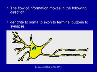
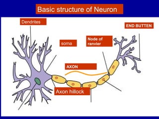
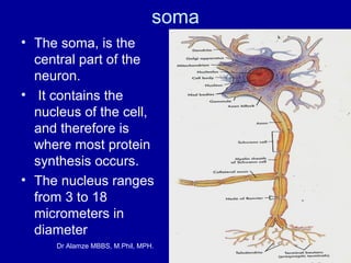
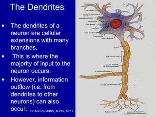
Neurons: The Basic Functional Unit

Dr Alamze MBBS, M.Phil, MPH.
Neuron

• A neuron is the functioning unit of the
nervous system;
• are electrically excitable cells in the nervous
system that function to process and transmit
information.
• In vertebrate animals, neurons are the core
components of the brain, spinal cord and
peripheral nerves.
• Although there are many types of neurons
among the estimated ten billion in the human
brain, each typically shares the same
Dr Alamze MBBS, M.Phil, MPH.
characteristics
• The flow of information moves in the following
direction:
• dendrite to soma to axon to terminal buttons to
synapse.

Dr Alamze MBBS, M.Phil, MPH.
Basic structure of Neuron
Dendrites

END BUTTEN

soma

Node of
ranvier

AXON

Axon hillock

Dr Alamze MBBS, M.Phil, MPH.
soma
• The soma, is the
central part of the
neuron.
• It contains the
nucleus of the cell,
and therefore is
where most protein
synthesis occurs.
• The nucleus ranges
from 3 to 18
micrometers in
diameter
Dr Alamze MBBS, M.Phil, MPH.
The Dendrites
•

•

•

The dendrites of a
neuron are cellular
extensions with many
branches,
This is where the
majority of input to the
neuron occurs.
However, information
outflow (i.e. from
dendrites to other
neurons) can also
occur. Dr Alamze MBBS, M.Phil, MPH.
axon
• The axon is a finer, cablelike projection which can
extend tens, hundreds, or
even tens of thousands of
times the diameter of the
soma in length.
• The axon carries nerve
signals away from the
soma (and also carry
some types of information
back to it).
• Many neurons have only
one axon, but this axon
may - and usually will undergo extensive
branching, enabling
communication with
many target cells.
Dr Alamze MBBS, M.Phil, MPH.
Axon Hillock.
• The part of the axon where it
emerges from the soma is called the
'axon hillock'.
• Besides being an anatomical
structure, the axon hillock is also the
part of the neuron that has the
greatest density of voltagedependent sodium channels.
• This makes it the most easily-excited
part of the neuron and the spike
initiation zone for the axon
• in neurological terms it has the
greatest hyperpolarized action
Dr Alamze MBBS, M.Phil, MPH.

•'axon hillok'.
Classification
• Structural classification
• Classification on basis of function;
Classification by action on other neurons
Classification by neurotransmitter released

Dr Alamze MBBS, M.Phil, MPH.
Structural classification
• Unipolar or
Pseudounipolar :
dendrite and axon
emerging from same
process.

Dr Alamze MBBS, M.Phil, MPH.
Bipolar:
• single axon
and single
dendrite on
opposite ends
of the soma.

Dr Alamze MBBS, M.Phil, MPH.
Structural classification
• Multipolar: more than
two dendrites

Dr Alamze MBBS, M.Phil, MPH.
• Axons and dendrites in the
central nervous system are
typically only about a micrometer
thick,
• while some in the peripheral
nervous system are much thicker.
• The soma is usually about 10–25
micrometers in diameter and
often is not much larger than the
cell nucleus it contains.
• The longest axon of a human
motoneuron can be over a meter
long, reaching from the base of
the spine to the toes.
• Sensory neurons have axons that
run from the toes to the dorsal
columns, over 1.5 meters in
adults.
•
Dr Alamze MBBS, M.Phil, MPH.
Classification on function;
• receptor or sensory neurons,
• motor neurons
• and interneurons.

Dr Alamze MBBS, M.Phil, MPH.
Sensory or afferent NEURONS
• (carrying toward the
brain)
• These neurons are
specialized & sensitive to
a particular physical
stimulation
• such as light (vision),
• sound (audition),
• chemical (olfaction),
• or pressure (touch).
Dr Alamze MBBS, M.Phil, MPH.
Motor or efferent neurons
• Motor or efferent
(carrying away from the
brain) neurons receive
impulses from other
neurons and transmit this
information to muscles or
glands.
• These neurons are of two
types, alpha motor
neurons and gamma
motor neurons.
Dr Alamze MBBS, M.Phil, MPH.
Alpha Motor Neurons.
•

•

•

The alpha motor neurons give
rise to large type A alpha (Aα)
motor nerve fibers, averaging
14 micrometers in diameter;
these fibers branch many
times after they enter the
muscle and innervate the large
skeletal muscle fibers.
Stimulation of a single alpha
nerve fiber excites from three
to several hundred skeletal
muscle fibers, which are
collectively called the motor
unit.

Dr Alamze MBBS, M.Phil, MPH.
Gamma Motor Neurons.
• gamma motor neurons
are located in the spinal
cord anterior horns.
These gamma motor
neurons transmit
impulses through much
smaller type A gamma
(Aγ) motor nerve fibers,
averaging 5 micrometers
in diameter, which go to
small, special skeletal
muscle fibers called
intrafusal fibers,
Dr Alamze MBBS, M.Phil, MPH.
Interneurons or intrinsic neurons
• Interneurons or
intrinsic neurons form
the largest group in
the nervous system.
• They form
connections between
themselves and
sensory neurons
before transmission of
control to motor
neurons
Dr Alamze MBBS, M.Phil, MPH.
Classification by action on other neurons
• Excitatory neurons excite their target
neurons. Excitatory neurons in the brain are
often glutamatergic.
• Inhibitory neurons inhibit their target neurons.
Inhibitory neurons are often interneurons.
These neurons use such neurotransmitters as
dopamine, acetylcholine, serotonin and others.

Dr Alamze MBBS, M.Phil, MPH.
Classification by discharge
patterns
•
Neurons can be classified according to
their electrophysiological characteristics

Dr Alamze MBBS, M.Phil, MPH.
Tonic or regular spiking
Some neurons are typically constantly (or
tonically) active.
Example: interneurons in neurostriatum.

Dr Alamze MBBS, M.Phil, MPH.
Phasic or bursting
• Neurons that fire in bursts are called
phasic.
• Fast spiking. Some neurons are notable
for their fast firing rates, for example some
types of cortical inhibitory interneurons,
cells in globus pallidus.

Dr Alamze MBBS, M.Phil, MPH.
Thin-spike.
• Action potentials of some neurons are
more narrow compared to the others. For
example, interneurons in prefrontal cortex
are thin-spike neurons.

Dr Alamze MBBS, M.Phil, MPH.
Classification by
neurotransmitter released
•
•
•
•

Some examples are cholinergic,
GABAergic,
Glutamatergic
and dopaminergic neurons.

Dr Alamze MBBS, M.Phil, MPH.
Glial cells

• In addition to neurons, the nervous system is
populated with another category of cells, glial
cells.
• Glial cells are approximately 10 times more
plentiful than neurons.
• But since they are approximately one-tenth the
size, they take up equal space.
• Glia is a Greek term meaning glue.
• glial cells served as the putty that held the
neurons together
Dr Alamze MBBS, M.Phil, MPH.
Myelin sheaths
• Oligodendrocytes and Schwann cells form
the myelin sheaths that insulate axons in
the central and peripheral nervous
systems, respectively.

Dr Alamze MBBS, M.Phil, MPH.
synapses

• The human brain has a huge number of
synapses.
• Each of the 1012 neurons (1,000 billion, i.e.
1 trillion) has on average 7,000 synaptic
connections to other neurons.
• It has been estimated that the brain of a
three-year-old child has about 10 16
synapses (10,000 trillion).
• This number declines with age, stabilizing
by adulthood.
• Estimates vary for an adult, ranging from
1015 to 5 x 10¹⁵ synapses (1,000 to 5,000
Dr Alamze MBBS, M.Phil, MPH.
trillion
Dr Alamze MBBS, M.Phil, MPH.
synaptic transmission
•
•
•

Neurons communicate via chemical and electrical synapses, in a process
known as synaptic transmission.
The fundamental process that triggers synaptic
transmission is the
action potential,
a propagating electrical signal that is generated by exploiting the electrically
excitable membrane of the neuron.

Dr Alamze MBBS, M.Phil, MPH.
stained pyramidal neurons in cerebral cortex.

Dr Alamze MBBS, M.Phil, MPH.

Neuron

  • 2.
    Neurons: The BasicFunctional Unit Dr Alamze MBBS, M.Phil, MPH.
  • 3.
    Neuron • A neuronis the functioning unit of the nervous system; • are electrically excitable cells in the nervous system that function to process and transmit information. • In vertebrate animals, neurons are the core components of the brain, spinal cord and peripheral nerves. • Although there are many types of neurons among the estimated ten billion in the human brain, each typically shares the same Dr Alamze MBBS, M.Phil, MPH. characteristics
  • 4.
    • The flowof information moves in the following direction: • dendrite to soma to axon to terminal buttons to synapse. Dr Alamze MBBS, M.Phil, MPH.
  • 5.
    Basic structure ofNeuron Dendrites END BUTTEN soma Node of ranvier AXON Axon hillock Dr Alamze MBBS, M.Phil, MPH.
  • 6.
    soma • The soma,is the central part of the neuron. • It contains the nucleus of the cell, and therefore is where most protein synthesis occurs. • The nucleus ranges from 3 to 18 micrometers in diameter Dr Alamze MBBS, M.Phil, MPH.
  • 7.
    The Dendrites • • • The dendritesof a neuron are cellular extensions with many branches, This is where the majority of input to the neuron occurs. However, information outflow (i.e. from dendrites to other neurons) can also occur. Dr Alamze MBBS, M.Phil, MPH.
  • 8.
    axon • The axonis a finer, cablelike projection which can extend tens, hundreds, or even tens of thousands of times the diameter of the soma in length. • The axon carries nerve signals away from the soma (and also carry some types of information back to it). • Many neurons have only one axon, but this axon may - and usually will undergo extensive branching, enabling communication with many target cells. Dr Alamze MBBS, M.Phil, MPH.
  • 9.
    Axon Hillock. • Thepart of the axon where it emerges from the soma is called the 'axon hillock'. • Besides being an anatomical structure, the axon hillock is also the part of the neuron that has the greatest density of voltagedependent sodium channels. • This makes it the most easily-excited part of the neuron and the spike initiation zone for the axon • in neurological terms it has the greatest hyperpolarized action Dr Alamze MBBS, M.Phil, MPH. •'axon hillok'.
  • 10.
    Classification • Structural classification •Classification on basis of function; Classification by action on other neurons Classification by neurotransmitter released Dr Alamze MBBS, M.Phil, MPH.
  • 11.
    Structural classification • Unipolaror Pseudounipolar : dendrite and axon emerging from same process. Dr Alamze MBBS, M.Phil, MPH.
  • 12.
    Bipolar: • single axon andsingle dendrite on opposite ends of the soma. Dr Alamze MBBS, M.Phil, MPH.
  • 13.
    Structural classification • Multipolar:more than two dendrites Dr Alamze MBBS, M.Phil, MPH.
  • 14.
    • Axons anddendrites in the central nervous system are typically only about a micrometer thick, • while some in the peripheral nervous system are much thicker. • The soma is usually about 10–25 micrometers in diameter and often is not much larger than the cell nucleus it contains. • The longest axon of a human motoneuron can be over a meter long, reaching from the base of the spine to the toes. • Sensory neurons have axons that run from the toes to the dorsal columns, over 1.5 meters in adults. • Dr Alamze MBBS, M.Phil, MPH.
  • 15.
    Classification on function; •receptor or sensory neurons, • motor neurons • and interneurons. Dr Alamze MBBS, M.Phil, MPH.
  • 16.
    Sensory or afferentNEURONS • (carrying toward the brain) • These neurons are specialized & sensitive to a particular physical stimulation • such as light (vision), • sound (audition), • chemical (olfaction), • or pressure (touch). Dr Alamze MBBS, M.Phil, MPH.
  • 17.
    Motor or efferentneurons • Motor or efferent (carrying away from the brain) neurons receive impulses from other neurons and transmit this information to muscles or glands. • These neurons are of two types, alpha motor neurons and gamma motor neurons. Dr Alamze MBBS, M.Phil, MPH.
  • 18.
    Alpha Motor Neurons. • • • Thealpha motor neurons give rise to large type A alpha (Aα) motor nerve fibers, averaging 14 micrometers in diameter; these fibers branch many times after they enter the muscle and innervate the large skeletal muscle fibers. Stimulation of a single alpha nerve fiber excites from three to several hundred skeletal muscle fibers, which are collectively called the motor unit. Dr Alamze MBBS, M.Phil, MPH.
  • 19.
    Gamma Motor Neurons. •gamma motor neurons are located in the spinal cord anterior horns. These gamma motor neurons transmit impulses through much smaller type A gamma (Aγ) motor nerve fibers, averaging 5 micrometers in diameter, which go to small, special skeletal muscle fibers called intrafusal fibers, Dr Alamze MBBS, M.Phil, MPH.
  • 20.
    Interneurons or intrinsicneurons • Interneurons or intrinsic neurons form the largest group in the nervous system. • They form connections between themselves and sensory neurons before transmission of control to motor neurons Dr Alamze MBBS, M.Phil, MPH.
  • 21.
    Classification by actionon other neurons • Excitatory neurons excite their target neurons. Excitatory neurons in the brain are often glutamatergic. • Inhibitory neurons inhibit their target neurons. Inhibitory neurons are often interneurons. These neurons use such neurotransmitters as dopamine, acetylcholine, serotonin and others. Dr Alamze MBBS, M.Phil, MPH.
  • 22.
    Classification by discharge patterns • Neuronscan be classified according to their electrophysiological characteristics Dr Alamze MBBS, M.Phil, MPH.
  • 23.
    Tonic or regularspiking Some neurons are typically constantly (or tonically) active. Example: interneurons in neurostriatum. Dr Alamze MBBS, M.Phil, MPH.
  • 24.
    Phasic or bursting •Neurons that fire in bursts are called phasic. • Fast spiking. Some neurons are notable for their fast firing rates, for example some types of cortical inhibitory interneurons, cells in globus pallidus. Dr Alamze MBBS, M.Phil, MPH.
  • 25.
    Thin-spike. • Action potentialsof some neurons are more narrow compared to the others. For example, interneurons in prefrontal cortex are thin-spike neurons. Dr Alamze MBBS, M.Phil, MPH.
  • 26.
    Classification by neurotransmitter released • • • • Someexamples are cholinergic, GABAergic, Glutamatergic and dopaminergic neurons. Dr Alamze MBBS, M.Phil, MPH.
  • 27.
    Glial cells • Inaddition to neurons, the nervous system is populated with another category of cells, glial cells. • Glial cells are approximately 10 times more plentiful than neurons. • But since they are approximately one-tenth the size, they take up equal space. • Glia is a Greek term meaning glue. • glial cells served as the putty that held the neurons together Dr Alamze MBBS, M.Phil, MPH.
  • 28.
    Myelin sheaths • Oligodendrocytesand Schwann cells form the myelin sheaths that insulate axons in the central and peripheral nervous systems, respectively. Dr Alamze MBBS, M.Phil, MPH.
  • 29.
    synapses • The humanbrain has a huge number of synapses. • Each of the 1012 neurons (1,000 billion, i.e. 1 trillion) has on average 7,000 synaptic connections to other neurons. • It has been estimated that the brain of a three-year-old child has about 10 16 synapses (10,000 trillion). • This number declines with age, stabilizing by adulthood. • Estimates vary for an adult, ranging from 1015 to 5 x 10¹⁵ synapses (1,000 to 5,000 Dr Alamze MBBS, M.Phil, MPH. trillion
  • 30.
    Dr Alamze MBBS,M.Phil, MPH.
  • 31.
    synaptic transmission • • • Neurons communicatevia chemical and electrical synapses, in a process known as synaptic transmission. The fundamental process that triggers synaptic transmission is the action potential, a propagating electrical signal that is generated by exploiting the electrically excitable membrane of the neuron. Dr Alamze MBBS, M.Phil, MPH.
  • 32.
    Dr Alamze MBBS,M.Phil, MPH.
  • 34.
    Dr Alamze MBBS,M.Phil, MPH.
  • 35.
    stained pyramidal neuronsin cerebral cortex. Dr Alamze MBBS, M.Phil, MPH.