
Recommended
More Related Content
Similar to chemistry of carbohydrates part 3. Dr Naim.pdf
Similar to chemistry of carbohydrates part 3. Dr Naim.pdf (20)
More from MuhammadAmmaz
More from MuhammadAmmaz (7)
Recently uploaded
Recently uploaded (20)
chemistry of carbohydrates part 3. Dr Naim.pdf
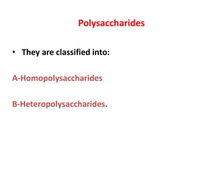
- 1. Polysaccharides • They are classified into: A-Homopolysaccharides B-Heteropolysaccharides.
- 2. A-Homopolysaccharides • They yield only one type of monosaccharides on hydrolysis and they are named according to the type of that monosaccharide, e.g., • Hexosans + H2O Hexoses • Pentosans + H2O Pentoses
- 3. Hexosans: 6 carbon sugars I. Glucosans: • They produce only glucose on hydrolysis. • They include; starch, dextrins, dextrans, glycogen and cellulose • Plants store glucose as amylose or amylopectin, glucose polymers collectively called starch.
- 4. A. Starch: • It is the stored form of carbohydrate of plants. It never exists in animals. • It is present in cereals such as wheat and rice and tubers such as potatoes. • It is in the form of starch granules. The core of the granule is amylose (20%) and the shell is amylopectin (80%). • Due to its high molecular weight it forms colloidal solution in hot water. S t a r c h g r a n u l e A m y lo s e A m y lo p e c t i n
- 5. O H H H OH H OH CH2OH H O H H OH H OH CH2OH H O Amylose 1 4 n O O 1 4 1. Amylose:. Straight chain compound present in the form glucose units linked by -1,4-glucosidic bond of a helix formed of a large number of - glucose. It forms the inner part of starch granules H O OH H OH H OH CH2OH H O H H OH H OH CH2OH H O H H H O O H OH H OH CH2OH H H H O H OH H OH CH2OH H OH H H O O H OH H OH CH2OH H O H 1 6 5 4 3 1 2 amylose
- 6. . • 2. Amylopectin: • It forms the outer coat of starch granule and is insoluble in water. • It is branched chains formed of a large number of -glucose units linked by -1,4-glucosidic linkage along the branch and by -1,6-glucosidic linkage at the branching point that occur every 25-30 glucose units. Due to its high molecular weight, it forms a colloidal solution. • Starch can be hydrolyzed by HCl or amylase.
- 7. Amylopectin is a glucose polymer with mainly (14) linkages, but it also has branches formed by (16) linkages. Branches are generally longer than shown above. The branches produce a compact structure & provide multiple chain ends at which enzymatic cleavage can occur. H O OH H OH H OH CH2OH H O H H OH H OH CH2OH H O H H H O O H OH H OH CH2 H H H O H OH H OH CH2OH H OH H H O O H OH H OH CH2OH H O H O 1 4 6 H O H OH H OH CH2OH H H H O H OH H OH CH2OH H H O 1 OH 3 4 5 2 amylopectin
- 8. . O H H H O H H O H CH 2O H H O H H O H H O H CH 2O H H O Amylopectin 1 4 O 1 4 O H H H O H H O H CH 2O H H O H H O H H O H CH 2O H H O 1 4 O O 1 4 O H H H O H H O H CH 2 H O H H O H H O H CH 2O H H O 1 4 n O 1 4 6 O
- 9. B. Dextrins: • Products of hydrolysis of starch and include amylodextrin, erythrodextrin, achrodextrin which form color with iodine • They have sweet taste. • They are easily digested than starch as in corn and rice syrup.
- 10. . C. Dextran: • A compound formed of -glucose units linked by -1,4, -1,3- and -1,6-linkage present in the form of a network that is synthesized by certain bacteria having sucrose in its media. It has a great biochemical importance, 1. It is used as plasma substitute to restore blood pressure in cases of shock. 2. Iron used for treatment of iron deficiency anemia is used as dextran ferrous sulfate intramuscular injection. 3. Sodium dextran sulfate is an anticoagulant.
- 11. . D. Glycogen: • It is the stored form of carbohydrate in animal, particularly in muscles and liver. • Its structure is similar to amylopectin a branched tree with -1,4-glucosidic linkage along the branch and -1,6-glucosidic linkage at the branching point. • The glycogen tree is shorter and more branched (a branch point every 8-10 glucose units) than amylopectin. • It is digestible because human amylases hydrolyze -glucosidic linkage.
- 12. . The glycogen molecule. A: General structure. The chains are either branched or unbranched and are arranged in 12 concentric layers (only four are shown in the figure). The branched chains (each has two branches) are found in the inner layers and the unbranched chains in the outer layer. (G, glycogenin, the primer molecule for glycogen synthesis.) Liver glycogen helps to maintain blood glucose level
- 13. Glycogen, the glucose storage polymer in animals, is similar in structure to amylopectin. But glycogen has more (16) branches. The highly branched structure permits rapid glucose release from glycogen stores, e.g., in muscle during exercise. The ability to rapidly mobilize glucose is more essential to animals than to plants. H O OH H OH H OH CH2OH H O H H OH H OH CH2OH H O H H H O O H OH H OH CH2 H H H O H OH H OH CH2OH H OH H H O O H OH H OH CH2OH H O H O 1 4 6 H O H OH H OH CH2OH H H H O H OH H OH CH2OH H H O 1 OH 3 4 5 2 glycogen
- 14. E. Cellulose: • It is a structural polysaccharide and forms the skeleton of plant cells and does not enter in animals cell structures. • It is a straight chain molecule formed of a large number of -glucose units linked by -1,4- glucosidic linkage. • It is water insoluble and enters in structure of cotton and paper • It is the major food for herbivorous animal where it is fermented into volatile fatty acids. • It gives cellobiose on hydrolysis with HCl.
- 15. . • It is indigestible but is very essential in food for: 1. Prevention of constipation by increasing the bulk of stools. 2. Its fermentation by large intestinal bacteria give volatile fatty acids that is anticancer for colon cells and gives also some water soluble vitamins. 3. It adsorbs toxins present in foods and prevents its absorption into the body. cellulose H O OH H OH H OH CH 2OH H O H OH H OH CH 2OH H O H H O O H OH H OH CH 2OH H H O H OH H OH CH 2OH H H OH H O O H OH H OH CH 2OH H O H H H H 1 6 5 4 3 1 2
- 16. Starch Glycogen Cellulose 1.Nature: Stored form of carbohydrate in plants. Stored form of carbohydrates in animals. Structural form of carbohydrate in plant cells but prevents constipation in human. 1.Source: Cereals, e.g., wheat, rice, and tubers, e.g., potatoes. Muscles and liver Linen and cotton are nearly pure cellulose. 1.Solubility: Amylose is water soluble and amylopectin is insoluble. Water soluble forming colloidal solution. Water insoluble. 1.Nature of the chains: Amylose is helical straight chain (-glucose units linked by -1,4-glucosidic bonds). Amylopectin is branched chain (-glucose units linked by -1,4- and -1,6-glucosidic bonds). Branched chain similar to amylopectin but its trees are shorter and have more branches than amylopectin tree. Straight chain (large number of -glucose units linked by -1,4- glucosidic bonds). 1.Reaction with iodine: Amylose gives blue color and amylopectin gives red color. Gives red color. No color. 1.Digestibility: Is hydrolyzed by HCl or amylase into dextrins and maltose. Digestible by amylase into dextrins and maltose. Non-digestiblebut HCl hydrolysis gives cellobiose.
- 17. . II. Fructosans: • They are formed of fructose as a building unit such as Inulin. Inulin: • It is formed of fructose only and present in onions. • It is not metabolizible in human body, therefore, it is used in evaluation of kidney function as a part of inulin clearance test.
- 18. . III. Galactosans: - They are formed of galactose as the building units such as agar-agar. - Biochemical importance: 1. It is used for growth of bacteria and mammalian cells in culture. 2. It imbibes water and increases intestinal contents to prevent and treat constipation. 3. Some electrophoresis gels is formed of it.
- 19. . IV. N-acetyl-glucosan: • It is a homopolysaccharide formed of -N-acetyl- glucosamine units such as chitin of insects. • Chitin: • It is a homopolysaccharide formed of -N-acetyl- glucosamine units linked by -1,4-glucosidic linkage present in the exoskeleton of insects.
- 20. . O H H H NH-CO- CH3 H OH CH2OH H O H H H NH-CO- CH3 H OH CH2OH H O O O Chitin -N-acetyl-glucosamine -N-acetyl-glucosamine n 1 4 1 4 Polymer of N-acetylglucosamine.
- 21. . B-Heteropolysaccharides • They are polysaccharides that on hydrolysis produce several types of sugars. There are two types: 1. Non-nitrogenous heteropolysaccharides 2. Nitrogenous heteropolysaccharides.
- 22. . A-Non-nitrogenous heteropolysaccharides: They do not contain sugar amines, such as pectin and plant gums. Also called Mucilages 1-Plant gums : • They are exudates of plants that do not contain amino sugars. • They contain pentoses, hexoses and uronic acids, e.g., Gum Arabic which is rich in arabinose. • They are emulsifying agents.
- 23. . • Mucilages: These are of plant origin and include agar, vegetable gums and pectins. • Hemiclellulose is a polymer of aldopentose [beta– D xylose]. It occurs with pectin [D–galacturonic acid] residues and a protein extensin in plant cell walls. • This heteropolysaccharide acts as a cementing material, provides shape and support to the plant tissues.
- 24. . 2. Pectin: • They are present in pulp of fruits and are responsible for settling of jams. • They are formed of pentoses, hexoses, uronic acids mainly galacturonic acid. • They are water soluble formed from a water insoluble compound called pectose present in raw fruits which is transformed into pectin by the action of sunlight, heat and pectase enzyme when fruits are ripened.
- 25. . • Biochemical importance of pectin: 1. Emulsifying reagents. 2. Demulcents. 3. Responsible for settling of jams. 4. They increase in size when they absorb water forming a jell and so they are used in the treatment of infantile diarrhea.
- 26. . • 3. Agar: • Vegetable mucilage, sulfuric acid ester of a complex galactose polysaccharide. • Non digestable • Peristalsis after ingestion, so as a laxative • Medium to grow bacteria
- 27. . B-Nitrogenous heteropolysaccharides: They contain sugar amines and are of two types: 1. Neutral nitrogenous heteropolysaccharides 2. Acidic nitrogenous heteropolysaccharides
- 28. 1-Neutral nitrogenous heteropolysaccharides Glycoproteins or mucoproteins: • They do not contain uronic acids or sulfate groups (which give acidic characteristics). • They are formed of a large protein core to which are attached smaller branched or unbranched chains of carbohydrate. • The carbohydrate present include: Hexoses: mannose, galactose and glucose Pentoses: xylose and arabinose. Amino sugars: glucosamine and galactosamine. Deoxysugars: L-fucose, L-rhamnose and sialic acid.
- 29. • Glycoproteins have branched oligosaccharide chains covalently attached to polypeptide backbone. • These are present in animals, human, viruses and bacteria. • Their carbohydrate content ranges form 4–85% by weight • Most of the monosaccharides are found in glycoproteins. The functions of oligosaccharide part is:
- 30. . • i. It changes the physiochemical properties [solubility, viscosity, charge and etc.] • ii. Protects against proteolysis by viruses and bacteria. • iii. Affects embryonic development, differentiation and cell–cell recognition. • iv. Oligosaccharide chain contains biological information, which is dependent upon constituent sugars, their sequence and their conformation.
- 31. . • Structure: • The common core has branched oligosaccharide of 3Man and 2GlcNAc residues. • Many other sugars may be added to it to form various glycoproteins. • Mannose rich oligosaccharides part is replaced by mixed sugars in some glycoproteins
- 32. . Distribution: Glycoproteins are widely distributed in mammalian tissues. They include: 1. Mucins of epithelium lining of gastrointestinal, urogenital and respiratory tracts are lubricant and protective. 2. Cell membranes where they play an important part in cell-cell attachment. 3. Blood group substances A and B antigens. 4. Mineral and vitamin transporting protein, e.g., transferrin.
- 33. . 5. Immunoglobulins (antibodies), IgG, IgM, IgA…. 6. Some hormones, e.g., anterior pituitary hormones: LH and FSH. 7. Some enzymes such as peptidases and alkaline phosphatase. 8. Intrinsic factor that is responsible for vitamin B12 absorption. 9. Structural function as a part of collagen of connective tissue.
- 34. 2. Acidic nitrogenous heteropolysaccharides A-Sulfur-free mucopolysaccharides: • Their sugar units are not sulfates, e.g., hyaluronic acid. B- Sulfur-containing mucopolysaccharides: • Their sugar units are sulfated ,e.g.,chondroiten sulfate , Heparin
- 35. STRUCTURE OF GLYCOSAMINOGLYCANS • Glycosaminoglycans are long, unbranched, heteropolysaccharide chains generally composed of a repeating disaccharide unit [acidic sugar-amino sugar]n • (glucosamine or galactosamine which may or may not be sulphated) and uronic acid (glucuronic or Iduronic acid). • keratan Sulfate – Galactose rather than an acidic sugar is present.
- 36. • The amino sugar is either D- glucosamine or D- galactosamine, in which the amino group is usually acetylated thus eliminating its positive charge • The amino sugar may also be sulfated on carbon 4 or 6 or on a nonacetylated nitrogen • The acidic sugar is either D- glucuronic acid or its carbon- 5 epimer L-iduronic acid • (Negative charge)
- 37. Glycosaminoglycan overview • Negatively charged heteropolysaccharide chains. • Associated with a small amount of protein, forming proteoglycans (95% CHO) • Bind large amounts of water(special ability) producing the gel-like matrix -- ground substance • EXTRACELLULAR MATRIX (ECM): • Glycosaminoglycans, Fibrous structural proteins eg. (Collagen, Elastin) and Adhesive proteins (fibronectin)
- 38. . • GLYCOSAMINOGLYCANS Flexible support for the ECM Interacting with the structural and adhesive proteins Molecular sieve Viscous, Lubricating properties of mucous secretions (original name: mucopolysaccharides)
- 39. . • ii. Disaccharide part is covalently attached to a protein molecule and the compound formed is called proteoglycan. • iii. They act as packing material in structural elements of tissues, bones, elastin, and collagen. • vi. They absorb large amount of water and act as lubricants.
- 40. . • v. Proteoglycan from cartilage has keratan–SO4 and chondrotin–SO4 chains, which are covalently linked to a core protein. • Some 140 core proteins are non–covalently linked at an interval of 300Å to a long filament of hyaluronate supported by small link proteins. • Proteoglycans from different sources have different structures.
- 41. . • Resilience of glycosaminoglycans large number of negative charges, repel each other they “slide” past each other,. produces the “slippery” consistency of mucous secretions and synovial fluid. water is “squeezed out,” and the GAGs are forced to occupy a smaller volume. GAGs spring back to their original, hydrated volume because of the repulsion of their negative charges. resilience of synovial fluid and the vitreous humor of the eye
- 42. C. Proteoglycans • Proteoglycans are found in the ECM and on the outer surface of cells. • 1. Structure of proteoglycan monomers: • A proteoglycan monomer found in cartilage consists of a core protein to which up to 100 linear chains of GAGs are covalently attached. • These chains, which may each be composed of up to 200 disaccharide units, extend out from the core protein and remain separated from each other because of charge repulsion. The resulting structure resembles a “bottle brush”
- 43. . • GAG-Protein linkage: • This covalent linkage is most commonly through a trihexoside. (galactose-galactose- xylose) • Aggregate formation: Many proteoglycan monomers associate with one molecule of hyaluronic acid to form proteoglycan aggregates. The association is not covalent but occurs primarily through ionic interactions between the core protein and the hyaluronic acid.
- 44. . • .
- 45. Proteoglycans are glycosaminoglycans that are covalently linked to serine residues of specific core proteins. The glycosaminoglycan chain is synthesized by sequential addition of sugar residues to the core protein. heparan sulfate glycosaminoglycan cytosol core protein transmembrane -helix
- 46. . • Functions in Body • Proteoglycans in extra-cellular matrix are criss crossed by fibrous matrix proteins (collagen, elastin, fibronectin).. Strength • Tisue organizers, development of specialised tissues, mediate activities of various growth factors. • Regulate extracellular assembly of collagen fibres • Major component of cartilage –strength
- 47. . • Heparan sulphate binds extracellular ligands— interaction with specific receptors on cell surface • Fibronectin can bind fibrin, heparan sulfate collagen ad integrins --- information transfer • Aggrecan –interacts with collagen – strength • Special pattern of sulfated and non-sulfated sugar units – recognition by ligands • Carboxyl and sulfated groups – negative charge
- 48. . • Repel each other – slippery and vicious • Found in cartilage, skin, blood vessels, cornea, tendons, ligaments, loose CT, heart valves. • All GAGs (except hyaluronic acid), occur in combination with extracellular proteins through covalent linkages---proteoglycan monomers
- 49. . Hyaluronic acid: - • It is formed of -D-N-acetyl glucosamine linked to -D-glucuronic acid by alternating -1,3- and -1,4- glycosidic linkages. O H H H OH H OH CO OH O H OH H H NH- CO- CH 3 H CH 2OH O O 1 3 1 4 O H H n -Glucuronicacid -N-acetyl-Glucosamine Hyaluronicacid
- 51. The glycosidic linkages are (13) & (14). Up to 50,000 repeats of these repeating units. H O H H OH H OH COO H H O OH H H NHCOCH 3 H CH2OH H O O D-glucuronate O 1 2 3 4 5 6 1 2 3 4 5 6 N-acetyl-D-glucosamine hyaluronate
- 52. . Biochemical importance: 1. The molecule is coiled and entwined making a very firm gel which prevents bacterial invasion of the skin. High viscosity 2. It is present in connective tissue matrix, vitreous humor of the eye, in the skin, synovial fluid, around the ovum, and in the umbilical cord to preserve the full-form of these structures. 3. It acts as a cementing substance between cells and as a lubricating material of joints.
- 53. . 4. Hyaluronic acid imbibes water and forms a incompressible substance due to the presence of several OH groups which creates negative charges causing repulsion between carbohydrate units enabling the molecule to perform its function as lubrication in joint synovial fluids an.
- 54. . • Hyaluronidase Enzyme or Spreading factor: • It is the enzyme that hydrolyzes hyaluronic acid. • It is present in sperms to help penetration of the ovum and fertilization. • It is present in some virulent strains of bacteria that are able spread through infected wounds. • It is used in medicine to treat fibrosis and to dissolve mucus.
- 55. . • B. Sulfur-containing mucopolysaccharides 1-Chondroitin sulfate: it is of three types as follows: • Chondroitin sulfate A: • It is formed of -N-acetyl-galactosamine-4-sulfate and -glucuronic acid linked by alternating -1,3- and -1,4-glycosidic linkages. O H H H OH H OH COOH O HO3SO H H H NH-CO-CH3 H CH2OH O O 1 3 1 4 O H H n -Glucuronic acid -N-acetyl-Galactosamine, 4-sulfate Chondroitin Sulfate A
- 56. . • Chondroitin sulfate B: • It is formed of -N-acetyl galactosamine-4- sulfate and -L-iduronic acid linked by alternating -1,3- and -1,4-glycosidic linkages. • L-iduronic acid is the C5 epimer of D-glucuronic acid, i.e., COOH group at C5 is inside the ring, O H H H OH H OH H O HO3SO H H H NH-CO-CH3 H CH2OH O O 1 3 1 4 O H COOH n -L-Iduronic acid -N-acetyl-Galactosamine, 4-sulfate Chondroitin Sulfate B
- 57. . • Chondroitin sulfate C: • It is formed of -N-acetyl galactosamine-6- sulfate and -glucuronic acid linked by alternating -1,3- and -1,4-glycosidic linkages. • Present in cornea of the eye, tendons, ligaments, bones, cartilage and connective tissue matrix. • They absorb water, form incompressible substances by means of their ionizable OH and sulfate groups, creating negative charges leading to repulsion between the molecules.
- 58. ….. O H H H OH H OH COOH O OH H H H NH-CO-CH3 H CH2OSO3H O O 1 3 1 4 O Chondroitin Sulfate C H H n -Glucuronic acid -N-acetyl-Galactosamine, 6-sulfate
- 60. . 2. Heparin: Structure: It is formed of a long repeat of sulfated -glucosamine and sulfated -L-iduronic acid linked by alternating - and -1,4-glycosidic linkages, synthesized on a core protein. Source: It is produced by mast cells (kidney, lung, liver skin). O H H H NH-SO3H H OH CH2OSO3H H O H H H OSO3H H OH H COOH O O O n Heparin 1 4 1 4 -Glucosamine, N- and 6-sulfated -L-Iduronic acid, 2-sulfate
- 61. ..
- 62. . • Function: 1. It is an anticoagulant and prevents intravascular clotting. It interferes with the activated clotting factors (thrombin, IX, X, XI and XII) in coagulation, through activation of antithrombin III that inhibits the intrinsic pathway of blood clotting. Therefore, it is used in cases of increased coagulability, e.g., cardiac ischemia or deep venous thrombosis. 2. It binds activates lipoprotein lipase enzyme (the plasma clearing factor) to clear the turbid plasma from the absorbed lipids after meals.
- 63. 3. It regulates the action of the heparin-binding growth factors. 4. It has a structural role in extracellular matrix. It affects cell-cell and cell-matrix interaction to modulate development, cell proliferation, apoptosis and differentiation. 5. Large number of sulfate groups ---highest negative charge density !! .
- 64. III. Kertan–SO4–I and II: • The repeating unit is [Gal ……. GlcNAc–SO4]n. • Sulphate is present at position 6 of GlcNAc or sometimes linked to Gal. • Type I predominates in cornea and type II along with chondroitin–SO4 is attached to hyaluronic acid in loose connective tissues. • Present in cornea, cartilage, bone, and dead cells horny structures like horns, hoofs, hair, nails, claws.
- 65. .
- 66. V. Dermatan–SO4: • The repeating unit is composed of [IdUA – GalNAc] n –[GlcUA – GalNAc]n. • It has both IdUA and GlcUA attached to GalNAc.. • Pliability of skin • Blood vessels and heart valves.
- 68. Mucopolysaccharidoses • Mucopolysaccharidoses is a genetic defect due to the deficiency of particular Iysosomal enzymes that degrade particular GAGs. • This leads to the accumulation of GAGs in various tissues. There occurs mental retardation. • The specific GAGs are also excreted in urine. • In arthritis, Proteoglycans may act as an autoantigen and contribute its effects to the pathology.
- 69. Important Monosaccharides • D-Glucose is the most common monosaccharide - Primary fuel for our cells, required for many tissues - Main sources are fruits, vegetables, corn syrup and honey - Blood glucose is maintained within a fairly small range - Some glucose is stored as glycogen, excess is stored as fat During urine formation, glucose is nearly completely reabsorbed and only traces escape in urine
- 70. …… • Its normal value in blood, at random ranges from 65–110 mg/dL but in abnormal conditions known as Diabetes Mellitus, its quantity is elevated to several hundred mg/dl. • It is the fuel for brain and provides energy to brain and other tissue • The estimation of glucose in various biological fluids is performed for diagnostic purposes.
- 71. .. • Glucose is converted to other sugars and their derivatives which are then used for the synthesis of heteropolysaccharides, glycolipids etc. • The other sugars of specific functions are formed from glucose and are incorporated into heteroplysaccharides of structure material.
- 72. . • D-Fructose is the sweetest carbohydrate, is levulose. - Converted by an enzyme into glucose for respiration - Main sources are fruits and honey - hydrolysis of sucrose, • In the seminal fluid, it is present in appreciable amounts
- 73. .. D–Galactose: • It is an aldohexose, dextrorotatory, mutarotation. • It is a part of milk sugar, lactose. • Galactose in the form of galactosamine is a part of sulphonated mucopolysacharide such as chondroitin [chondroitin sulphuric acid and protein]. • Galactose is also a component of galactocerebrosides present in brain. • It is an epimer of glucose and differs in configuration around ‘C’ No.4 of glucose.
- 74. .. • In liver, galactose is converted into glucose. • In a disease known as Galactosemia, the body cannot utilize it and galactose increases in the blood and aqueous humor. • Galactose in the lens of the eye is converted to galactitol and forms cataract. • Galactosemia in children is treated by feeding them galactose free diet. • The galactose necessary for the biosynthesis of cell membrane, cerebrosides, glycoproteins etc. is formed from glucose.
- 75. .. D–Mannose: • Aldohexose and is an epimer of glucose, differing in configuration around C atom No. 2 of glucose. • Mannose is a carbohydrate component of albumin, globulins and mucoids. • Mannose is richly present in the common core of glycoproteins. • Its derivative IdUA is a component of heparin.
- 76. CARBOHYDRATES OCCUR IN CELL MEMBRANES & IN LIPOPROTEINS • In addition to the lipid of cell membranes, approximately 5% is carbohydrate in glycoproteins and glycolipids. • Carbohydrates are also present in apo B of lipoproteins. • They are present on the outer surface of the plasma membrane (the glycocalyx). • Glycophorin is a major integral membrane glycoprotein of human erythrocytes and spans the lipid membrane.
- 77. ..