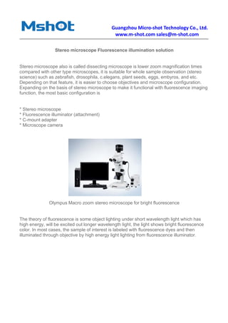
Download to read offline
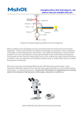


Guangzhou Micro-Shot Technology Co., Ltd. specializes in solutions for upgrading stereo microscopes to include fluorescence illumination, providing a cost-effective alternative to expensive brands like Zeiss and Leica. Their product, the Mshot MZX-BG-LED stereo fluorescence illuminator, allows for easy installation and operation, enabling observation of various samples with fluorescence dyes. The company has a 15-year history of serving customers in need of upgrading their stereo microscopes.


