Evolution of the sporophyte in
Brophytes
Presented by: Prajapati Dimpal B.
M.sc sem-1
CBO-402
Content
Introduction
Theory of Sterilization
Reduction Theory
Introduction
The sporophyte of liverworts and mosses show mostly the
same fundamental plan.
It is made up of an anchoring and absorbing foot,a stalk- like
seta and a capsule, which contains spores and elaters.
The following two contrasting views have been put forword
with regard to the evolution of sporophytes in bryophytes:-
1. Evolution of sporophytes by progressive sterilization of
potential sporogenous tissue.
2. Evolution of sporophyte due to progressive reduction or
simplification.
Theory of sterilization
Sporogenous Tissue:-
• This view , also known as 'theory of progressive
evolution 'or 'theory of sterilization ',was put
forward by Bower (1908-35) and supported by
cavers (1910) and Campbell (1940).
• According to this view ,the sporophytes of
complex forms (e.g.,Funaria ,Sphagnum
,Pogonatum) have evolved due to progressive
sterilization of the potential fertile tissue of the
simpler forms (e.g.,Riccia, Marchantia).
First stage
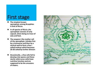
 The simplest known
sporophyte among Bryophtes
is that of Riccia.
 In all species of Riccia ,the
sporophyte consists of only
capsule ,there being no trace of
seta and foot.
 The oospore ( the mother cell
of the sporophyte ) divides first
by a transverse and then by a
vertical wall to form a four -
celled embryo which becomes
20-30 celled by further division
.
 Occasionally , a few cells fail to
develop into spores and these
sterile cells( nurse cells) have
nutritive function and are
supposed to be the ancestors
of elaters.
Second Stage
In Corsinia , the oospore divides by a
transverse wall into a hypobasal and an
epibasal cell.
The former gives rise to a small foot.
The derivatives of the epibasal cell from an
outer amphithecium and an inner
endothecium.
Thus in the sporophyte of Corsinia the
sterilization has gone a step further and in
addition to nurse cells, a sterile foot is also
present.
Third Stage
 Further sterilization is
seen in spharocarpus
where the sporophyte
has a sterile bulbous
foot and a narrow seta,
in addition to fertile
capsule.
 The amphithecium
forms a single layered
jacket of the capsule
and the endothecium
,the sporogenous
tissue and the sterile
nurse cells.
Forth Stage
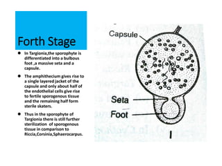
 In Targionia,the sporophyte is
differentiated into a bulbous
foot ,a massive seta and a
capsule.
 The amphithecium gives rise to
a single layered jacket of the
capsule and only about half of
the endothelial cells give rise
to fertile sporogenous tissue
and the remaining half form
sterile skaters.
 Thus in the sporophyte of
Targionia there is still further
sterilization of sporogenous
tissue in comparison to
Riccia,Corsinia,Sphaerocarpus.
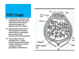
Fifth Stage
 In Marchantia , the lower half
(hypobasal cells) of the octant
embryo froms the foot,and
seta and the upper half
(epibasal cells ) the capsule.
 Approximately 50 percent of
the sporogenous cells form
spores and the remaining 50
percent elaters with
characteristic thickening bands.
 Thus,a bulbous foot,an
elongated seta , a single
layered capsule wall and large
number of elaters are sterile
constituents of the
sporophytes of Marchantia.
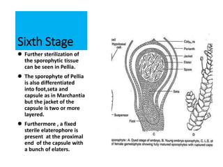
Sixth Stage
 Further sterilization of
the sporophytic tissue
can be seen in Pellia.
 The sporophyte of Pellia
is also differentiated
into foot,seta and
capsule as in Marchantia
but the jacket of the
capsule is two or more
layered.
 Furthermore , a fixed
sterile elaterophore is
present at the proximal
end of the capsule with
a bunch of elaters.
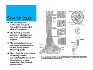
Seventh Stage
 The sporophyte of
Anthoceros is of great
interest from the point of
view of evolution .
 The mature sporophyte
consists of a bulbous foot
and horn or bristle -like
capsule.
 The elaters of Anthoceros
are known as pseudoelaters
as they do not possess
thickening bands.
 Thus , from the point of view
of sterility and self-
dependence , the sporophyte
of Anthoceros is more
specialized than those of
Hepaticopsida.
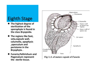
Eighth Stage
 The highest degree of
sterilization of the
sporophyte is found in
the class Bryopsida.
 The regions like foot,
seta,capsule wall,
columella, apophysis
,operculum and
peristome in the
Bryophyta .
 Funaria,Politrichum and
Pogonatum represent
the sterile tissue.
Thus the sporophyte of Riccia is the simplest
amongst the bryophytes, with a very high
proportion of fertile tissue and the sterile tissue is
very small.
On the other hand , the members of the class
Bryopsida (e.g.,Funaria,Polytrichum , Pogonatum )
have the most complex sporophyte with a very high
degree of sterility.
The evolution of sporophyte in bryophytes as such
is considered to have taken place by progressive
sterilization of the fertile tissue .
Reduction Theory
There is however, an -opposite school of
thought led by Kashyap,Church,Goebel and
Evans.
They hold that so serlated the evolution of the
sporophyte has been in the downward direction
.
The series they believe furnishes an example of
retrogressive evolution.
There is ample evidence of reduction rather
than progressive elaboration of the sporophytes
of this group.
The significant steps in the
reduction series are:-
1. Simplification of the dehiscence apparatus.
2. Reduction of the green photosynthetic
tissue in the capsule wall.
3. Associated with the above is the
disappearance of stomata and intercellular
spaces.
Side by side with the above changes in the
gradually elimination of the seta and
subsequently the disappearance of the foot.
All these changes are accompanied by the
progressive increase in the fertility of the
sporogenous cells.
Evidence from comparative morphology and
experimentaly genetics support the view that
the simple sporophyte of Riccia is an advanced
but a reduced structure.
Reference
Botany For Degree Students Bryophytes by:-
B. R. Vashistha
A. K.Sinha
V. P. Singh
https://en.m.wikipedia.org
https://www.slideshare.net
Thank You