SYMBLEPHORON
MODERATOR:
DR PENCHALAIAH., M.S
PRESENTER:
DR T.SRINIVASULU
1st Year PG.
SYMBLEPHARON
Definition: A symblepharon is an adhesion between the eyelid and
the eyeball.
 It can involve just one lid or both.
 This adhesion of Sub-epithelial (beneath) scarring can lead to
other lid complications such as aberrant (deviated) lash growth
and entropion.
SYMBLEPHARON
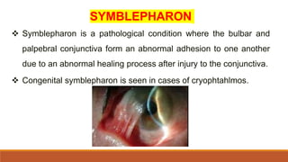
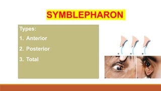
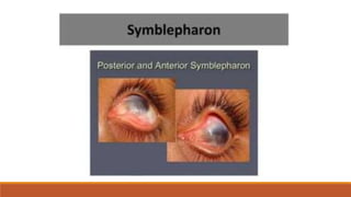
 Symblepharon is a pathological condition where the bulbar and
palpebral conjunctiva form an abnormal adhesion to one another
due to an abnormal healing process after injury to the conjunctiva.
 Congenital symblepharon is seen in cases of cryophtahlmos.
SYMBLEPHARON
Types:
1. Anterior
2. Posterior
3. Total
 Anterior symblepharon, when the eyelid edge or a part
of it is attached to the eyeball.
 Posterior symblepharon, when the cul-de-sac or
conjunctival fornix is involved.
 Total symblepharon, when the whole eyelid surface is
attached to the globe and the fornix is obliterated
 Signs and symptoms of symblepharon include fibrous
adhesions between the palpebral conjunctiva and the
bulbar conjunctiva, eye redness, burning and tearing,
photophobia, diplopia, restricted extraocular motility,
lagophthalmos, entropion, and cosmetic
Anatomically symblepharon is a partial or complete adhesion of the palpebral
conjunctiva of the eyelid to the bulbar conjunctiva of the eyeball.
ANATOMY
The conjunctival tissue starts from the limbus and ends in the lid margin. According
to the anatomic location, the conjunctiva can be subdivided into the bulbar and
palpebral portions, palpebral conjunctiva: The part of the conjunctiva, a clear
membrane, that coats the inside of the eyelids. The palpebral conjunctiva is as
opposed to the ocular (or bulbar) conjunctiva. bulbar conjunctiva: bulbar
conjunctiva, the part of the conjunctiva that covers the outer surface of the eye.
Synonym: pseudopterygium / cicatricial pterygium
ETIOLOGY
Symblepharon can be acquired due to a number of
inflammatory or traumatic etiologies.
Immune mediated inflammatory conditions include:
 Steven-Johnson Syndrome/Toxic Epidermal Necrolysis
 Ocular cicatricial pemphigoid
 Sarcoid
 Granulomatosis with Polyangiitis
 Chronic Graft-versus-host disease
 Paraneoplastic Mucous Membrane Pemphigoid
 Lichen Planus
 Recessive Dystrophic Epidermolysis Bullosa
 Infections from both bacteria and viruses can also cause symblepharon,
such as in the case of chronic chlamydial eye infections or Epidemic
Keratoconjunctivitis.
 Trauma to the eye may cause enough damage leading to symblepharon.
 This can be seen in cases of chemical, particularly alkali, or thermal burns,
such as firework injuries.
 Congenital symblepharon has been documented in cases of
cryptophthalmos.
EPIDEMIOLOGY
 Epidemiologic data on this condition is not readily available.
 This condition occurs in a number of contexts. For each underlying
pathology, the prevalence of symblepharon varies.
 That data is available elsewhere and is outside the scope of this
section.
PATHOPHYSIOLOGY
 Symblepharon occurs from an abnormal healing process
after injury to the conjunctiva.
 Whatever the inciting injury, the loss of epithelial cells
from both the bulbar and palpebral conjunctiva allow an
abnormal adhesion to form between the bulbar and
palpebral conjunctiva.
PHYSICAL EXAMINATION
 Symblepharon has variable severity and tissue involvement.
 On a physical exam, there may be only small adhesion between the two layers of
conjunctiva.
 The fornix of the eye may become obliterated, cicatricial entropion may form, or there
may be permanent lagophthalmos with exposure of the cornea.
 Obliteration of the fornix can cause insufficient tear reservoir and blinking.
 This in turn leads to eventual keratinization of the ocular surface. Entropion can cause
ocular trauma to the surface of the eye as the eyelashes rub on the outer surface. With
greater tissue involvement, decreased extraocular movement may be seen.
Depending on the severity, the symblepharon may or may not involve the cornea.
ANKYLOBLEPHARON It refers to the adhesions between margins of the upper and
lower lids. Etiology: Congenital anomaly and aquired adhesions after healing of
chemical Burn, ulcers and traumatic wounds of the lid margins. Clinically
Ankyloblepharon may be complete or incomplete. It is usually associate with
Symblephoron. Treat ment:. Lids should be separated by excision of adhesions
between the lid margins and kept apart during healing process. When adhesions
extend to the angles, epithelial grafts should be given to prevent recurrence.
LABORATORY TEST
 Laboratory studies are not currently used to diagnose
symblepharon.
 Laboratory studies are often necessary to diagnose the
underlying pathology
MANAGEMENT
MEDICAL THERAPY
 Medical management of symblepharon aims to prevent or decrease symblepharon
formation and to treat the underlying pathology.
 A number of inflammatory conditions can lead to symblepharon.
 Utilizing immune modulating therapy to suppress inflammation may improve outcomes,
as seen when using Rituximab when treating Severe Refractory Paraneoplastic Mucous
Membrane Pemphigoid.
 Steroids and other immuno-suppressive drugs such as azathioprine,
cyclophosphamide, or mycophenolate may also be used.
 The symptoms of dry eye caused by symblepharon can be managed using
preservative free artificial tears and eye lubricants.
Oculoplastics, or oculoplastic surgery, includes a wide variety of
surgical procedures that deal with the orbit (eye socket), eyelids, tear
ducts, and the face. It also deals with the reconstruction of the eye
and associated structures
Prevention:
 Minimize the cause definitely able to stay away the disease, such
as
 Chemical burns
 Trauma
 Disease (conjunctivitis)
SURGERY
 Surgery for symblepharon typically involves tissue grafting to the
affected areas, and reconstruction of a normal fornix.
 Surgical techniques including cicatrix lysis and intraoperative mitomycin
C (MMC) application are paired with reconstruction using tissue grafts
from either oral mucosal transplantation, conjunctival autografting, or
amniotic membrane transplantation.
 Cultivated limbal stem cell transplantation is another surgical procedure
to treat symblepharon that can be used in cases of severe burns.
 With stem cell transplantation, care must be taken to not transplant
during active inflammation.
COMPLICATIONS
Symblepharon can lead to a number of complications.
The adhesion can encroach on the limbus and grow over the cornea,
leading to vision loss.
The adhesions can also decrease eye movement, cause diplopia, and
prevent the normal functioning of the eyelids through mechanical
forces.
PROGNOSIS
Symblepharon has variable severity.
In some cases, symblepharon may be mild and cause no symptoms or
damage to the eye.
Depending on the severity, etiology, and management of the symblepharon,
there is a variable prognosis.
In some cases, there may be permanent blindness, in others there may be
full resolution with good visual outcome.
symblephoron types etiology management.pptx

symblephoron types etiology management.pptx

  • 1.
  • 2.
    SYMBLEPHARON Definition: A symblepharonis an adhesion between the eyelid and the eyeball.  It can involve just one lid or both.  This adhesion of Sub-epithelial (beneath) scarring can lead to other lid complications such as aberrant (deviated) lash growth and entropion.
  • 3.
    SYMBLEPHARON  Symblepharon isa pathological condition where the bulbar and palpebral conjunctiva form an abnormal adhesion to one another due to an abnormal healing process after injury to the conjunctiva.  Congenital symblepharon is seen in cases of cryophtahlmos.
  • 4.
  • 7.
     Anterior symblepharon,when the eyelid edge or a part of it is attached to the eyeball.  Posterior symblepharon, when the cul-de-sac or conjunctival fornix is involved.  Total symblepharon, when the whole eyelid surface is attached to the globe and the fornix is obliterated
  • 8.
     Signs andsymptoms of symblepharon include fibrous adhesions between the palpebral conjunctiva and the bulbar conjunctiva, eye redness, burning and tearing, photophobia, diplopia, restricted extraocular motility, lagophthalmos, entropion, and cosmetic
  • 9.
    Anatomically symblepharon isa partial or complete adhesion of the palpebral conjunctiva of the eyelid to the bulbar conjunctiva of the eyeball. ANATOMY The conjunctival tissue starts from the limbus and ends in the lid margin. According to the anatomic location, the conjunctiva can be subdivided into the bulbar and palpebral portions, palpebral conjunctiva: The part of the conjunctiva, a clear membrane, that coats the inside of the eyelids. The palpebral conjunctiva is as opposed to the ocular (or bulbar) conjunctiva. bulbar conjunctiva: bulbar conjunctiva, the part of the conjunctiva that covers the outer surface of the eye. Synonym: pseudopterygium / cicatricial pterygium
  • 10.
    ETIOLOGY Symblepharon can beacquired due to a number of inflammatory or traumatic etiologies. Immune mediated inflammatory conditions include:  Steven-Johnson Syndrome/Toxic Epidermal Necrolysis  Ocular cicatricial pemphigoid  Sarcoid  Granulomatosis with Polyangiitis  Chronic Graft-versus-host disease  Paraneoplastic Mucous Membrane Pemphigoid  Lichen Planus  Recessive Dystrophic Epidermolysis Bullosa
  • 11.
     Infections fromboth bacteria and viruses can also cause symblepharon, such as in the case of chronic chlamydial eye infections or Epidemic Keratoconjunctivitis.  Trauma to the eye may cause enough damage leading to symblepharon.  This can be seen in cases of chemical, particularly alkali, or thermal burns, such as firework injuries.  Congenital symblepharon has been documented in cases of cryptophthalmos.
  • 12.
    EPIDEMIOLOGY  Epidemiologic dataon this condition is not readily available.  This condition occurs in a number of contexts. For each underlying pathology, the prevalence of symblepharon varies.  That data is available elsewhere and is outside the scope of this section.
  • 13.
    PATHOPHYSIOLOGY  Symblepharon occursfrom an abnormal healing process after injury to the conjunctiva.  Whatever the inciting injury, the loss of epithelial cells from both the bulbar and palpebral conjunctiva allow an abnormal adhesion to form between the bulbar and palpebral conjunctiva.
  • 14.
    PHYSICAL EXAMINATION  Symblepharonhas variable severity and tissue involvement.  On a physical exam, there may be only small adhesion between the two layers of conjunctiva.  The fornix of the eye may become obliterated, cicatricial entropion may form, or there may be permanent lagophthalmos with exposure of the cornea.  Obliteration of the fornix can cause insufficient tear reservoir and blinking.  This in turn leads to eventual keratinization of the ocular surface. Entropion can cause ocular trauma to the surface of the eye as the eyelashes rub on the outer surface. With greater tissue involvement, decreased extraocular movement may be seen.
  • 15.
    Depending on theseverity, the symblepharon may or may not involve the cornea. ANKYLOBLEPHARON It refers to the adhesions between margins of the upper and lower lids. Etiology: Congenital anomaly and aquired adhesions after healing of chemical Burn, ulcers and traumatic wounds of the lid margins. Clinically Ankyloblepharon may be complete or incomplete. It is usually associate with Symblephoron. Treat ment:. Lids should be separated by excision of adhesions between the lid margins and kept apart during healing process. When adhesions extend to the angles, epithelial grafts should be given to prevent recurrence.
  • 16.
    LABORATORY TEST  Laboratorystudies are not currently used to diagnose symblepharon.  Laboratory studies are often necessary to diagnose the underlying pathology
  • 17.
    MANAGEMENT MEDICAL THERAPY  Medicalmanagement of symblepharon aims to prevent or decrease symblepharon formation and to treat the underlying pathology.  A number of inflammatory conditions can lead to symblepharon.  Utilizing immune modulating therapy to suppress inflammation may improve outcomes, as seen when using Rituximab when treating Severe Refractory Paraneoplastic Mucous Membrane Pemphigoid.
  • 18.
     Steroids andother immuno-suppressive drugs such as azathioprine, cyclophosphamide, or mycophenolate may also be used.  The symptoms of dry eye caused by symblepharon can be managed using preservative free artificial tears and eye lubricants.
  • 19.
    Oculoplastics, or oculoplasticsurgery, includes a wide variety of surgical procedures that deal with the orbit (eye socket), eyelids, tear ducts, and the face. It also deals with the reconstruction of the eye and associated structures Prevention:  Minimize the cause definitely able to stay away the disease, such as  Chemical burns  Trauma  Disease (conjunctivitis)
  • 21.
    SURGERY  Surgery forsymblepharon typically involves tissue grafting to the affected areas, and reconstruction of a normal fornix.  Surgical techniques including cicatrix lysis and intraoperative mitomycin C (MMC) application are paired with reconstruction using tissue grafts from either oral mucosal transplantation, conjunctival autografting, or amniotic membrane transplantation.  Cultivated limbal stem cell transplantation is another surgical procedure to treat symblepharon that can be used in cases of severe burns.  With stem cell transplantation, care must be taken to not transplant during active inflammation.
  • 22.
    COMPLICATIONS Symblepharon can leadto a number of complications. The adhesion can encroach on the limbus and grow over the cornea, leading to vision loss. The adhesions can also decrease eye movement, cause diplopia, and prevent the normal functioning of the eyelids through mechanical forces.
  • 23.
    PROGNOSIS Symblepharon has variableseverity. In some cases, symblepharon may be mild and cause no symptoms or damage to the eye. Depending on the severity, etiology, and management of the symblepharon, there is a variable prognosis. In some cases, there may be permanent blindness, in others there may be full resolution with good visual outcome.