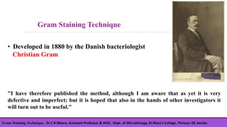
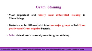
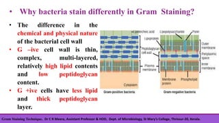
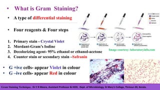
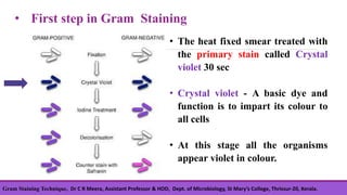
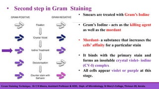
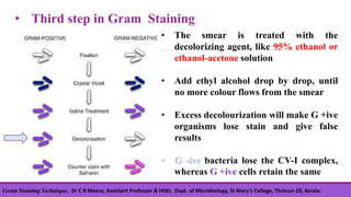
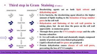
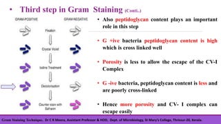
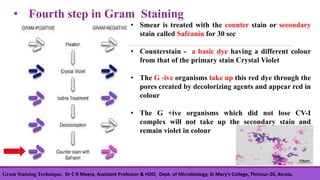
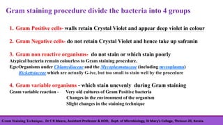
GRAM STAINING TECHNIQUE
THINGS YOU MUST KNOW
Dr C R Meera
Assistant Professor & HOD
Department of Microbiology
St. Mary’s College, Thrissur-20,
Kerala, India
Gram Staining Technique, Dr C R Meera, Assistant Professor & HOD, Dept. of Microbiology, St Mary’s College, Thrissur-20, Kerala.
• Developed in 1880 by the Danish bacteriologist
Christian Gram
"I have therefore published the method, although I am aware that as yet it is very
defective and imperfect; but it is hoped that also in the hands of other investigators it
will turn out to be useful."
Gram Staining Technique
• Most important and widely used differential staining in
Microbiology
• Bacteria can be differentiated into two major groups called Gram
positive and Gram negative bacteria.
• 24 hr. old cultures are usually used for gram staining
Gram Staining
Gram Staining Technique, Dr C R Meera, Assistant Professor & HOD, Dept. of Microbiology, St Mary’s College, Thrissur-20, Kerala.
• Why bacteria stain differently in Gram Staining?
• The difference in the
chemical and physical nature
of the bacterial cell wall
• G –ive cell wall is thin,
complex, multi-layered,
relatively high lipid contents
and low peptidoglycan
content.
• G +ive cells have less lipid
and thick peptidoglycan
layer.
Gram Staining Technique, Dr C R Meera, Assistant Professor & HOD, Dept. of Microbiology, St Mary’s College, Thrissur-20, Kerala.
• What is Gram Staining?
• A type of differential staining
• Four reagents & Four steps
1. Primary stain - Crystal Violet
2. Mordant-Gram’s Iodine
3. Decolorizing agent- 95% ethanol or ethanol-acetone
4. Counter stain or secondary stain –Safranin
• G +ive cells- appear Violet in colour
• G –ive cells- appear Red in colour
Image courtesy: laboratoryinfo.com
Gram Staining Technique, Dr C R Meera, Assistant Professor & HOD, Dept. of Microbiology, St Mary’s College, Thrissur-20, Kerala.
• The heat fixed smear treated with
the primary stain called Crystal
violet 30 sec
• Crystal violet - A basic dye and
function is to impart its colour to
all cells
• At this stage all the organisms
appear violet in colour.
• First step in Gram Staining
Gram Staining Technique, Dr C R Meera, Assistant Professor & HOD, Dept. of Microbiology, St Mary’s College, Thrissur-20, Kerala.
• Second step in Gram Staining
• Smears are treated with Gram’s Iodine
• Gram’s Iodine - acts as the killing agent
as well as the mordant
• Mordant- a substance that increases the
cells’ affinity for a particular stain
• It binds with the primary stain and
forms an insoluble crystal violet- iodine
(CV-I) complex
• All cells appear violet or purple at this
stage.
Gram Staining Technique, Dr C R Meera, Assistant Professor & HOD, Dept. of Microbiology, St Mary’s College, Thrissur-20, Kerala.
• Third step in Gram Staining
• The smear is treated with the
decolorizing agent, like 95% ethanol or
ethanol-acetone solution
• Add ethyl alcohol drop by drop, until
no more colour flows from the smear
• Excess decolourization will make G +ive
organisms lose stain and give false
results
• G -ive bacteria lose the CV-I complex,
whereas G +ive cells retain the same
Gram Staining Technique, Dr C R Meera, Assistant Professor & HOD, Dept. of Microbiology, St Mary’s College, Thrissur-20, Kerala.
• Third step in Gram Staining (Conti..)
• Decolorizing agents act as both lipid solvent and
dehydrating agent
• G-ive bacteria, the decolorizing agent dissolves the higher
amount of lipids leading to the formation of large number
pores in the cell wall
• Dehydration and flattening of the cell wall proteins is
taking place, but do not close the pores on the cell wall
appreciably as numerous pores
• Through these pores the CV-I complex escape and the cells
become colourless
• G +ive cell walls are thick and chemically simple, composed
mainly of protein and cross-linked polypeptides
• Lipid is dissolved and few pores are produced
• Protein dehydration causes closure of cell wall pores,
preventing the loss of CV-I complex
Gram Staining Technique, Dr C R Meera, Assistant Professor & HOD, Dept. of Microbiology, St Mary’s College, Thrissur-20, Kerala.
• Also peptidoglycan content plays an important
role in this step
• G +ive bacteria peptidoglycan content is high
which is cross linked well
• Porosity is less to allow the escape of the CV-I
Complex
• G -ive bacteria, peptidoglycan content is less and
are poorly cross-linked
• Hence more porosity and CV- I complex can
escape easily
• Third step in Gram Staining (Conti..)
Gram Staining Technique, Dr C R Meera, Assistant Professor & HOD, Dept. of Microbiology, St Mary’s College, Thrissur-20, Kerala.
• Smear is treated with the counter stain or secondary
stain called Safranin for 30 sec
• Counterstain - a basic dye having a different colour
from that of the primary stain Crystal Violet
• The G -ive organisms take up this red dye through the
pores created by decolorizing agents and appear red in
colour
• The G +ive organisms which did not lose CV-I
complex will not take up the secondary stain and
remain violet in colour
• Fourth step in Gram Staining
Gram Staining Technique, Dr C R Meera, Assistant Professor & HOD, Dept. of Microbiology, St Mary’s College, Thrissur-20, Kerala.
1. Gram Positive cells- walls retain Crystal Violet and appear deep violet in colour
2. Gram Negative cells- do not retain Crystal Violet and hence take up safranin
3. Gram non reactive organisms- do not stain or which stain poorly
Atypical bacteria remain colourless to Gram staining procedure.
Egs:Organisms under Chlamydiaceae and the Mycoplasmataceae (including mycoplasma)
Rickettsiaceae which are actually G-ive, but too small to stain well by the procedure
4. Gram variable organisms - which stain unevenly during Gram staining
Gram variable reaction - Very old cultures of Gram Positive bacteria
Changes in the environment of the organism
Slight changes in the staining technique
Gram staining procedure divide the bacteria into 4 groups
Gram Staining Technique, Dr C R Meera, Assistant Professor & HOD, Dept. of Microbiology, St Mary’s College, Thrissur-20, Kerala.
Gram Staining Technique

Gram Staining Technique

  • 1.
    GRAM STAINING TECHNIQUE THINGSYOU MUST KNOW Dr C R Meera Assistant Professor & HOD Department of Microbiology St. Mary’s College, Thrissur-20, Kerala, India
  • 2.
    Gram Staining Technique,Dr C R Meera, Assistant Professor & HOD, Dept. of Microbiology, St Mary’s College, Thrissur-20, Kerala. • Developed in 1880 by the Danish bacteriologist Christian Gram "I have therefore published the method, although I am aware that as yet it is very defective and imperfect; but it is hoped that also in the hands of other investigators it will turn out to be useful." Gram Staining Technique
  • 3.
    • Most importantand widely used differential staining in Microbiology • Bacteria can be differentiated into two major groups called Gram positive and Gram negative bacteria. • 24 hr. old cultures are usually used for gram staining Gram Staining Gram Staining Technique, Dr C R Meera, Assistant Professor & HOD, Dept. of Microbiology, St Mary’s College, Thrissur-20, Kerala.
  • 4.
    • Why bacteriastain differently in Gram Staining? • The difference in the chemical and physical nature of the bacterial cell wall • G –ive cell wall is thin, complex, multi-layered, relatively high lipid contents and low peptidoglycan content. • G +ive cells have less lipid and thick peptidoglycan layer. Gram Staining Technique, Dr C R Meera, Assistant Professor & HOD, Dept. of Microbiology, St Mary’s College, Thrissur-20, Kerala.
  • 5.
    • What isGram Staining? • A type of differential staining • Four reagents & Four steps 1. Primary stain - Crystal Violet 2. Mordant-Gram’s Iodine 3. Decolorizing agent- 95% ethanol or ethanol-acetone 4. Counter stain or secondary stain –Safranin • G +ive cells- appear Violet in colour • G –ive cells- appear Red in colour Image courtesy: laboratoryinfo.com Gram Staining Technique, Dr C R Meera, Assistant Professor & HOD, Dept. of Microbiology, St Mary’s College, Thrissur-20, Kerala.
  • 6.
    • The heatfixed smear treated with the primary stain called Crystal violet 30 sec • Crystal violet - A basic dye and function is to impart its colour to all cells • At this stage all the organisms appear violet in colour. • First step in Gram Staining Gram Staining Technique, Dr C R Meera, Assistant Professor & HOD, Dept. of Microbiology, St Mary’s College, Thrissur-20, Kerala.
  • 7.
    • Second stepin Gram Staining • Smears are treated with Gram’s Iodine • Gram’s Iodine - acts as the killing agent as well as the mordant • Mordant- a substance that increases the cells’ affinity for a particular stain • It binds with the primary stain and forms an insoluble crystal violet- iodine (CV-I) complex • All cells appear violet or purple at this stage. Gram Staining Technique, Dr C R Meera, Assistant Professor & HOD, Dept. of Microbiology, St Mary’s College, Thrissur-20, Kerala.
  • 8.
    • Third stepin Gram Staining • The smear is treated with the decolorizing agent, like 95% ethanol or ethanol-acetone solution • Add ethyl alcohol drop by drop, until no more colour flows from the smear • Excess decolourization will make G +ive organisms lose stain and give false results • G -ive bacteria lose the CV-I complex, whereas G +ive cells retain the same Gram Staining Technique, Dr C R Meera, Assistant Professor & HOD, Dept. of Microbiology, St Mary’s College, Thrissur-20, Kerala.
  • 9.
    • Third stepin Gram Staining (Conti..) • Decolorizing agents act as both lipid solvent and dehydrating agent • G-ive bacteria, the decolorizing agent dissolves the higher amount of lipids leading to the formation of large number pores in the cell wall • Dehydration and flattening of the cell wall proteins is taking place, but do not close the pores on the cell wall appreciably as numerous pores • Through these pores the CV-I complex escape and the cells become colourless • G +ive cell walls are thick and chemically simple, composed mainly of protein and cross-linked polypeptides • Lipid is dissolved and few pores are produced • Protein dehydration causes closure of cell wall pores, preventing the loss of CV-I complex Gram Staining Technique, Dr C R Meera, Assistant Professor & HOD, Dept. of Microbiology, St Mary’s College, Thrissur-20, Kerala.
  • 10.
    • Also peptidoglycancontent plays an important role in this step • G +ive bacteria peptidoglycan content is high which is cross linked well • Porosity is less to allow the escape of the CV-I Complex • G -ive bacteria, peptidoglycan content is less and are poorly cross-linked • Hence more porosity and CV- I complex can escape easily • Third step in Gram Staining (Conti..) Gram Staining Technique, Dr C R Meera, Assistant Professor & HOD, Dept. of Microbiology, St Mary’s College, Thrissur-20, Kerala.
  • 11.
    • Smear istreated with the counter stain or secondary stain called Safranin for 30 sec • Counterstain - a basic dye having a different colour from that of the primary stain Crystal Violet • The G -ive organisms take up this red dye through the pores created by decolorizing agents and appear red in colour • The G +ive organisms which did not lose CV-I complex will not take up the secondary stain and remain violet in colour • Fourth step in Gram Staining Gram Staining Technique, Dr C R Meera, Assistant Professor & HOD, Dept. of Microbiology, St Mary’s College, Thrissur-20, Kerala.
  • 12.
    1. Gram Positivecells- walls retain Crystal Violet and appear deep violet in colour 2. Gram Negative cells- do not retain Crystal Violet and hence take up safranin 3. Gram non reactive organisms- do not stain or which stain poorly Atypical bacteria remain colourless to Gram staining procedure. Egs:Organisms under Chlamydiaceae and the Mycoplasmataceae (including mycoplasma) Rickettsiaceae which are actually G-ive, but too small to stain well by the procedure 4. Gram variable organisms - which stain unevenly during Gram staining Gram variable reaction - Very old cultures of Gram Positive bacteria Changes in the environment of the organism Slight changes in the staining technique Gram staining procedure divide the bacteria into 4 groups Gram Staining Technique, Dr C R Meera, Assistant Professor & HOD, Dept. of Microbiology, St Mary’s College, Thrissur-20, Kerala.