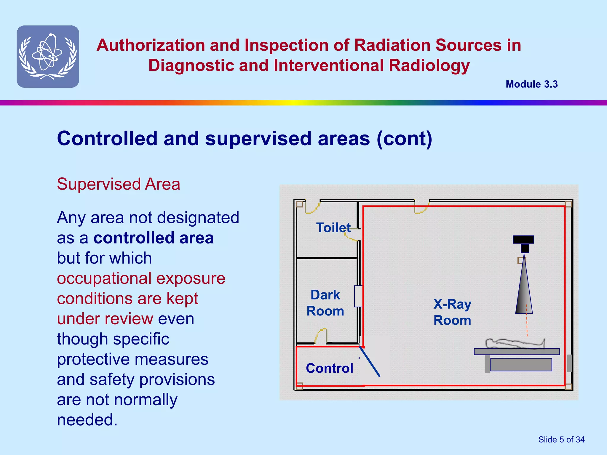
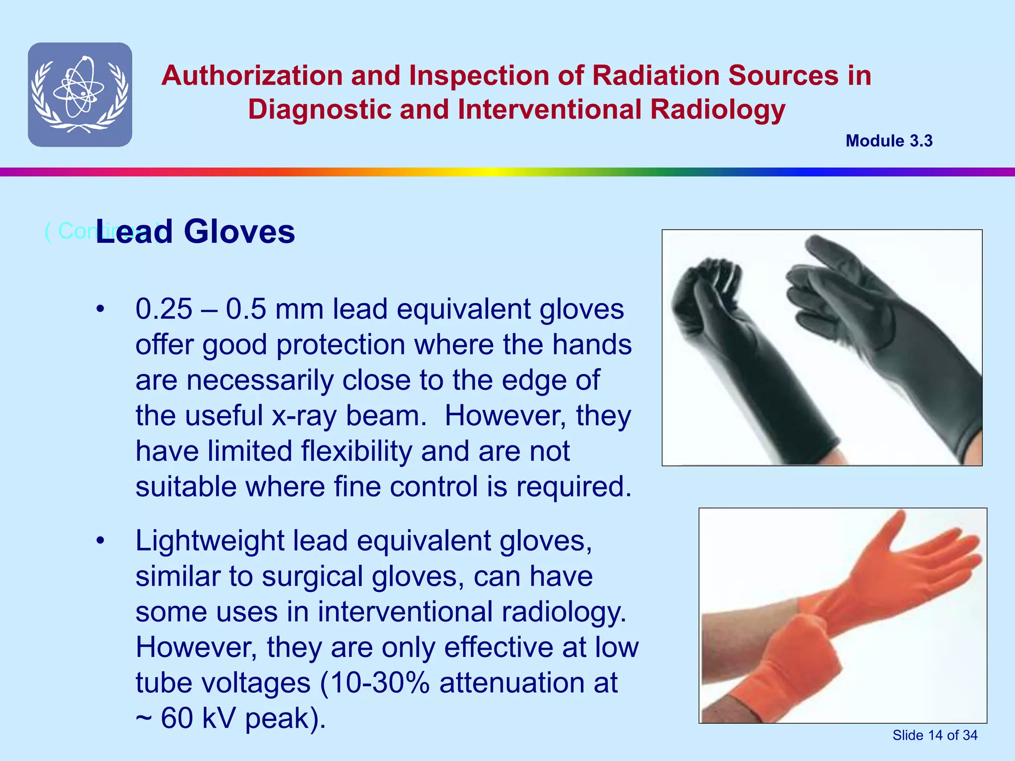
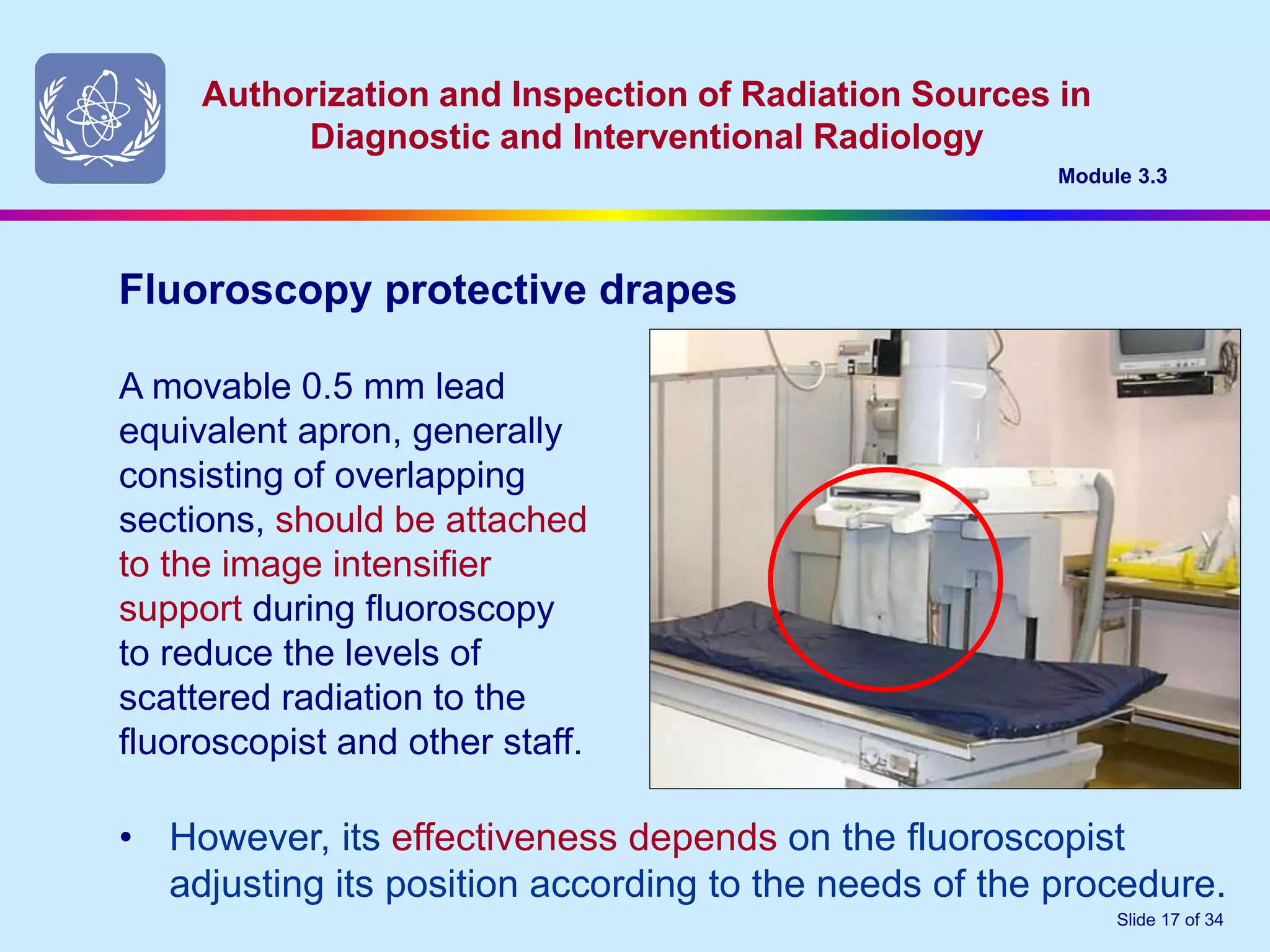
1. Authorization and Inspection of Radiation Sources in
Diagnostic and Interventional Radiology
Module 3.3
Slide 1 of 34
KITIMBO GODFREY
SENIOR RADIOGRAPHER
INCHARGE CT SCAN UNIT.
RADIATION SAFETY OFFICER
RADIOLOGY DEPT.MULAGO HOSPITAL.
2. Authorization and Inspection of Radiation Sources in
Diagnostic and Interventional Radiology
Module 3.3
Slide 2 of 34
Staff Radiation Protection
3. Authorization and Inspection of Radiation Sources in
Diagnostic and Interventional Radiology
Module 3.3
Slide 3 of 34
Objective
• To foster a safety culture that is encouraged by
management and supported by staff, including the
assessment of controlled and supervised areas, the
provision and use of appropriate protective devices and
the provision of personal radiation monitoring.
4. Authorization and Inspection of Radiation Sources in
Diagnostic and Interventional Radiology
Module 3.3
Slide 4 of 34
Controlled Area
Controlled and supervised areas
(a) controlling normal
exposures during normal
working conditions; and
(b) preventing or limiting the
extent of potential
exposures.
A controlled area is any
area in which specific
protection measures and
safety provisions are or
could be required for:
Toilet
Dark
Room
X-Ray
Room
Control
5. Authorization and Inspection of Radiation Sources in
Diagnostic and Interventional Radiology
Module 3.3
Slide 5 of 34
Controlled and supervised areas (cont)
Supervised Area
Any area not designated
as a controlled area
but for which
occupational exposure
conditions are kept
under review even
though specific
protective measures
and safety provisions
are not normally
needed.
Toilet
Dark
Room
X-Ray
Room
Control
6. Authorization and Inspection of Radiation Sources in
Diagnostic and Interventional Radiology
Module 3.3
Slide 6 of 34
Workplace monitoring shall be carried out :
• before initiating operation of a new installation;
Workplace Monitoring
• when there is a change in the structural (or other)
shielding that may affect radiation levels in surrounding
areas; and
• following maintenance or repair of x-ray equipment that
may impact on the x ray tube output by increasing
radiation levels.
7. Authorization and Inspection of Radiation Sources in
Diagnostic and Interventional Radiology
Module 3.3
Slide 7 of 34
• the position of the tube
and the direction of the
primary beam
B
C
D
Toilet
Dark
Room X-Ray Room
A
CONTROL
In carrying out a workplace
area monitoring, consider:
Workplace Monitoring (cont)
• the position of the
patient
• adjacent areas that
bound the controlled
area
• locations at which measurements might be taken.
8. Authorization and Inspection of Radiation Sources in
Diagnostic and Interventional Radiology
Module 3.3
Slide 8 of 34
The licensee shall ensure that all persons engaged in activities
that involve occupational radiation exposure are provided
with:-
The licensee
• appropriate protective clothing, and
• individual monitoring devices from an approved service
provider
However, protective clothing must not be used as a
substitute for proper protective measures.
9. Authorization and Inspection of Radiation Sources in
Diagnostic and Interventional Radiology
Module 3.3
Slide 9 of 34
• No person under the age of 16 years shall be subjected
to occupational exposure.
Age limitations
• No person under the age of 18 years shall be allowed
to work in a controlled area unless supervised, and
only for the purpose of training.
10. Authorization and Inspection of Radiation Sources in
Diagnostic and Interventional Radiology
Module 3.3
Slide 10 of 34
Protective clothing and related devices that may be used in a
radiology facility include:-
• Lead equivalent aprons
• Lead equivalent gloves
• Thyroid shields
• Protective eye – wear
Protective clothing and related devices
11. Authorization and Inspection of Radiation Sources in
Diagnostic and Interventional Radiology
Module 3.3
Slide 11 of 34
• should be stored on hangers and not folded.
Lead aprons
• those with suspected damage should be
reported immediately and the apron taken
out of use until tested and declared safe.
• should be assigned an ID code and tested
at regular intervals (e.g. 12-18 months) for
shielding integrity.
• should be equivalent to not less than 0.25 mm lead (up to 100
kV peak) and 0.35mm lead above 100 kV peak. Interventional
radiology staff should always use 0.5 mm lead equivalent wrap-
around aprons
12. Authorization and Inspection of Radiation Sources in
Diagnostic and Interventional Radiology
Module 3.3
Slide 12 of 34
• With potentially high radiation doses and
dose rates during interventional radiology,
the specialist medical practitioner i.e.
radiologist, cardiologist, neuro-radiologist,
etc., should use thyroid protection.
Thyroid Protection
• However, proper use of a suspended protective barrier
between the patient and the interventionist can reduce the
need for separate thyroid protection.
13. Authorization and Inspection of Radiation Sources in
Diagnostic and Interventional Radiology
Module 3.3
Slide 13 of 34
• In some interventional radiology
procedures, the lens of the
interventionist’s eyes may receive
annual radiation doses which approach,
or might exceed, 150 mSv.
( Continue )
Protective eyewear
• Lead equivalent glasses or face shields
should be used In these cases and
should include side protection.
• However, the proper use of a suspended protective barrier
between the patient and the interventionist may reduce the
need for separate eye protection.
14. Authorization and Inspection of Radiation Sources in
Diagnostic and Interventional Radiology
Module 3.3
Slide 14 of 34
• 0.25 – 0.5 mm lead equivalent gloves
offer good protection where the hands
are necessarily close to the edge of
the useful x-ray beam. However, they
have limited flexibility and are not
suitable where fine control is required.
( Continue )
Lead Gloves
• Lightweight lead equivalent gloves,
similar to surgical gloves, can have
some uses in interventional radiology.
However, they are only effective at low
tube voltages (10-30% attenuation at
~ 60 kV peak).
15. Authorization and Inspection of Radiation Sources in
Diagnostic and Interventional Radiology
Module 3.3
Slide 15 of 34
• Ceiling suspended protective screens.
Other protective devices
• Protective lead drapes
mounted on the patient table.
16. Authorization and Inspection of Radiation Sources in
Diagnostic and Interventional Radiology
Module 3.3
Slide 16 of 34
• Lead equivalent glass or acrylic
windows in operator’s protective
screens allow observation of the
patient during the x-ray procedure.
They should be marked with their
lead equivalence and the
maximum tube voltage (kV peak)
at which this applies.
Viewing windows
• These barriers are intended to protect the operator from
scattered radiation. The useful x-ray beam should not be
pointed in their direction. Ideally, the x-ray tube and patient
should be approximately 1.5 – 2 metres distant.
17. Authorization and Inspection of Radiation Sources in
Diagnostic and Interventional Radiology
Module 3.3
Slide 17 of 34
A movable 0.5 mm lead
equivalent apron, generally
consisting of overlapping
sections, should be attached
to the image intensifier
support during fluoroscopy
to reduce the levels of
scattered radiation to the
fluoroscopist and other staff.
Fluoroscopy protective drapes
• However, its effectiveness depends on the fluoroscopist
adjusting its position according to the needs of the procedure.
18. Authorization and Inspection of Radiation Sources in
Diagnostic and Interventional Radiology
Module 3.3
Slide 18 of 34
• All lead equivalent vinyl material (aprons, gloves, etc) should
comply with relevant international standards. They should be
tested at purchase and regularly thereafter, at least every 2
years.
Protective Devices – Quality Control
• Incorrect storage may lead to cracks in the shielding but this
may not be detectable by visual inspection alone
• A simple test is to examine the devices using fluoroscopy (at
about 60 kVp).
Faulty devices should be discarded immediately
19. Authorization and Inspection of Radiation Sources in
Diagnostic and Interventional Radiology
Module 3.3
Slide 19 of 34
• Radiologists;
• Radiographers;
• Qualified experts and persons servicing x-ray equipment;
• Radiation Safety Officer;
• Nurses and others as appropriate;
• Specialist medical practitioners (e.g. anaesthetists, cardiologists,
surgeons, etc.;
• Ancillary workers who occasionally work in controlled areas may
also need to be monitored
Individual monitoring devices shall be provided to every staff
member who might normally be exposed to radiation in controlled
areas. This may include:-
Personal Monitoring
20. Authorization and Inspection of Radiation Sources in
Diagnostic and Interventional Radiology
Module 3.3
Slide 20 of 34
The Radiation Protection Officer shall:-
• identify personnel who need individual monitoring;
• identify the person responsible for issuing and exchanging
dosimeters;
• issue, as appropriate, extremity dosimeters for special tasks;
• advise individuals of their dose results and maintain records;
Personal Monitoring Devices
• the location on the body where dosimeters will be worn;
• the monitoring period;
• the procedure for dealing with lost individual dosimeters
Subject to the requirements of the Regulatory Authority, determine:-
21. Authorization and Inspection of Radiation Sources in
Diagnostic and Interventional Radiology
Module 3.3
Slide 21 of 34
Each dosimeter shall be used only by the person to whom it is
assigned
Personal Monitoring Devices (cont)
Staff shall always wear their personal dosimeters while working
in controlled areas. The dosimeter should be worn on the front
of the upper torso of their body between the shoulders and the
waist, usually under any lead protective apron.
22. Authorization and Inspection of Radiation Sources in
Diagnostic and Interventional Radiology
Module 3.3
Slide 22 of 34
In some radiology departments, three dosimeters may be
issued to interventional medical practitioners. One to be worn
under the lead apron, one outside the apron, and a third
dosimeter may be attached to a finger to obtain an estimate of
the dose equivalent to the hands.
Where personnel work at more than one facility, separate
dosimeters are usually issued so that the location of any
unusual doses can be determined. However, records should
account for the doses received at individual locations and the
appropriate dose constraint applied.
Personal Monitoring Devices (cont)
23. Authorization and Inspection of Radiation Sources in
Diagnostic and Interventional Radiology
Module 3.3
Slide 23 of 34
Personal Monitoring Devices (cont)
When wearing a protective apron and two dosimeters
are used; one under and one outside the lead apron,
the effective dose can be estimated as follows:
Effective dose = 0.5HW + 0.025 HN
where:
HW = dose at waist level worn under the apron
HN = dose at neck level worn outside the apron.
24. Authorization and Inspection of Radiation Sources in
Diagnostic and Interventional Radiology
Module 3.3
Slide 24 of 34
• The monitoring period for occupationally exposed staff
should be one month but shall not exceed three months;
• Dose reports should be provided to users within one month
(or as may be specified by the Regulatory Authority).
Personal Monitoring Devices (cont)
25. Authorization and Inspection of Radiation Sources in
Diagnostic and Interventional Radiology
Module 3.3
Slide 25 of 34
• the RSO should perform a dose assessment and record this
evaluation of the dose and add it to the worker’s dose
record. Where there is a national dose registry, they should
be advised of the dose estimate.
Personal Monitoring Devices (cont)
If an individual’s dosimeter is lost:-
The most reliable method for estimating an individual’s dose is
to use his or her recent dose history. In those cases where the
individual performs non-routine work, it may be appropriate to
use co-workers’ doses as a basis for the dose estimate.
26. Authorization and Inspection of Radiation Sources in
Diagnostic and Interventional Radiology
DISTRIBUTION OF TLDS
Each radiation worker will get 1 TLD
ISSUE OF TLDS
The prescribed wearing periods will be three (3)months except for radiotherapy and
mines where it will be (1)month
The dispatch will be made about 12 to 14 days before the commencement of each
period.
Receipt of the TLDs is assumed unless the AEC is notified at this time .
27. Authorization and Inspection of Radiation Sources in
Diagnostic and Interventional Radiology
Module 3.3
Slide 27 of 34
Prior to issue ,TLDs will be individually numbered for ease of identification by the
individuals wearing them in the facility and in the processing system used in the
AEC to evaluate the doses received .
Care must be taken to ensure that the correct monitor number is issued to the
wearer listed against that monitor number on the Monitor Details Forms .
The number of TLDs issued in a batch will be equal to the number of persons to be
monitored. (except where an individual wears more than one monitor) plus a
control.
AEC will send the TLDs to the facilities/ users.
28. Authorization and Inspection of Radiation Sources in
Diagnostic and Interventional Radiology
Module 3.3
Slide 28 of 34
WEARING OF TLDS
TLDs will normally be issued to individual wearers who are registered
with the AEC .
If unnamed TLDs are issued , the wearer`s name and wearer
identification number should be written against the TLDs number
allotted to the wearer on the TLD forms.
29. Authorization and Inspection of Radiation Sources in
Diagnostic and Interventional Radiology
Module 3.3
Slide 29 of 34
If doses to un shielded parts of the body, such as
fingers, head ,etc are to be determined , then TLDs
should be worn on or as close as possible to the
region of interest.
Whenever the TLD is worn on the body , this should be
indicated on the TLDs forms when returning them to
the AEC .
Out side of working hours, or when not in use, TLDs
should not be worn but kept in a known storage
place.
30. Authorization and Inspection of Radiation Sources in
Diagnostic and Interventional Radiology
Module 3.3
Slide 30 of 34
If doses to un shielded parts of the body, such as
fingers, head ,etc are to be determined , then TLDs
should be worn on or as close as possible to the
region of interest.
Whenever the TLD is worn on the body , this should be
indicated on the TLDs forms when returning them to
the AEC .
Out side of working hours, or when not in use, TLDs
should not be worn but kept in a known storage
place.
31. Authorization and Inspection of Radiation Sources in
Diagnostic and Interventional Radiology
Module 3.3
Slide 31 of 34
This is necessary to ensure environmental conditions do
not adversely affect the TLDs and to reduce the
possibility of their loss.
This also avoids the delays that frequently arise in
returning TLDs to the AEC due to an individual being on
leave at the end of wearing period and not having left the
monitor in the appropriate storage space .
32. Authorization and Inspection of Radiation Sources in
Diagnostic and Interventional Radiology
Module 3.3
Slide 32 of 34
In the event that an individual has to undergo medical procedures which use
radiation , the TLD should not be worn but placed in an area of low
background radiation .
Doses from such procedures are not to be included as part of the dose from
occupation exposure .
A monitor should be worn by one person only.
If it is shared by two or more people , it is subsequently impossible to
determine the dose received by each person .
33. Authorization and Inspection of Radiation Sources in
Diagnostic and Interventional Radiology
Module 3.3
Slide 33 of 34
RETURN OF TLDs
At the end of each period, the TLDs issued for that period, and
the original of the TLDform , should be returned to AEC .
A duplicate copy of the TLD form should be retained for your
reference as some of the information on it is not repeated on
the dose forms that are referred to later .
Any delays in the return may lead to errors in the assessment of
doses of the TLDs , due to the possibilities of TLDs being
exposed un knowingly to radiation or to changes in their
characteristics
34. Authorization and Inspection of Radiation Sources in
Diagnostic and Interventional Radiology
Module 3.3
Slide 34 of 34
Prompt return also makes it possible to determine if any usually high doses
have been received, and action can then be taken to investigate such
doses in order to avoid recurrence in the future.
All TLDs should be returned after the wearing period even if they have not
been used.
A charge will be made for TLD cards that are not returned to the AEC in14
working days .
Facilities or users will bring the TLD s back to AEC
35. Authorization and Inspection of Radiation Sources in
Diagnostic and Interventional Radiology
Module 3.3
Slide 35 of 34
RADIATION DOSE FORMS AND RECORDS
Reports on the doses received by the TLD s will be issued to a
facility as soon as possible after assessment
In the event of a very high dose being received, advise to this
effect will be made by telephone to the facility .
It is recommended that a qualified Expert and or RSO
investigates the reasons for such doses in order to reduce
the possibility of any further high doses arising from the same
source .
36. Authorization and Inspection of Radiation Sources in
Diagnostic and Interventional Radiology
Module 3.3
Slide 36 of 34
The dose form lists the doses received for the
specified wearing period for all the staff of the
facility .
Every authorised person shall keep records of
individual workers up to the age of 75 years
and for at least not less than 30years.
37. Authorization and Inspection of Radiation Sources in
Diagnostic and Interventional Radiology
Module 3.3
Slide 37 of 34
There will be a charge for non returned or irreparably damaged
TLD cards .
Payments should be made before the issue of TLDs where
applicable .
Cheques and money orders should be made payable to the
atomic energy council account .
If the person dosimetry service is discontinued, all TLDs and
their accessories must be returned to AEC
38. Authorization and Inspection of Radiation Sources in
Diagnostic and Interventional Radiology
Module 3.3
Slide 38 of 34
ENQUIRIES
Requests for changes in requirements and enquiries
relating to assessment of reported doses , etc
should be addressed to radiation protection officer
(dosimetry) ; atomic energy council, ministry of
energy and mineral development , amber house ,
kampala road.
39. Authorization and Inspection of Radiation Sources in
Diagnostic and Interventional Radiology
Module 3.3
Slide 39 of 34
THANK YOU FOR LISTENING ACTIVELY.