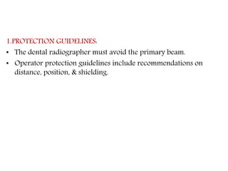
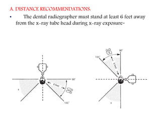
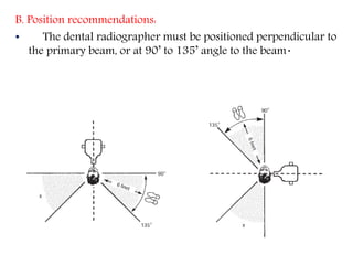
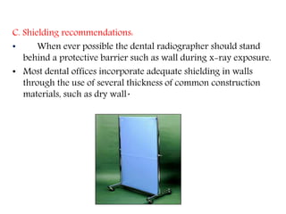
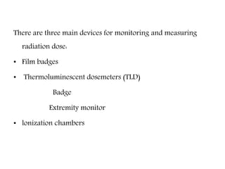
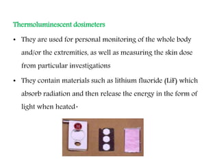
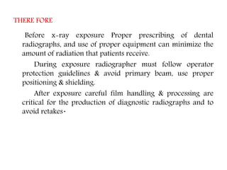
RADIATION SAFETY AND PROTECTION
PART-2
OPERATOR PROTECTION
• Operator protection measures include following protection
guide lines & using radiation monitoring devices.
1. Protection guidelines.
2. Radiation monitoring Devices.
1.PROTECTION GUIDELINES:
• The dental radiographer must avoid the primary beam.
• Operator protection guidelines include recommendations on
distance, position, & shielding.
A. DISTANCE RECOMMENDATIONS:
• The dental radiographer must stand at least 6 feet away
from the x-ray tube head during x-ray exposure.
B. Position recommendations:
• The dental radiographer must be positioned perpendicular to
the primary beam, or at 90’ to 135’ angle to the beam.
C. Shielding recommendations:
• When ever possible the dental radiographer should stand
behind a protective barrier such as wall during x-ray exposure.
• Most dental offices incorporate adequate shielding in walls
through the use of several thickness of common construction
materials, such as dry wall.
2. RADIATION MONITORING:
a. Equipment monitoring:
leakage radiation is any radiation with the exception of primary
beam, that is emitted from dental tube head.
• dental x-ray equipment must be monitored for leakage
radiation through the use of film device that can be obtained
through the state health department or manufacturers of dental
x-ray equipment.
b. Personnel monitoring:
• -the amount of x-radiation that reaches the body of
dental radiographer can be measured through the use of
personnel monitoring devices.
There are three main devices for monitoring and measuring
radiation dose:
• Film badges
• Thermoluminescent dosemeters (TLD)
Badge
Extremity monitor
• lonization chambers
Film badges
Film badges
The main features of film badges are:
• They consist of a blue plastic frame containing a variety of different
metal filters and a small radiographic film which reacts to radiation
• They are worn on the outside of the clothes, usually at the waist
level, for 1-3 months before being processed
• They are the most common form of personal monitoring device
currently in use.
Advantages
• Provides a permanent record of dose received.
• May be checked and reassessed at a later date.
• Can measure the type and energy of radiation encountered.
• Simple, robust and relatively inexpensive.
Disadvantages
• No immediate indication of exposure — all information is
retrospective.
• Processing is required which may lead to errors.
• The badges are prone to filter loss.
Thermoluminescent dosimeters
• They are used for personal monitoring of the whole body
and/or the extremities, as well as measuring the skin dose
from particular investigations
• They contain materials such as lithium fluoride (LiF) which
absorb radiation and then release the energy in the form of
light when heated.
• The intensity of the emitted light is proportional to the
radiation energy absorbed originally
• Personal monitors consist of a yellow or orange plastic
holder, worn like the film badge for 1-3 months.
Advantages
• The lithium fluoride is re-usable.
• Read-out measurements are easily automated and rapidly
produced.
• Suitable for a wide variety of dose measurements.
Disadvantages
• Read-out is destructive, giving no permanent record, results
cannot be checked or reassessed.
• Only limited information provided on the type and energy of
the radiation.
• Dose gradients are not detectable.
• Relatively expensive.
Ionization chambers
• They are used for personal monitoring (thimble chamber) and
by physicists (free-air chamber) to measure radiation
exposure
• Radiation produces ionization of the air molecules inside the
closed chamber, which results in a measurable discharge and
hence a direct read-out
• They are available in many different sizes and forms.
Advantages
• The most accurate method of measuring radiation dose.
• Direct read-out gives immediate information.
Disadvantages
• They give no permanent record of exposure.
• No indication of the type or energy of the radiation.
• Personal ionization monitors are not very sensitive to low-energy
radiation.
• They are fragile and easily damaged.
• PROTECTION OF THE ENVIRONMENT:
• Primary beam should never be directed at any one other than the
pateint.
• Patient should be positioned such that the x-ray beam is aimed at
the wall of the room & not through a door or other opening
where people maybe located.
• Walls made of 3” concrete, 3”x16” of steel or 1mm of lead will
suffice to protect adjacent rooms, even if the work load in the
radiology department is high.
• Windows: it is necessary for the operator to see the patient as he
is being irradiated, so a window is provided this window should
be situated such that the primary beam is not directed on it. Lead
glass should be used.
• Doors: of the radiology room should function as secondary
barriers. They may have lead incorporated in them.
• Quality assurance: defined as any planned activity to ensure
that a dental office will consistently produce high quality
images with the minimum exposure to patient & personnel.
• Continuing education: practitioners should stay informed of
new information on radiation safety issues as well as
developments in equipment, materials and techniques &
adopt appropriate items to improve radiographic practise.
• Radiation monitoring .
RADIATION EXPOSURE GUIDE LINES
• These guide lines include safety legislation & exposure limits
for the general public & for person's who are occupationally
exposed to radiation.
1. Radiation safety legislation:
• This has been established at both state & federal level to
protect the patient , operator & general public from radiation
hazards‘.
• At the federal level the radiation control for health & safety
act was enacted in 1968 to standardize the performance of
x-ray equipment.
• Also the federal Consumer-Patient Radiation Health & Safety
Act was enacted in 1981 to address the issues of education &
certification of persons using radiographic equipment
PATIENT EXPOSURE AND DOSE
• Patient dose from dental radiography is usually reported as
the amount of radiation received by a target organ.
• One of the most common measurements is skin or surface
exposure.
• The surface exposure, obtained by direct measurement, is the
simplest way to record a patient's exposure to x rays
The mean active bone marrow dose
Intraoral full-mouth survey of 21 films exposed
• with round collimation is 0.142 mSv;
• rectangular collimation - 0.06mSv.
Panoramic radiography - 0.01 mSv per film.
• The dose to the thyroid from oral radiography is fairly low.
• A 21-film complete mouth examination results in a thyroid
dose of O.94mGy one sixth that resulting from a
radiographic examination of the cervical spine.
• Likewise, the thyroid dose from panoramic radiography is
about 0.074mGy, 1 % that from a cervical spinal
examination.
Gonad Dose
• Dental x-ray examinations result in a genetically
insignificant dose of only 0.001 mGy.
2. Maximum permissible dose:
• The maximum permissible dose is defined by national council
on radiation protection & measurements (NCRP) as the
maximum dose equivalent that a body can receive in a
specific period of time.
• The recent report in 2003 states the current MPD for
occupationally exposed persons or persons who work with
radiation is 5.0 rems/year non-occupationally exposed
persons is 0.1rem/year.
• The MPD for an occupationally exposed pregnant women is
same as that for non occupationally exposed person or
0.1rem/year.
These dose limits apply only to exposure from manmade sources
and do not apply to either natural radiation or radiation
exposure that patients receive in the course of dental and
medical treatment
3. MAXIMUM ACCUMULATED DOSE:
• occupationally exposed workers must not exceed an
accumulated lifetime radiation. This is referred to as
maximum accumulated dose (MAD).
• MAD=(N-18) X 5rems/year.
• MAD=(N-18) X 0.05Sv/year.
ALARA CONCEPT
• The ALARA concept states that all exposure to radiation
must be kept to a minimum , or “as low as reasonably
achievable,”
• To provide protection for both patients & operators, every
possible method of reducing exposure to radiation should be
employed to minimize risk.
THERE FORE
Before x-ray exposure Proper prescribing of dental
radiographs, and use of proper equipment can minimize the
amount of radiation that patients receive.
During exposure radiographer must follow operator
protection guidelines & avoid primary beam, use proper
positioning & shielding.
After exposure careful film handling & processing are
critical for the production of diagnostic radiographs and to
avoid retakes.
THANK YOU..

RADIATION SAFETY AND PROTECTION-2.pptx

  • 1.
    RADIATION SAFETY ANDPROTECTION PART-2
  • 2.
    OPERATOR PROTECTION • Operatorprotection measures include following protection guide lines & using radiation monitoring devices. 1. Protection guidelines. 2. Radiation monitoring Devices.
  • 3.
    1.PROTECTION GUIDELINES: • Thedental radiographer must avoid the primary beam. • Operator protection guidelines include recommendations on distance, position, & shielding.
  • 4.
    A. DISTANCE RECOMMENDATIONS: •The dental radiographer must stand at least 6 feet away from the x-ray tube head during x-ray exposure.
  • 5.
    B. Position recommendations: •The dental radiographer must be positioned perpendicular to the primary beam, or at 90’ to 135’ angle to the beam.
  • 6.
    C. Shielding recommendations: •When ever possible the dental radiographer should stand behind a protective barrier such as wall during x-ray exposure. • Most dental offices incorporate adequate shielding in walls through the use of several thickness of common construction materials, such as dry wall.
  • 7.
    2. RADIATION MONITORING: a.Equipment monitoring: leakage radiation is any radiation with the exception of primary beam, that is emitted from dental tube head. • dental x-ray equipment must be monitored for leakage radiation through the use of film device that can be obtained through the state health department or manufacturers of dental x-ray equipment.
  • 8.
    b. Personnel monitoring: •-the amount of x-radiation that reaches the body of dental radiographer can be measured through the use of personnel monitoring devices.
  • 9.
    There are threemain devices for monitoring and measuring radiation dose: • Film badges • Thermoluminescent dosemeters (TLD) Badge Extremity monitor • lonization chambers
  • 10.
  • 11.
    Film badges The mainfeatures of film badges are: • They consist of a blue plastic frame containing a variety of different metal filters and a small radiographic film which reacts to radiation • They are worn on the outside of the clothes, usually at the waist level, for 1-3 months before being processed • They are the most common form of personal monitoring device currently in use.
  • 12.
    Advantages • Provides apermanent record of dose received. • May be checked and reassessed at a later date. • Can measure the type and energy of radiation encountered. • Simple, robust and relatively inexpensive.
  • 13.
    Disadvantages • No immediateindication of exposure — all information is retrospective. • Processing is required which may lead to errors. • The badges are prone to filter loss.
  • 14.
    Thermoluminescent dosimeters • Theyare used for personal monitoring of the whole body and/or the extremities, as well as measuring the skin dose from particular investigations • They contain materials such as lithium fluoride (LiF) which absorb radiation and then release the energy in the form of light when heated.
  • 15.
    • The intensityof the emitted light is proportional to the radiation energy absorbed originally • Personal monitors consist of a yellow or orange plastic holder, worn like the film badge for 1-3 months.
  • 16.
    Advantages • The lithiumfluoride is re-usable. • Read-out measurements are easily automated and rapidly produced. • Suitable for a wide variety of dose measurements.
  • 17.
    Disadvantages • Read-out isdestructive, giving no permanent record, results cannot be checked or reassessed. • Only limited information provided on the type and energy of the radiation. • Dose gradients are not detectable. • Relatively expensive.
  • 18.
    Ionization chambers • Theyare used for personal monitoring (thimble chamber) and by physicists (free-air chamber) to measure radiation exposure • Radiation produces ionization of the air molecules inside the closed chamber, which results in a measurable discharge and hence a direct read-out • They are available in many different sizes and forms.
  • 19.
    Advantages • The mostaccurate method of measuring radiation dose. • Direct read-out gives immediate information. Disadvantages • They give no permanent record of exposure. • No indication of the type or energy of the radiation. • Personal ionization monitors are not very sensitive to low-energy radiation. • They are fragile and easily damaged.
  • 20.
    • PROTECTION OFTHE ENVIRONMENT: • Primary beam should never be directed at any one other than the pateint. • Patient should be positioned such that the x-ray beam is aimed at the wall of the room & not through a door or other opening where people maybe located. • Walls made of 3” concrete, 3”x16” of steel or 1mm of lead will suffice to protect adjacent rooms, even if the work load in the radiology department is high. • Windows: it is necessary for the operator to see the patient as he is being irradiated, so a window is provided this window should be situated such that the primary beam is not directed on it. Lead glass should be used. • Doors: of the radiology room should function as secondary barriers. They may have lead incorporated in them.
  • 21.
    • Quality assurance:defined as any planned activity to ensure that a dental office will consistently produce high quality images with the minimum exposure to patient & personnel. • Continuing education: practitioners should stay informed of new information on radiation safety issues as well as developments in equipment, materials and techniques & adopt appropriate items to improve radiographic practise. • Radiation monitoring .
  • 22.
    RADIATION EXPOSURE GUIDELINES • These guide lines include safety legislation & exposure limits for the general public & for person's who are occupationally exposed to radiation. 1. Radiation safety legislation: • This has been established at both state & federal level to protect the patient , operator & general public from radiation hazards‘. • At the federal level the radiation control for health & safety act was enacted in 1968 to standardize the performance of x-ray equipment.
  • 23.
    • Also thefederal Consumer-Patient Radiation Health & Safety Act was enacted in 1981 to address the issues of education & certification of persons using radiographic equipment
  • 24.
    PATIENT EXPOSURE ANDDOSE • Patient dose from dental radiography is usually reported as the amount of radiation received by a target organ. • One of the most common measurements is skin or surface exposure. • The surface exposure, obtained by direct measurement, is the simplest way to record a patient's exposure to x rays
  • 25.
    The mean activebone marrow dose Intraoral full-mouth survey of 21 films exposed • with round collimation is 0.142 mSv; • rectangular collimation - 0.06mSv. Panoramic radiography - 0.01 mSv per film.
  • 26.
    • The doseto the thyroid from oral radiography is fairly low. • A 21-film complete mouth examination results in a thyroid dose of O.94mGy one sixth that resulting from a radiographic examination of the cervical spine. • Likewise, the thyroid dose from panoramic radiography is about 0.074mGy, 1 % that from a cervical spinal examination.
  • 27.
    Gonad Dose • Dentalx-ray examinations result in a genetically insignificant dose of only 0.001 mGy.
  • 28.
    2. Maximum permissibledose: • The maximum permissible dose is defined by national council on radiation protection & measurements (NCRP) as the maximum dose equivalent that a body can receive in a specific period of time. • The recent report in 2003 states the current MPD for occupationally exposed persons or persons who work with radiation is 5.0 rems/year non-occupationally exposed persons is 0.1rem/year. • The MPD for an occupationally exposed pregnant women is same as that for non occupationally exposed person or 0.1rem/year.
  • 29.
    These dose limitsapply only to exposure from manmade sources and do not apply to either natural radiation or radiation exposure that patients receive in the course of dental and medical treatment
  • 32.
    3. MAXIMUM ACCUMULATEDDOSE: • occupationally exposed workers must not exceed an accumulated lifetime radiation. This is referred to as maximum accumulated dose (MAD). • MAD=(N-18) X 5rems/year. • MAD=(N-18) X 0.05Sv/year.
  • 33.
    ALARA CONCEPT • TheALARA concept states that all exposure to radiation must be kept to a minimum , or “as low as reasonably achievable,” • To provide protection for both patients & operators, every possible method of reducing exposure to radiation should be employed to minimize risk.
  • 34.
    THERE FORE Before x-rayexposure Proper prescribing of dental radiographs, and use of proper equipment can minimize the amount of radiation that patients receive. During exposure radiographer must follow operator protection guidelines & avoid primary beam, use proper positioning & shielding. After exposure careful film handling & processing are critical for the production of diagnostic radiographs and to avoid retakes.
  • 35.

Editor's Notes

  • #6 As primary beam travels in straight line
  • #31 NCRP- NATINAL COUNCIL ON RADIATION PROTECTION AND MEASUREMENTS ICRP- INTERNATIONAL COMMISSION ON RADIATION PROTECTIO AND MONITORING