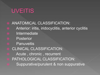
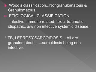
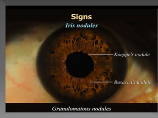
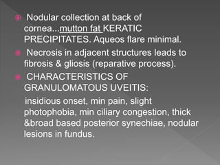
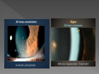
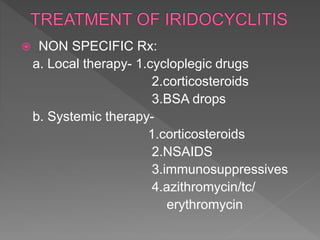
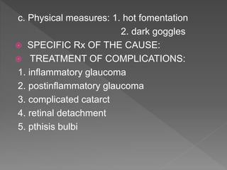
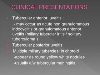
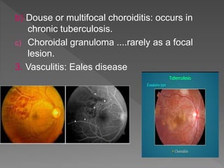
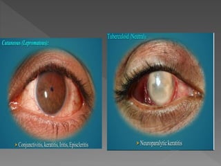
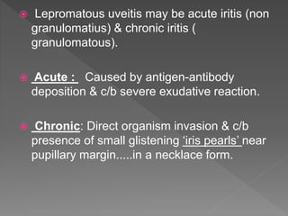
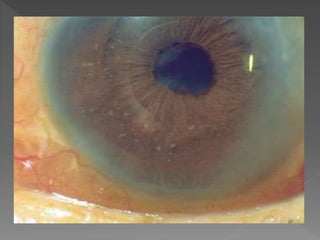
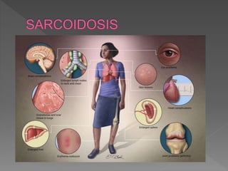
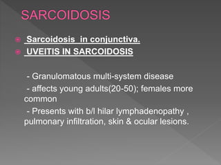
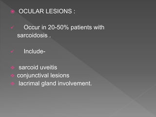
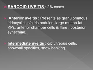
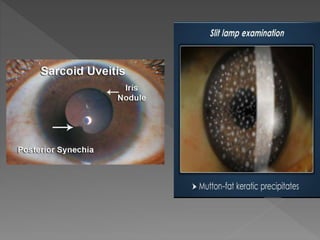
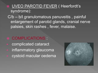
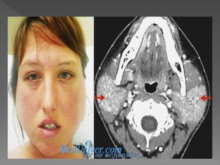
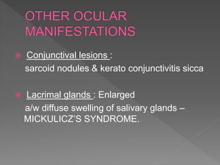
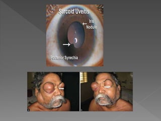
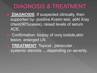
 TUBERCULOSIS
 LEPROSY
 SARCOIDOSIS
 PHLYCTENULAR KERATO
CONJUNCTIVITIS:
- Etiology: Delayed hypersensitivity type 4 CM
response to endogenous microbial
proteins(tuberculous)
* Presents as whitish raised nodule on bulbar
conjunctiva with hyperaemia of surrounding
conjunctiva in a child living in bad hygiene
conditions
 PATHOLOGY:
1. Stage of nodule formation
2. Stage of ulceration
3. Stage of granulation
4. Stage of healing
 CLINICAL FEATURES:
Mild discomfort in eye
Irritation
Reflex watering
 SIGNS: SIMPLE, NECROTIC, MILIARY.
 PHLYCTENULAR KERATITIS:
1. Ulcerative phlyctenular keratitis:
a. Sacrofulous ulcer
b. Fascicular ulcer
c. Miliary ulcer
2. Diffuse infiltrative keratitis
 CLINICAL COURSE:
self limiting & phlycten disappears in 8-10 days
.....no trace left. Recurrences are very common.
 ANATOMICAL CLASSIFICATION:
 Anterior: iritis, iridocyclitis, anterior cyclitis
 Intermediate
 Posterior
 Panuveitis
 CLINICAL CLASSIFICATION:
 Acute , chronic , recurrent
 PATHOLOGICAL CLASSIFICATION:
 Suppurative/purulent & non suppurative
 Wood’s classification...Nongranulomatous &
Granulomatous
 ETIOLOGICAL CLASSIFICATION:
Infective, immune related, toxic, traumatic ,
idiopathic, a/w non infective systemic disease.
* TB, LEPROSY,SARCOIDOSIS ...All are
granulomatous ......sarcoidosis being non
infective.
 In response to irritant foreign
body..inorganic/organic material introduced
from outside; hemorrhagic/necrotic tissue
within eye; organisms- non pyogenic & non
virulent.
 C/b granuloma formation...eventually
giantcells aggregate to form nodules.
 Iris nodules near pupillary
border.......KOEPPE’S NODULES
 Near collarette....BUSACCA NODULES
 Nodular collection at back of
cornea...mutton fat KERATIC
PRECIPITATES. Aqueos flare minimal.
 Necrosis in adjacent structures leads to
fibrosis & gliosis (reparative process).
 CHARACTERISTICS OF
GRANULOMATOUS UVEITIS:
insidious onset, min pain, slight
photophobia, min ciliary congestion, thick
&broad based posterior synechiae, nodular
lesions in fundus.
 NON SPECIFIC Rx:
a. Local therapy- 1.cycloplegic drugs
2.corticosteroids
3.BSA drops
b. Systemic therapy-
1.corticosteroids
2.NSAIDS
3.immunosuppressives
4.azithromycin/tc/
erythromycin
c. Physical measures: 1. hot fomentation
2. dark goggles
 SPECIFIC Rx OF THE CAUSE:
 TREATMENT OF COMPLICATIONS:
1. inflammatory glaucoma
2. postinflammatory glaucoma
3. complicated catarct
4. retinal detachment
5. pthisis bulbi
 TUBERCULAR UVEITIS:
-Chronic granulomatous infection
-human or bovine tubercle bacilli
-both anterior & posterior uveitis
-common cause of uveitis
- accoumts for 1% of uveitis in developed
countries.
- In India, still a common cause.
1. Tubercular anterior uveitis :
- may occur as acute non granulomatous
iridocyclitis or granulomatous anterior
uveitis (miliary tuberclar iritis / solitary
tuberculoma )
2. Tubercular posterior uveitis:
a) Multiple miliary tubercles: in choroid
-appear as round yellow white nodules
-usually a/w tubercular meningitis.
b) Douse or multifocal choroiditis: occurs in
chronic tuberculosis.
c) Choroidal granuloma ....rarely as a focal
lesion.
3. Vasculitis: Eales disease
 INTERSTITIAL KERATITIS:
- Form of deep keratitis
- Inflammation of corneal stroma w/o primary
involvement of epithelium/endothelium.
-More frequently unilateral & sectorial(lower part of
cornea).
• CLINICAL FEATURES:
• Initial progressive stage
• Florid stage
• Stage of regression
 Scleritis & Episcleritis also occur .
 DIAGNOSIS:
1. X-Ray Chest
2. Mantoux test
3. TLC,DLC,ESR
4. Intractable uveitis – unresponsive to
steroids.
5. Isoniazid test (dramatic response of iritis to
isoniazid).
 TREATMENT:
1. Local therapy: Atropine
Antibiotics
Topical steroids
2. Specific therapy: ANTI TUBERCULAR
DRUGS.
* General measures : Vitamins A, C ,D in case of
children along with high protein supplemented
diet (phlyctenular keratoconjuctivitis) .
 HANSEN’S DISEASE
 Mycobacterium leprae
 Lepromatous & tuberculoid forms
 Conjunctivitis
 Neurotrophic keratopathy.....loss of corneal
nerve sensations.
 Involves anterior uvea
 Lepromatous uveitis may be acute iritis (non
granulomatius) & chronic iritis (
granulomatous).
 Acute : Caused by antigen-antibody
deposition & c/b severe exudative reaction.
 Chronic: Direct organism invasion & c/b
presence of small glistening ‘iris pearls’ near
pupillary margin.....in a necklace form.
 small pearls enlarge & coalesce to form
large pearls .
 rarely lepromata nodule may be seen.
 TREATMENT:
 Local therapy of iridocyclitis
 Antileprotic Rx – Dapsone 50-100 mg daily
 Sarcoidosis in conjunctiva.
 UVEITIS IN SARCOIDOSIS
- Granulomatous multi-system disease
- affects young adults(20-50); females more
common
- Presents with b/l hilar lymphadenopathy ,
pulmonary infiltration, skin & ocular lesions.
 OCULAR LESIONS :
 Occur in 20-50% patients with
sarcoidosis .
 Include-
 sarcoid uveitis
 conjunctival lesions
 lacrimal gland involvement.
 SARCOID UVEITIS : 2% cases
 Anterior uveitis : Presents as granulomatous
iridocyclitis c/b iris nodules, large mutton fat
KPs, anterior chamber cells & flare , posterior
synechiae.
 Intermediate uveitis : c/b vitreous cells,
snowball opacities, snow banking.
 Posterior / pan uveitis : includes –
choroidal & retinal granulomas , cystoid
macular oedema, periphlebitis retinae with
sheathing....appear as Candle wax
droppings.
* Peripheral multifocal chorioretinitis: c/b small
punched out atrophic spots....highly
suggestive of sarcoidosis.
 UVEO PAROTID FEVER ( Heerfordt’s
syndrome):
C/b – b/l granulomatous panuveitis , painful
enlargement of parotid glands, cranial nerve
palsies, skin rashes , fever, malaise.
 COMPLICATIONS :
 complicated cataract
 inflammatory glaucoma
 cystoid macular oedema
 Conjunctival lesions :
sarcoid nodules & kerato conjunctivitis sicca
 Lacrimal glands : Enlarged
a/w diffuse swelling of salivary glands –
MICKULICZ’S SYNDROME.
 DIAGNOSIS: If suspected clinically, then
supported by- positive Kveim test, abN Xray
chest(90%cases), raised levels of serum
ACE.
 Confirmation- biopsy of conj.nodule,skin
lesion, enlarged LN.
 TREATMENT: Topical , periocular ,
systemic steroids ....depending on severity.
Sravani gembali.pptx[1]

Sravani gembali.pptx[1]

  • 2.
  • 3.
     PHLYCTENULAR KERATO CONJUNCTIVITIS: -Etiology: Delayed hypersensitivity type 4 CM response to endogenous microbial proteins(tuberculous) * Presents as whitish raised nodule on bulbar conjunctiva with hyperaemia of surrounding conjunctiva in a child living in bad hygiene conditions
  • 4.
     PATHOLOGY: 1. Stageof nodule formation 2. Stage of ulceration 3. Stage of granulation 4. Stage of healing  CLINICAL FEATURES: Mild discomfort in eye Irritation Reflex watering  SIGNS: SIMPLE, NECROTIC, MILIARY.
  • 5.
     PHLYCTENULAR KERATITIS: 1.Ulcerative phlyctenular keratitis: a. Sacrofulous ulcer b. Fascicular ulcer c. Miliary ulcer 2. Diffuse infiltrative keratitis  CLINICAL COURSE: self limiting & phlycten disappears in 8-10 days .....no trace left. Recurrences are very common.
  • 6.
     ANATOMICAL CLASSIFICATION: Anterior: iritis, iridocyclitis, anterior cyclitis  Intermediate  Posterior  Panuveitis  CLINICAL CLASSIFICATION:  Acute , chronic , recurrent  PATHOLOGICAL CLASSIFICATION:  Suppurative/purulent & non suppurative
  • 10.
     Wood’s classification...Nongranulomatous& Granulomatous  ETIOLOGICAL CLASSIFICATION: Infective, immune related, toxic, traumatic , idiopathic, a/w non infective systemic disease. * TB, LEPROSY,SARCOIDOSIS ...All are granulomatous ......sarcoidosis being non infective.
  • 11.
     In responseto irritant foreign body..inorganic/organic material introduced from outside; hemorrhagic/necrotic tissue within eye; organisms- non pyogenic & non virulent.  C/b granuloma formation...eventually giantcells aggregate to form nodules.  Iris nodules near pupillary border.......KOEPPE’S NODULES  Near collarette....BUSACCA NODULES
  • 13.
     Nodular collectionat back of cornea...mutton fat KERATIC PRECIPITATES. Aqueos flare minimal.  Necrosis in adjacent structures leads to fibrosis & gliosis (reparative process).  CHARACTERISTICS OF GRANULOMATOUS UVEITIS: insidious onset, min pain, slight photophobia, min ciliary congestion, thick &broad based posterior synechiae, nodular lesions in fundus.
  • 15.
     NON SPECIFICRx: a. Local therapy- 1.cycloplegic drugs 2.corticosteroids 3.BSA drops b. Systemic therapy- 1.corticosteroids 2.NSAIDS 3.immunosuppressives 4.azithromycin/tc/ erythromycin
  • 16.
    c. Physical measures:1. hot fomentation 2. dark goggles  SPECIFIC Rx OF THE CAUSE:  TREATMENT OF COMPLICATIONS: 1. inflammatory glaucoma 2. postinflammatory glaucoma 3. complicated catarct 4. retinal detachment 5. pthisis bulbi
  • 17.
     TUBERCULAR UVEITIS: -Chronicgranulomatous infection -human or bovine tubercle bacilli -both anterior & posterior uveitis -common cause of uveitis - accoumts for 1% of uveitis in developed countries. - In India, still a common cause.
  • 18.
    1. Tubercular anterioruveitis : - may occur as acute non granulomatous iridocyclitis or granulomatous anterior uveitis (miliary tuberclar iritis / solitary tuberculoma ) 2. Tubercular posterior uveitis: a) Multiple miliary tubercles: in choroid -appear as round yellow white nodules -usually a/w tubercular meningitis.
  • 20.
    b) Douse ormultifocal choroiditis: occurs in chronic tuberculosis. c) Choroidal granuloma ....rarely as a focal lesion. 3. Vasculitis: Eales disease
  • 21.
     INTERSTITIAL KERATITIS: -Form of deep keratitis - Inflammation of corneal stroma w/o primary involvement of epithelium/endothelium. -More frequently unilateral & sectorial(lower part of cornea). • CLINICAL FEATURES: • Initial progressive stage • Florid stage • Stage of regression  Scleritis & Episcleritis also occur .
  • 22.
     DIAGNOSIS: 1. X-RayChest 2. Mantoux test 3. TLC,DLC,ESR 4. Intractable uveitis – unresponsive to steroids. 5. Isoniazid test (dramatic response of iritis to isoniazid).
  • 23.
     TREATMENT: 1. Localtherapy: Atropine Antibiotics Topical steroids 2. Specific therapy: ANTI TUBERCULAR DRUGS. * General measures : Vitamins A, C ,D in case of children along with high protein supplemented diet (phlyctenular keratoconjuctivitis) .
  • 24.
     HANSEN’S DISEASE Mycobacterium leprae  Lepromatous & tuberculoid forms  Conjunctivitis  Neurotrophic keratopathy.....loss of corneal nerve sensations.  Involves anterior uvea
  • 26.
     Lepromatous uveitismay be acute iritis (non granulomatius) & chronic iritis ( granulomatous).  Acute : Caused by antigen-antibody deposition & c/b severe exudative reaction.  Chronic: Direct organism invasion & c/b presence of small glistening ‘iris pearls’ near pupillary margin.....in a necklace form.
  • 28.
     small pearlsenlarge & coalesce to form large pearls .  rarely lepromata nodule may be seen.  TREATMENT:  Local therapy of iridocyclitis  Antileprotic Rx – Dapsone 50-100 mg daily
  • 30.
     Sarcoidosis inconjunctiva.  UVEITIS IN SARCOIDOSIS - Granulomatous multi-system disease - affects young adults(20-50); females more common - Presents with b/l hilar lymphadenopathy , pulmonary infiltration, skin & ocular lesions.
  • 31.
     OCULAR LESIONS:  Occur in 20-50% patients with sarcoidosis .  Include-  sarcoid uveitis  conjunctival lesions  lacrimal gland involvement.
  • 32.
     SARCOID UVEITIS: 2% cases  Anterior uveitis : Presents as granulomatous iridocyclitis c/b iris nodules, large mutton fat KPs, anterior chamber cells & flare , posterior synechiae.  Intermediate uveitis : c/b vitreous cells, snowball opacities, snow banking.
  • 35.
     Posterior /pan uveitis : includes – choroidal & retinal granulomas , cystoid macular oedema, periphlebitis retinae with sheathing....appear as Candle wax droppings. * Peripheral multifocal chorioretinitis: c/b small punched out atrophic spots....highly suggestive of sarcoidosis.
  • 37.
     UVEO PAROTIDFEVER ( Heerfordt’s syndrome): C/b – b/l granulomatous panuveitis , painful enlargement of parotid glands, cranial nerve palsies, skin rashes , fever, malaise.  COMPLICATIONS :  complicated cataract  inflammatory glaucoma  cystoid macular oedema
  • 39.
     Conjunctival lesions: sarcoid nodules & kerato conjunctivitis sicca  Lacrimal glands : Enlarged a/w diffuse swelling of salivary glands – MICKULICZ’S SYNDROME.
  • 41.
     DIAGNOSIS: Ifsuspected clinically, then supported by- positive Kveim test, abN Xray chest(90%cases), raised levels of serum ACE.  Confirmation- biopsy of conj.nodule,skin lesion, enlarged LN.  TREATMENT: Topical , periocular , systemic steroids ....depending on severity.