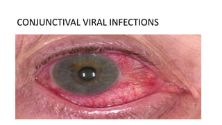
6. Signs
• Eyelids :
Edema, ranging from mild to severe.
• Lymphadenopathy : Common tender pre-auricular nodes.
• Conjunctiva:
Hyperemia, Follicles.
7. • Severe Inflammation:
May be associated with conjunctival haemorrhage, chemosis,
membranes(rare) and pseudomembranes.
Sometimes conjunctival scarring.
• Keratitis(Adenoviral):
Epithelial microcysts in the early stage.
8. • Punctate epithelial keratitis:
Usually occur in 7-10 days of onset of symptoms.
Resolving in 2 weeks.
• Anterior Stromal infiltrates/SEI: may persist for months or years.
• Anterior uveitis: Usually mild.
10. Spread of infection:-
• Facilitated by :
i)Virus can survive on dry surfaces for weeks.
ii)Viral shedding may occur for many days before clinical features are
apparent.
• Transmission by:
i)Contact with respiratory or ocular secretions.
ii)Via contaminated fomites such as towels.
iii)Route of transmission is usually eye-hands-eyes.
• In clinical settings, eye-instruments-eye.
13. Pharyngoconjunctival fever (PCF)
• Acute follicular conjunctivitis with pharyngitis.
• Occour as outbreaks in children summer camps(causing swimming
pool conjunctivitis).
• Associated with adenovirus type3 and 7.
• Cornea : superficial punctate keratitis. (30%)
14. Epidemic keratoconjunctivitis (EKC)
• Most severe presentation.
• Caused by adenoviruses type 8,19 and 37.
• Occour mainly in adults.
• Mostly associated with keratitis and preauricular lymphadenopathy.
• Markedly contagious.
• Incubation period after infection (8 days) & virus shed from the
inflamed eye for 2-3 weeks.
• Keratitis occurs in 80% cases.
16. Herpes simplex Virus:-
• Causes Follicular conjunctivitis particularly in primary disease.
• Usually unilateral.
• Often Associated skin lesions.
• Minute, micro dendrites may be mistaken for punctate epithelial
keratitis, but corneal sensation is reduced in HSV.
17. Zoster ophthalmicus
• Caused by infection of gasserian ganglion of fifth cranial nerve by
Varicella-zoster virus.
• Constitute almost 10% cases of zoster.
• Mucopurulent conjunctivitis.
18. Acute Haemorrhagic conjunctivitis:-
• Caused by Enterovirus A and coxsackie virus(Picorna
virus family).
• Extremely contagious.
• In acute inflammation conjunctiva characterised by conjunctival
haemorrhage, hyperaemia and mild follicular hyperplasia.
• Usually occurs in tropical areas.
• Rapid onset, resolves within 1-2 weeks.
19. MOLUSCUM CONTAGIOSM
• ds- DNA pox virus.
• Typically affects otherwise healthy individuals.
• Peak incidence between 2 to 4 years of age.
• Lesions on lid margins may shed virus into tear film giving rise to
secondary ipsilateral chronic follicular conjunctivitis.
20. NEWCASTLE DISEASE (CONJUNCTIVITIS)
• Rare type
• Caused by avian parainfluenza virus – called as Newcastle disease
virus (or Ranikhet virus in India)
• Occours in pollutary workers.
• Mild self limiting conjunctivitis.
21. Cytomegalovirus
• Cause mild unilateral catarrhal conjunctivitis.
• Most frequent ocular opportunistic infection with patients with AIDS.
24. • PCR
• Viral culture: 100% specific but slow and requires specific transport
medium.
• Immunochromatography.
• Serology: detection of IgG and IgM
25. ADENOVIRAL CONJUNCTIVITIS TREATMENT :-
• Supportive treatment for amelioration of symptoms is the only
treatment required and includes:
• Artificial tears preferably preservative free.
• Topical Anti Histamines and vasoconstrictors.
• Cold Compresses
• Discontinuation of contact lens wear.
26. • Removal of membranes/pseudo membranes.
• Topical antibiotics.
• Povidone-Iodine: kills free adenoviruses.
• Topical Steroids: For severe membranous or pseudo-membranous
conjunctivitis and SEIs.
27. Reduction of Transmission Risk:-
• Hand hygiene.
• Avoiding eye rubbing and towel sharing.
• Disinfection of instruments and clinical surfaces after examining an
infected person.
29. Herpes Simplex Treatment:-
• Usually self limiting.
• Topical antiviral drugs control the infection effectively and prevent
recurrences.
• Supportive measures are similar with Adenoviral.
30. Moluscom Treatment
• Usually the lesion is self-limiting in immunocompetent patient.
• Removal is needed to address secondary Conjunctivitis or for
Cosmetic reasons.
• Expression by making a nick in the skin by a needle is usually
effective.