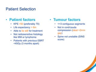
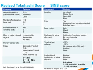
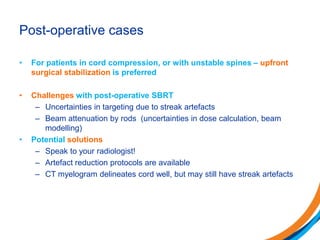
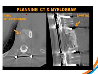
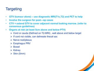
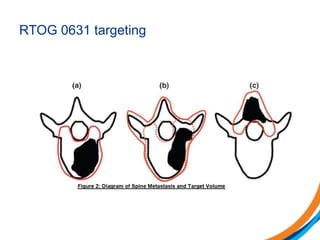
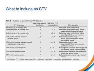
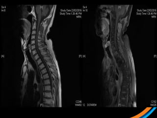
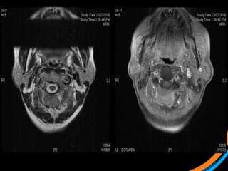
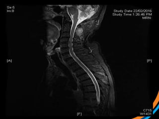
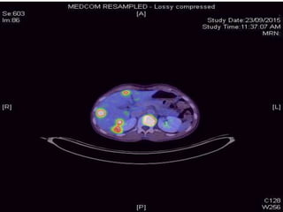
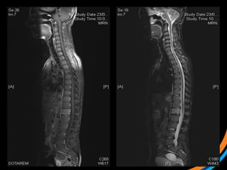
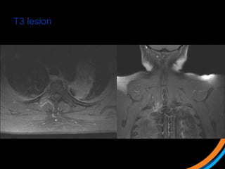
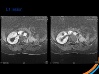
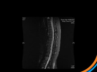
Education
Clinical Care
Research
Spine SBRT 101 : practical tips
3rd International SBRT symposium
2016
Dr. Bala Vellayappan
Assoc. Consultant
MBBS (Singapore), GDFM,
FRANZCR
Outline
• Background and rationale
• Determining eligibility (Patient selection)
• Required imaging
• Simulation
• Targeting
• Planning, dose selection
• OAR tolerances
• Plan evaluation
• Delivery and IGRT
• Case studies
Background
• Exponential rise in the use of SBRT
• Patients are living longer due to more effective
systemic therapy, and therefore durable control
becomes important
• There is a dose-response curve in palliation. Higher
doses  more durable pain relief
• SBRT affords high doses in short schedules (win-
win)
Rationale
• Advances in techniques, equipment and positional
verification high doses in tight spaces
• 35Gy/ 5# = BED 60Gy
• 24Gy/2# = BED 53Gy
• Large dose fractions = immune-mediated cell-killing
and endothelial apoptosis 1,2
1. Radiobiological mechanisms of stereotactic body radiation therapy and stereotactic radiation surgery.
Kim et al Radiat Oncol J. 2015 Dec; 33,265
2. Radiation-Induced Vascular Damage in Tumors: Implications of Vascular Damage in Ablative
Hypofractionated Radiotherapy. Park et al. Radiation Research: March 2012, (177),311
Patient Selection
• Patient factors
• KPS >50 (preferably 70)
• Life expectancy > 6m
• Able to lie still for treatment
• Not radiosensitive histology
like MM or lymphoma
• Patients with previous EBRT
<45Gy (3 months apart)
• Tumour factors
• <=3 contiguous segments
• Not in cord/cauda
compression (ideal >2mm
gap)
• Spine not unstable (SINS
score)
Revised Tokuhashi Score SINS score
Category Options
General Condition
(Performance status)
Poor
Moderate
Good
Number of extraspinal
bone met
>=3
1-2
0
Number of mets in
vertebral body
>=3
2
1
Mets to major internal
organs
Unremovable
Removable
No mets
Primary cancer site
Palsy Complete (Frankel
A,B)
Incomplete (Frankel
C,D)
None (Frankel E)
0-8 : 6m
9-11 : 6 – 12m
12-15 >1 y
Ref : Tokuhashi Y, et al. Spine 2005 2186-91
Component Options
Location Junctional
Mobile Spine
Semi-rigid
Rigid
Pain Yes
Occasional pain but not mechanical
Pain-free lesion
Bone Lesion Lytic
Mixed
Blastic
Radiographic spinal
alignment
Subluxation/translation present
De novo deformity
Normal
Vertebral body
collapse
>50%
<50%
No collapse with >50% body
involved
None
Posterolateral
involvement of spinal
elements
Bilateral
Unilateral
None
0-6 : stable
7 – 12: potentially unstable
13 – 18 : unstable
Ref: Fisher et al Spine 2010 1221
Denis’ three column concept
• One column injury is stable
• Two column injury is unstable
• Three column injury is
invariably unstable
Spinal instability as defined by the three-column spine concept in acute spinal trauma. Clin Orthop Relat Res. 1984
Oct;(189):65-76.
All cord compressions are not equal…
Bilsky et al J Neurosurg: Spine V13 2010
MSKCC NOMS algorithm
• NEUROLOGICAL ASSESSMENT
• ONCOLOGICAL ASSESSMENT
• MECHANICAL ASSESSMENT
• SYSTEMIC ASSESSMENT
Laufer et al. The Oncologist 2013; 18 : 744
Post-operative cases
• For patients in cord compression, or with unstable spines – upfront
surgical stabilization is preferred
• Challenges with post-operative SBRT
– Uncertainties in targeting due to streak artefacts
– Beam attenuation by rods (uncertainties in dose calculation, beam
modelling)
• Potential solutions
– Speak to your radiologist!
– Artefact reduction protocols are available
– CT myelogram delineates cord well, but may still have streak artefacts
Which machine should I use?
Is there one machine that is better?
ASTRO 2016
VMAT
Cyberknife
Tomotherapy – fixed jaw
Tomotherapy – dynamic jaws
You have decided to
deliver SBRT..now
what?
Patient preparation
• During consult –
discuss goals and
challenges of the
procedure with the
patient
• Assess pain score,
anxiety and
claustrophobia
• MRI whole spine with
contrast
– T1/T2 sag and axial
– Short tau inversion
recovery (STIR)
• Re-staging CT
• Targeted MRI (limited
sequences for RT
planning)
• +- CT myelogram
Required imaging
CT-Simulation
• Near-rigid immobilization
• Thick thermoplastic shell for lesions above T4 (5 point)
• BODYFix for other sites (not simple Vaclock)
• IV contrast optional (good to have)
• We don’t use 4DCT or fiducials
• <3mm cuts through region of interest
• Patient must be relaxed and pain-free *
• Recent MRI for fusion
• Good practice point : reposition patient before simulation
Rigid immobilisation
• T-shaped bags – more support
under the arms
• Full length bag - more support
for pelvis and legs
Targeting
• GTV (tumour alone) – use diagnostic MRI(T1c,T2) and PET to help
• Involve the surgeon for post—op cases
• CTV = extend GTV to cover adjacent normal looking marrow. (refer to
consensus guidelines)
• Organs at risk (at least 5cm above and below PTV)
 Cord or cauda (Defined on T2-MRI) , well above and below target
 If cord not visible, can delineate thecal sac
 Nerve root/plexus
 Esophagus PRV
 Bowel
 Kidney
 Skin (5mm)
Anatomy of the spine
RTOG 0631 targeting
Consensus
Cox et al Red Journal Vol 83 e597
What to include as CTV
Treatment planning
• Core group
• Dose selection
– 3 to 5 fraction SBRT is more forgiving, and still gives good results
– 24 – 27Gy in 3 fractions, 30 to 35Gy in 4 to 5 fractions
• Dose constraints
– TG 101
– RTOG 0631
– Follow what the experts do
• VMAT 2 arc
– Quicker than 9-13 field IMRT
– Non-coplanar usually doesn’t add much, and runs the risk of couch
collision
• Plan evaluation
– Know where to compromise
– Cord takes priority  there will be dose spillage outside target
Treatment delivery
• Equipment
– Commissioning and QA is very important
– Daily machine and indv. patient QA
• Manage the situation
– Setup, imaging, verification and delivery done swiftly
• Setup with lasers and DRO shift initially
• 3D Cone-beam CT
– Estimated to be 2-5minutes
• Tolerances should align with PTV/PRV margins (Desired 1mm/2degree)
• What to match?
• Treatment time
• Mid-CBCT ? Post CBCT?
• Pre-meds
– Dexamethasone 4mg prior to each fraction and few days after
– Adequate analgesia
– Anxiolytics as needed
Case study 1 (Patient 001)
• 70 Male, ECOG 0
• Metastatic Cholangioca since 2006
• Progressive C2 lesion causing neck pain
 1) Review the images
 2) Target the lesion
Case study 2 (Patient 003)
• 37 F, ECOG 0
• Met cervical Ca
• On PDL1 clinical trial
• Symptomatic L1 met s/p stabilisation
• For post-op SBRT
 1) Review the images
 2) Target the lesion
*Post-op targeting is more subjective. Post-op consensus guidelines likely to
be out in Red Journal soon
Case study 3 (Patient 009)
• 44, Female, ECOG 1
• RCC diagnosed 2007. Metastatic recurrence in 2012
• Prior RT to prevascular LN 50Gy/20# (2012)
• Started on Pazopanib, then everolimus
• Recent MRI spine shows met involving T3 and L1 vertebral body, with
involvement of superior endplate (L1)
 1) Review the images
 2) Target the lesion
T3 lesion
L1 lesion
Education
Clinical Care
Research
Thank you for your attention!
Email :
bala_vellayappan@nuhs.edu.sg

SPINE SBRT for beginners

  • 1.
    Education Clinical Care Research Spine SBRT101 : practical tips 3rd International SBRT symposium 2016 Dr. Bala Vellayappan Assoc. Consultant MBBS (Singapore), GDFM, FRANZCR
  • 2.
    Outline • Background andrationale • Determining eligibility (Patient selection) • Required imaging • Simulation • Targeting • Planning, dose selection • OAR tolerances • Plan evaluation • Delivery and IGRT • Case studies
  • 3.
    Background • Exponential risein the use of SBRT • Patients are living longer due to more effective systemic therapy, and therefore durable control becomes important • There is a dose-response curve in palliation. Higher doses  more durable pain relief • SBRT affords high doses in short schedules (win- win)
  • 4.
    Rationale • Advances intechniques, equipment and positional verification high doses in tight spaces • 35Gy/ 5# = BED 60Gy • 24Gy/2# = BED 53Gy • Large dose fractions = immune-mediated cell-killing and endothelial apoptosis 1,2 1. Radiobiological mechanisms of stereotactic body radiation therapy and stereotactic radiation surgery. Kim et al Radiat Oncol J. 2015 Dec; 33,265 2. Radiation-Induced Vascular Damage in Tumors: Implications of Vascular Damage in Ablative Hypofractionated Radiotherapy. Park et al. Radiation Research: March 2012, (177),311
  • 5.
    Patient Selection • Patientfactors • KPS >50 (preferably 70) • Life expectancy > 6m • Able to lie still for treatment • Not radiosensitive histology like MM or lymphoma • Patients with previous EBRT <45Gy (3 months apart) • Tumour factors • <=3 contiguous segments • Not in cord/cauda compression (ideal >2mm gap) • Spine not unstable (SINS score)
  • 6.
    Revised Tokuhashi ScoreSINS score Category Options General Condition (Performance status) Poor Moderate Good Number of extraspinal bone met >=3 1-2 0 Number of mets in vertebral body >=3 2 1 Mets to major internal organs Unremovable Removable No mets Primary cancer site Palsy Complete (Frankel A,B) Incomplete (Frankel C,D) None (Frankel E) 0-8 : 6m 9-11 : 6 – 12m 12-15 >1 y Ref : Tokuhashi Y, et al. Spine 2005 2186-91 Component Options Location Junctional Mobile Spine Semi-rigid Rigid Pain Yes Occasional pain but not mechanical Pain-free lesion Bone Lesion Lytic Mixed Blastic Radiographic spinal alignment Subluxation/translation present De novo deformity Normal Vertebral body collapse >50% <50% No collapse with >50% body involved None Posterolateral involvement of spinal elements Bilateral Unilateral None 0-6 : stable 7 – 12: potentially unstable 13 – 18 : unstable Ref: Fisher et al Spine 2010 1221
  • 7.
    Denis’ three columnconcept • One column injury is stable • Two column injury is unstable • Three column injury is invariably unstable Spinal instability as defined by the three-column spine concept in acute spinal trauma. Clin Orthop Relat Res. 1984 Oct;(189):65-76.
  • 8.
    All cord compressionsare not equal… Bilsky et al J Neurosurg: Spine V13 2010
  • 9.
    MSKCC NOMS algorithm •NEUROLOGICAL ASSESSMENT • ONCOLOGICAL ASSESSMENT • MECHANICAL ASSESSMENT • SYSTEMIC ASSESSMENT Laufer et al. The Oncologist 2013; 18 : 744
  • 10.
    Post-operative cases • Forpatients in cord compression, or with unstable spines – upfront surgical stabilization is preferred • Challenges with post-operative SBRT – Uncertainties in targeting due to streak artefacts – Beam attenuation by rods (uncertainties in dose calculation, beam modelling) • Potential solutions – Speak to your radiologist! – Artefact reduction protocols are available – CT myelogram delineates cord well, but may still have streak artefacts
  • 12.
  • 13.
    Is there onemachine that is better? ASTRO 2016
  • 14.
  • 15.
  • 16.
  • 17.
  • 18.
    You have decidedto deliver SBRT..now what?
  • 19.
    Patient preparation • Duringconsult – discuss goals and challenges of the procedure with the patient • Assess pain score, anxiety and claustrophobia • MRI whole spine with contrast – T1/T2 sag and axial – Short tau inversion recovery (STIR) • Re-staging CT • Targeted MRI (limited sequences for RT planning) • +- CT myelogram Required imaging
  • 20.
    CT-Simulation • Near-rigid immobilization •Thick thermoplastic shell for lesions above T4 (5 point) • BODYFix for other sites (not simple Vaclock) • IV contrast optional (good to have) • We don’t use 4DCT or fiducials • <3mm cuts through region of interest • Patient must be relaxed and pain-free * • Recent MRI for fusion • Good practice point : reposition patient before simulation
  • 21.
    Rigid immobilisation • T-shapedbags – more support under the arms • Full length bag - more support for pelvis and legs
  • 22.
    Targeting • GTV (tumouralone) – use diagnostic MRI(T1c,T2) and PET to help • Involve the surgeon for post—op cases • CTV = extend GTV to cover adjacent normal looking marrow. (refer to consensus guidelines) • Organs at risk (at least 5cm above and below PTV)  Cord or cauda (Defined on T2-MRI) , well above and below target  If cord not visible, can delineate thecal sac  Nerve root/plexus  Esophagus PRV  Bowel  Kidney  Skin (5mm)
  • 23.
  • 24.
  • 25.
    Consensus Cox et alRed Journal Vol 83 e597
  • 26.
  • 27.
    Treatment planning • Coregroup • Dose selection – 3 to 5 fraction SBRT is more forgiving, and still gives good results – 24 – 27Gy in 3 fractions, 30 to 35Gy in 4 to 5 fractions • Dose constraints – TG 101 – RTOG 0631 – Follow what the experts do • VMAT 2 arc – Quicker than 9-13 field IMRT – Non-coplanar usually doesn’t add much, and runs the risk of couch collision • Plan evaluation – Know where to compromise – Cord takes priority  there will be dose spillage outside target
  • 28.
    Treatment delivery • Equipment –Commissioning and QA is very important – Daily machine and indv. patient QA • Manage the situation – Setup, imaging, verification and delivery done swiftly • Setup with lasers and DRO shift initially • 3D Cone-beam CT – Estimated to be 2-5minutes • Tolerances should align with PTV/PRV margins (Desired 1mm/2degree) • What to match? • Treatment time • Mid-CBCT ? Post CBCT? • Pre-meds – Dexamethasone 4mg prior to each fraction and few days after – Adequate analgesia – Anxiolytics as needed
  • 29.
    Case study 1(Patient 001) • 70 Male, ECOG 0 • Metastatic Cholangioca since 2006 • Progressive C2 lesion causing neck pain  1) Review the images  2) Target the lesion
  • 33.
    Case study 2(Patient 003) • 37 F, ECOG 0 • Met cervical Ca • On PDL1 clinical trial • Symptomatic L1 met s/p stabilisation • For post-op SBRT  1) Review the images  2) Target the lesion *Post-op targeting is more subjective. Post-op consensus guidelines likely to be out in Red Journal soon
  • 38.
    Case study 3(Patient 009) • 44, Female, ECOG 1 • RCC diagnosed 2007. Metastatic recurrence in 2012 • Prior RT to prevascular LN 50Gy/20# (2012) • Started on Pazopanib, then everolimus • Recent MRI spine shows met involving T3 and L1 vertebral body, with involvement of superior endplate (L1)  1) Review the images  2) Target the lesion
  • 40.
  • 41.
  • 43.
    Education Clinical Care Research Thank youfor your attention! Email : bala_vellayappan@nuhs.edu.sg

Editor's Notes

  • #4 Conventional RT results in poor control rates at 1 year (50%) for mass-type lesions
  • #5 Improved imaging and localisation of tumours (and critical structures) We are delivering 2-3x the doses compared to palliative EBRT – potential for improved local control
  • #6 No progressive neurologic deficits
  • #28 Be weary of skin dose in thin patients and where lesions are in the spinous process
  • #31 Sag T1, Sag T1c
  • #32 Ax T2 Ax T1C
  • #33 Sag STIR
  • #35 Pre-treatment PET showing very avid disease in anterior vert body
  • #36 T1c and T2
  • #37 Post-instrumentation diagnostic CT
  • #38 Post RT PET at 7m The treatment effect is likely a combination of PDL1 and SBRT