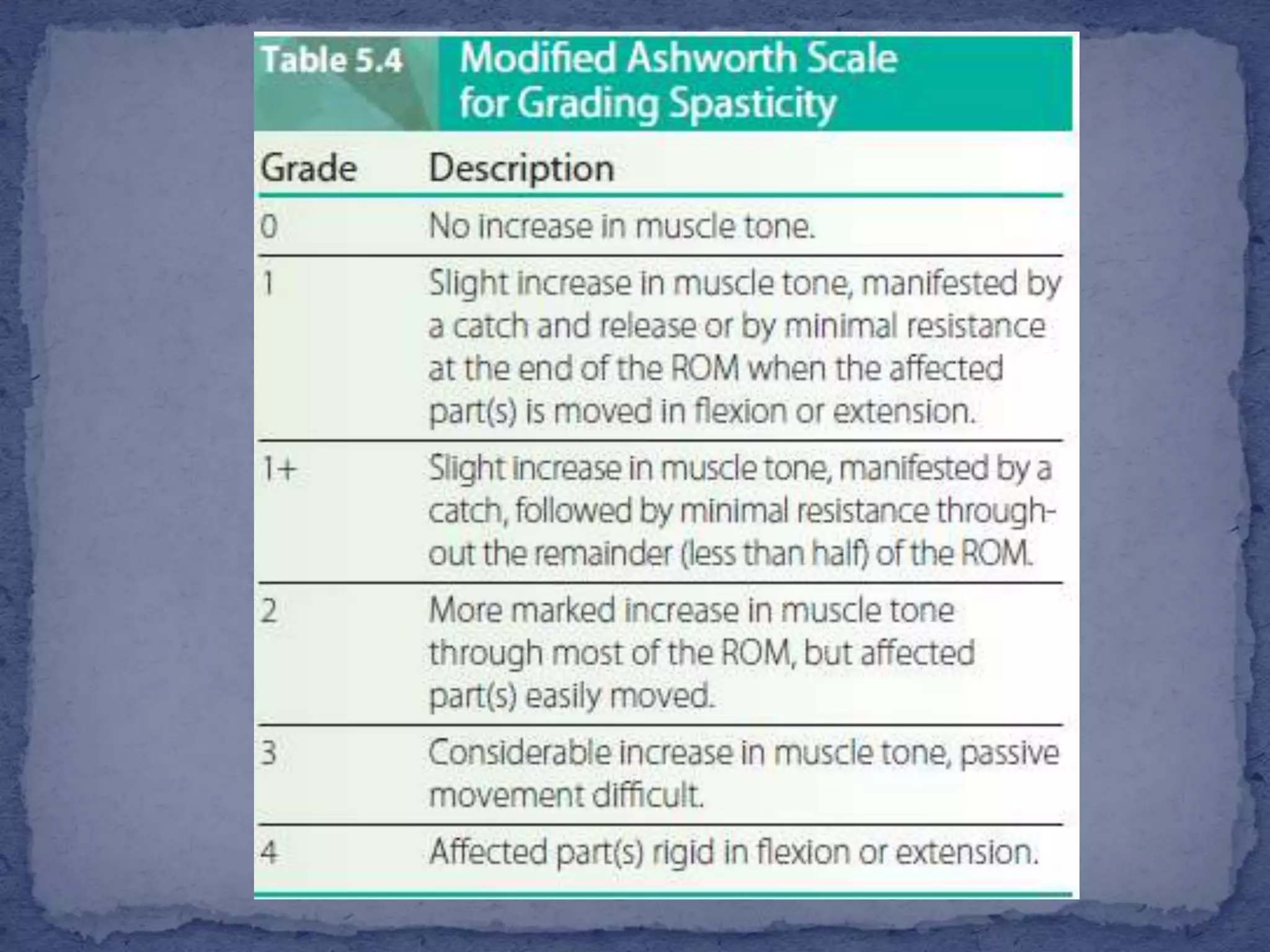
The document discusses various types of muscle tone abnormalities including hypertonia, hypotonia, spasticity, rigidity, dystonia, and their clinical presentations and assessments. Spasticity is characterized by velocity-dependent resistance to stretch and may result in contractures and functional limitations. Rigidity differs from spasticity in being independent of movement velocity. Various assessments of tone are discussed including observation of posture and movement, palpation of muscles, passive range of motion testing, scales like the Modified Ashworth Scale, and reflex testing.
2. Tone is defined as the resistance of muscle to passive
elongation or stretch. It represents a state of slight residual
contraction in normally innervated, resting muscle, or
steady-state contraction.
Postural tone -a pattern of muscular tension that exists
throughout the body and affects groups of muscles.
Tonal abnormalities are categorized as
hypertonia (increased above normal resting levels),
hypotonia (decreased below normal resting levels), or
dystonia (impaired or disordered tonicity).
3. Spasticity
Spasticity is a motor disorder characterized by velocity
dependent increase in muscle tone with increased resistance to
stretch;
the larger and quicker the stretch, the stronger the resistance of
the spastic muscle.
During rapid movement, initial high resistance (spastic catch)
may be followed by a sudden inhibition or letting go of the limb
(relaxation) in response to a stretch stimulus, termed clasp-knife
response.
Chronic spasticity is associated with contracture, abnormal
posturing and deformity, functional limitations, and disability.
4.
5. associated reactions, defined as involuntary
movements resulting from activity occurring in other
parts of the body (e.g., sneezing, yawning, squeezing
the hand).
Clonus is characterized by cyclical, spasmodic
alteration of muscular contraction and relaxation in
response to sustained stretch of a spastic muscle.
6. Babinski sign is dorsiflexion of the great toe with
fanning of the other toes on stimulation of the lateral
sole of the foot.
7.
8. Rigidity is a hypertonic state characterized by
constant resistance throughout ROM that is
independent of the velocity of movement (lead-pipe
rigidity). It is associated with lesions of the basal
ganglia system (extrapyramidal syndromes) and is seen
in Parkinson’s disease.
Cogwheel rigidity-a hypertonic state with
superimposed ratchet-like jerkiness and is commonly
seen in upper extremity movements
9. Severe brain injury can result in coma
with decorticate or decerebrate
rigidity.
Decorticate rigidity refers to
sustained contraction and posturing
of the upper limbs in flexion and
the lower limbs in extension.
elbows, wrists, and fingers - flexion
shoulders adducted tightly to the
sides
legs - extension, internal rotation,
and plantarflexion.
Decorticate rigidity is indicative of a
corticospinal tract lesion above the
superior colliculus.
10. Decerebrate rigidity (abnormal
extensor response) refers to sustained
contraction and posturing of the trunk
and limbs in a position of full
extension.
The elbows -extended
shoulders adducted,
forearms pronated,
and wrist and fingers flexed.
The legs are held in stiff extension
with plantarflexion.
decerebrate rigidity indicates a
corticospinal lesion in the brainstem
between the superior colliculus and
vestibular nucleus.
11. Dystonia is a prolonged involuntary movement
disorder characterized by twisting or writhing
repetitive movements and increased muscular tone.
Dystonic posturing refers to sustained abnormal
postures caused by cocontraction of muscles that may
last for several minutes, for hours or permanently.
12. Hypotonia and flaccidity -decreased or absent muscular
tone.
Resistance to passive movement is diminished,
stretch reflexes are dampened or absent,
and limbs are easily moved (floppy).
Hyperextensibility of joints is common.
Acute UMN lesions (e.g., hemiplegia, tetraplegia,
paraplegia) can produce temporary hypotonia, termed
spinal shock or cerebral shock.
It is followed by the development of spasticity and classic
UMN signs.
13. Initial observation of the
patient –
To reveal abnormal posturing of
the limbs or body.
With spasticity, posturing in
fixed, antigravity positions ;
for example, a spastic upper
extremity is typically held fixed
against the body with the
shoulder adducted, elbow flexed,
forearm supinated with
wrist/fingers flexed.
In the supine position, the lower
extremities are typically held in
extension, adduction with
plantarflexion, and inversion
14. Limbs that appear
floppy and lifeless
(e.g., a lower
extremity [LE]
rolled out to the
side in external
rotation) may
indicate
hypotonicity
15. Palpation
of the muscle belly yields additional information
about the resting state of muscle.
Consistency, firmness, and turgor
Hypotonic muscles will feel soft and flabby, whereas
hypertonic muscles will feel taut and harder than
normal.
16. Passive motion testing
reveals information about the responsiveness of muscles to
stretch.
The patient is instructed to relax, letting the therapist support
and move the limb.
the therapist should maintain firm and constant manual
contact, moving the limb in all motions.
When tone is normal, the limb moves easily and the therapist is
able to alter direction and speed without feeling abnormal
resistance.
Hypertonic limbs generally feel stiff and resistant to movement,
Flaccid limbs feel heavy and unresponsive
17.
18.
19. The Modified Ashworth Scale (MAS) is a clinical scale
used to assess muscle spasticity that is in commonly
used in many rehabilitation facilities and spasticity
clinics
20.
21. In the lower limbs, spasticity can be examined using the pendulum
test.
The patient is positioned in supine with knees flexed over the end of a
table.
The examiner passively extends the knee fully against gravity and then
allows the leg to drop and swing like a pendulum.
A normal and hypotonic limb will swing freely for several oscillations.
In patients with quadriceps or hamstring spasticity, the leg is resistant
to full extension and when dropped swings for only a few repetitions.
It quickly returns to the initial dependent starting position.
The pendulum test can be quantified using an isokinetic dynamometer,
an electrogoniometer, or computerized video equipment with high
test–retest reliability.
22.
23. A reflex is an involuntary, predictable, and
specific response to a stimulus dependent on an
intact reflex arc
(sensory receptor, afferent neurons, efferent
neurons,and responding muscles or gland).
24. To ensure adequate response, the muscle is positioned
in midrange and the patient is instructed to relax.
Stimulation can result in observable movement of the
joint (brisk or strong responses).
Weak responses may be evident only with palpation
(slight or sluggish responses with little or no joint
movement).
25. 0- Absent, no response
1+- Slight reflex, present but depressed, low normal
2+ -Normal, typical reflex
3+ -Brisk reflex, possibly but not necessarily abnormal
4+ -Very brisk reflex, abnormal, clonus
26. If DTRs are difficult to elicit, responses can be
enhanced by specific reinforcement maneuvers.
In the Jendrassik maneuver, the patient hooks together
the fingers of the hands and strongly pulls them apart.
While this pressure is maintained, LE reflexes are
tested.
27.
28.
29. utilize functionally linked muscles or synergies that
are constrained by the CNS to act cooperatively to
produce an action.
The CNS is able to reduce the degrees of freedom,
defined as the number of seperate independent
dimensions of movement that must be controlled by
engaging these cooperative units of muscle action.
30. Abnormal mass synergies are defined as obligatory.
highly stereotyped mass patterns of
movement.
Selective movement control (isolated joint
movements) becomes severely disordered or
disappears completely.
For example, patients with stroke may demonstrate
obligatory flexion synergies and extension synergies