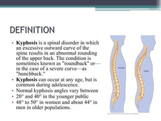
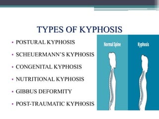
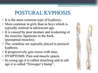
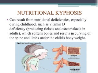
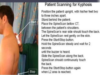
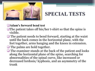
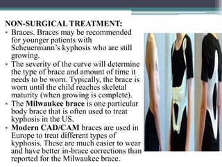
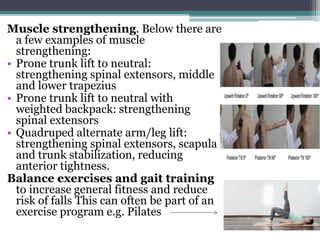
KYPHOSIS
Dr. Jasjyot Kaur Sabharwal (PT)
ANATOMY
• Spine is made up of three segments. When
viewed from the side, these segments form
three natural curves.
• The "c-shaped" curves of the neck (cervical
spine) and lower back (lumbar spine) are
called lordosis.
• The "reverse c-shaped" curve of the chest
(thoracic spine) is called kyphosis.
• This natural curvature of the spine is
important for balance and helps us to
stand upright. If any one of the curves
becomes too large or too small, it becomes
difficult to stand up straight and our
posture appears abnormal.
DEFINITION
• Kyphosis is a spinal disorder in which
an excessive outward curve of the
spine results in an abnormal rounding
of the upper back. The condition is
sometimes known as "roundback" or—
in the case of a severe curve—as
"hunchback."
• Kyphosis can occur at any age, but is
common during adolescence.
• Normal kyphosis angles vary between
• 20° and 40° in the younger public
• 48° to 50° in women and about 44° in
men in older populations.
TYPES OF KYPHOSIS
• POSTURAL KYPHOSIS
• SCHEUERMANN’S KYPHOSIS
• CONGENITAL KYPHOSIS
• NUTRITIONAL KYPHOSIS
• GIBBUS DEFORMITY
• POST-TRAUMATIC KYPHOSIS
POSTURAL KYPHOSIS
• It is the most common type of kyphosis.
• More common in girls than in boys which is
typically noticed in adolescent age.
• It is caused by poor posture and weakening of
the muscles, ligaments in the back.
(paraspinal muscles).
• The vertebrae are typically placed in postural
kyphosis.
• It progressively gets worse with time.
• SYMPTOMS: Pain and muscle spasm.
• In young age it is called slouching and in old
age it is called “Dowager’s hump”.
SCHEUERMANN KYPHOSIS
It is significantly worse cosmetically and can cause
varying degrees of pain, and can also affect different
areas of the spine (the most common being the mid-
thoracic area).
 Scheuermann's kyphosis is considered a form of
juvenile osteochondrosis of the spine, and is more
commonly called Scheuermann's disease.
It is found mostly in teenagers and presents a
significantly worse deformity than postural kyphosis.
A patient suffering from Scheuermann's kyphosis
cannot consciously correct posture.
The apex of the curve, located in the thoracic
vertebrae, is quite rigid. The patient may feel pain at
this apex, which can be aggravated by physical
activity and by long periods of standing or sitting.
This can have a significantly detrimental effect on their lives, as their
level of activity is curbed by their condition; they may feel isolated or
uneasy amongst peers if they are children, depending on the level of
deformity.
 Whereas in postural kyphosis, the vertebrae and discs appear normal,
in Scheuermann's kyphosis, they are irregular, often herniated, and
wedge-shaped over at least three adjacent levels.
Fatigue is a very common symptom, most likely because of the intense
muscle work that has to be put into standing or sitting properly. The
condition appears to run in families. Most patients who undergo
surgery to correct their kyphosis have Scheuermann's disease.
CONGENITAL KYPHOSIS
• It is the least common type of kyphosis.
• It result in infants whose spinal column has not developed correctly in the
womb.
• Vertebrae may be malformed or fused together and can cause further
progressive kyphosis as the child develops.
• Surgical treatment may be necessary at a very early stage and can help
maintain a normal curve in coordination with consistent follow-ups to
monitor changes.
• A congenital kyphosis can also suddenly appear in teenage years, more
commonly in children with cerebral palsy and other neurological disorders.
NUTRITIONAL KYPHOSIS
• Can result from nutritional deficiencies, especially
during childhood, such as vitamin D
deficiency (producing rickets and osteomalacia in
adults), which softens bones and results in curving of
the spine and limbs under the child's body weight.
• Gibbus deformity is a
form of structural
kyphosis, often
a sequela to tuberculosis.
• Post-traumatic
kyphosis can arise from
untreated or ineffectively
treated vertebral
fractures
ETIOLOGY
• Aging, especially if you have poor posture
• Muscle weakness in the upper back
• Scheuermann’s disease, which occurs in
children and has no known cause
• Arthritis or other bone degeneration diseases
• Osteoporosis, or the loss of bone strength due
to age
• Injury to the spine
• Slipped discs
• Scoliosis, or spinal curvature
• Infection in the spine
• Birth defects, such as spina bifida
• Tumours
• Diseases of the connective tissues
• Polio
• Paget disease
• Muscular dystrophy
EXAMINATION
• OBSERVATION:
• Begins with observing the patient e.g.
abnormalities during gait and undressing can be
noticed very quickly.
• Examined in sagittal plane in standing and
relaxed position “in order to assess the most
affected somatic areas and posture alterations.”
The standing position of the clinical examination
needs to be reproducible.
• Ankles and toes are placed in a neutral
anatomical posture.
• The lower limbs are stretched straight limiting
an excessive recurvatum.
• The trunk and upper limbs are relaxed, palms of
the hands on the lateral thighs, the eye is looking
horizontally”.
• Tragus to wall test can be used
• When the angle is between 40-45° we consider it
as a thoracal hyperkyphosis.
Equipment to measure the
thoracic kyphosis angles.
These are:
• Modified Cobb Angle
• The pantograph
• The Debrunner kyphometer: Arms of
the kyphometer are placed on C7 and
T12, we read the angle from the
protractor.
• The flexicurve index: Place a marker on C7 and the lumbar-
sacral joint space.
• Then use a flexicurve carefully and place it between C7 and the L-S
joint space.
• When you take the flexicurve away from the back it will conserve the
form of the whole back.
• Then put the flexicurve on a 10x10 paper and draw a vertical line
and then the TW and LW lines you need to measure
• Functional examination e.g. rib cage abnormalities such as pectus
excavatum or carnatum;
How to calculate Cobb
angle.
• Cobb suggested that the
angle of curvature be
measured by drawing
lines parallel to the
upper border of the
upper vertebral body
and the lower border of
the lowest vertebra of
the structural curve,
then erecting
perpendiculars from
these lines to cross each
other, the angle between
these perpendiculars
being the ‘angle of
curvature’
PANTOGRAPH
SPECIAL TESTS
Adam's forward bend test
The patient takes off his/her t-shirt so that the spine is
visible.
 The patient needs to bend forward, starting at the waist
until the back comes in the horizontal plane, with the
feet together, arms hanging and the knees in extension.
 The palms are hold together.
 The examiner stands at the back of the patient and looks
along the horizontal plane of the spine, searching for
abnormalities of the spinal curve, like increased or
decreased lordosis/ kyphosis, and an asymmetry of the
trunk
TREATMENT
MEDICAL TREATMENT:
• Medications:
• Non-steroidal anti-inflammatory
drugs such as:
• Advil and Motrin (ibuprofen)
• Aleve (naproxen) can help relieve
back pain.
NON-SURGICAL TREATMENT:
• Braces. Braces may be recommended
for younger patients with
Scheuermann’s kyphosis who are still
growing.
• The severity of the curve will determine
the type of brace and amount of time it
needs to be worn. Typically, the brace is
worn until the child reaches skeletal
maturity (when growing is complete).
• The Milwaukee brace is one particular
body brace that is often used to treat
kyphosis in the US.
• Modern CAD/CAM braces are used in
Europe to treat different types of
kyphosis. These are much easier to wear
and have better in-brace corrections than
reported for the Milwaukee brace.
SURGICAL
TREATMENT
• There are multiple surgical procedures
that can be applied, depending on:
• Flexibility: X-rays will be taken to
determine how flexible the spine is. If it’s
flexible, a posterior approach may be
adequate. If the patient is less flexible, an
anterior or combined approach will be
needed. These techniques are based on
bone fusion with screws and rods.
• Osteotomy (a surgical operation in
which a bone is cut to shorten, lengthen,
or change its alignment.).
• °.
• Osteoporosis: An osteoporotic kyphosis can be caused by a collapsed
vertebra.
• This can be treated with a kyphoplasty, whereby a balloon is inserted into
the affected vertebra and filled with liquid that hardens to restore the
vertebral height.
• Vertebroplasty
• They even have resulted in a decreased kyphosis angle. reduced pain,
improved physical function, vertebral height restoration of up to 90%, and
kyphosis angle correction ranging from 8.5° to 14
PHYSIOTHERAPY
TREATMENT
It most commonly occurs in thoracic spine:
• Thoracic joint mobilizations are required in
patients diagnosed with hyperkyphosis
because of the reduced thoracic motion. e.g.
Scapular, myofascial and spinal
mobilization techniques increase postural
alignment.
• Self-mobilization techniques :
Diaphragmatic breathing on foam roller for
expanding the rib cage.
Pursed lip breathing exercises.
• Teach these exercises at home.
• Stretching: To decrease tonus in the
muscles:
• Chest stretching on foam roller:
lengthening pectoralis muscles
• Prone hip extension/ knee flexion:
lengthening iliopsoas and rectus femoris
• Supine knee extension with hip at 90°
flexion: lengthening hamstrings.
• Pain management using modalities
such as
• Hot packs
• Cryotherapy
• Electrical stimulation such as
transcutaneous electrical nerve
stimulation (TENS).
Muscle strengthening. Below there are
a few examples of muscle
strengthening:
• Prone trunk lift to neutral:
strengthening spinal extensors, middle
and lower trapezius
• Prone trunk lift to neutral with
weighted backpack: strengthening
spinal extensors
• Quadruped alternate arm/leg lift:
strengthening spinal extensors, scapula
and trunk stabilization, reducing
anterior tightness.
Balance exercises and gait training
to increase general fitness and reduce
risk of falls This can often be part of an
exercise program e.g. Pilates
BRACING:
• A Milwaukee Brace is the most
common brace used by patients with
a thoracic hyperkyphosis. This brace
has posterior pads pushing anteriorly
on the kyphosis.
• The neck and the pelvis are
controlled by the other segments of
the brace. The patients should wear
this brace 23 hours a day for 1-2
years.
• Lyon Antikyphosis Brace
• Kyphologic Brace
• Another kind of brace using the two
3-point pressure system is the
‘Gschwend type’ brace. This brace
is mostly used in Germany
Taping
May also reduce kyphosis. Tape from the
anterior aspect of the acromioclavicular
joint, over the muscle bulk of the upper
trapezius, and diagonally over the
spinous process of T6
Spinal Orthosis
A is a spinal orthosis which can be used
for a thoracic hyperkyphosis.
 The patient should wear this for 2 hours
a day during 6 months.
 This will result in a decrease in kyphosis
angle, an improvement of standing
height, an increase in spinal extensor
strength and a decreased postural sway.
Yoga
PROGNOSIS
• Scheuermann kyphosis disorder in adolescents
stop once their growth stops.
• If the disorder occurs due to degenerative types,
then they may need a surgical correction.
COMPLICATIONS:
• Decreased lung capacity
• Disability pain
• Neurological symptoms including weakness in
legs or paralysis.
• Round back deformity.
THANK YOU

Kyphosis

  • 1.
  • 2.
    ANATOMY • Spine ismade up of three segments. When viewed from the side, these segments form three natural curves. • The "c-shaped" curves of the neck (cervical spine) and lower back (lumbar spine) are called lordosis. • The "reverse c-shaped" curve of the chest (thoracic spine) is called kyphosis. • This natural curvature of the spine is important for balance and helps us to stand upright. If any one of the curves becomes too large or too small, it becomes difficult to stand up straight and our posture appears abnormal.
  • 3.
    DEFINITION • Kyphosis isa spinal disorder in which an excessive outward curve of the spine results in an abnormal rounding of the upper back. The condition is sometimes known as "roundback" or— in the case of a severe curve—as "hunchback." • Kyphosis can occur at any age, but is common during adolescence. • Normal kyphosis angles vary between • 20° and 40° in the younger public • 48° to 50° in women and about 44° in men in older populations.
  • 4.
    TYPES OF KYPHOSIS •POSTURAL KYPHOSIS • SCHEUERMANN’S KYPHOSIS • CONGENITAL KYPHOSIS • NUTRITIONAL KYPHOSIS • GIBBUS DEFORMITY • POST-TRAUMATIC KYPHOSIS
  • 5.
    POSTURAL KYPHOSIS • Itis the most common type of kyphosis. • More common in girls than in boys which is typically noticed in adolescent age. • It is caused by poor posture and weakening of the muscles, ligaments in the back. (paraspinal muscles). • The vertebrae are typically placed in postural kyphosis. • It progressively gets worse with time. • SYMPTOMS: Pain and muscle spasm. • In young age it is called slouching and in old age it is called “Dowager’s hump”.
  • 6.
    SCHEUERMANN KYPHOSIS It issignificantly worse cosmetically and can cause varying degrees of pain, and can also affect different areas of the spine (the most common being the mid- thoracic area).  Scheuermann's kyphosis is considered a form of juvenile osteochondrosis of the spine, and is more commonly called Scheuermann's disease. It is found mostly in teenagers and presents a significantly worse deformity than postural kyphosis. A patient suffering from Scheuermann's kyphosis cannot consciously correct posture. The apex of the curve, located in the thoracic vertebrae, is quite rigid. The patient may feel pain at this apex, which can be aggravated by physical activity and by long periods of standing or sitting.
  • 7.
    This can havea significantly detrimental effect on their lives, as their level of activity is curbed by their condition; they may feel isolated or uneasy amongst peers if they are children, depending on the level of deformity.  Whereas in postural kyphosis, the vertebrae and discs appear normal, in Scheuermann's kyphosis, they are irregular, often herniated, and wedge-shaped over at least three adjacent levels. Fatigue is a very common symptom, most likely because of the intense muscle work that has to be put into standing or sitting properly. The condition appears to run in families. Most patients who undergo surgery to correct their kyphosis have Scheuermann's disease.
  • 8.
    CONGENITAL KYPHOSIS • Itis the least common type of kyphosis. • It result in infants whose spinal column has not developed correctly in the womb. • Vertebrae may be malformed or fused together and can cause further progressive kyphosis as the child develops. • Surgical treatment may be necessary at a very early stage and can help maintain a normal curve in coordination with consistent follow-ups to monitor changes. • A congenital kyphosis can also suddenly appear in teenage years, more commonly in children with cerebral palsy and other neurological disorders.
  • 9.
    NUTRITIONAL KYPHOSIS • Canresult from nutritional deficiencies, especially during childhood, such as vitamin D deficiency (producing rickets and osteomalacia in adults), which softens bones and results in curving of the spine and limbs under the child's body weight.
  • 10.
    • Gibbus deformityis a form of structural kyphosis, often a sequela to tuberculosis. • Post-traumatic kyphosis can arise from untreated or ineffectively treated vertebral fractures
  • 11.
    ETIOLOGY • Aging, especiallyif you have poor posture • Muscle weakness in the upper back • Scheuermann’s disease, which occurs in children and has no known cause • Arthritis or other bone degeneration diseases • Osteoporosis, or the loss of bone strength due to age • Injury to the spine • Slipped discs • Scoliosis, or spinal curvature • Infection in the spine • Birth defects, such as spina bifida • Tumours • Diseases of the connective tissues • Polio • Paget disease • Muscular dystrophy
  • 14.
    EXAMINATION • OBSERVATION: • Beginswith observing the patient e.g. abnormalities during gait and undressing can be noticed very quickly. • Examined in sagittal plane in standing and relaxed position “in order to assess the most affected somatic areas and posture alterations.” The standing position of the clinical examination needs to be reproducible. • Ankles and toes are placed in a neutral anatomical posture. • The lower limbs are stretched straight limiting an excessive recurvatum. • The trunk and upper limbs are relaxed, palms of the hands on the lateral thighs, the eye is looking horizontally”. • Tragus to wall test can be used • When the angle is between 40-45° we consider it as a thoracal hyperkyphosis.
  • 15.
    Equipment to measurethe thoracic kyphosis angles. These are: • Modified Cobb Angle • The pantograph • The Debrunner kyphometer: Arms of the kyphometer are placed on C7 and T12, we read the angle from the protractor.
  • 16.
    • The flexicurveindex: Place a marker on C7 and the lumbar- sacral joint space. • Then use a flexicurve carefully and place it between C7 and the L-S joint space. • When you take the flexicurve away from the back it will conserve the form of the whole back. • Then put the flexicurve on a 10x10 paper and draw a vertical line and then the TW and LW lines you need to measure • Functional examination e.g. rib cage abnormalities such as pectus excavatum or carnatum;
  • 17.
    How to calculateCobb angle. • Cobb suggested that the angle of curvature be measured by drawing lines parallel to the upper border of the upper vertebral body and the lower border of the lowest vertebra of the structural curve, then erecting perpendiculars from these lines to cross each other, the angle between these perpendiculars being the ‘angle of curvature’
  • 19.
  • 20.
    SPECIAL TESTS Adam's forwardbend test The patient takes off his/her t-shirt so that the spine is visible.  The patient needs to bend forward, starting at the waist until the back comes in the horizontal plane, with the feet together, arms hanging and the knees in extension.  The palms are hold together.  The examiner stands at the back of the patient and looks along the horizontal plane of the spine, searching for abnormalities of the spinal curve, like increased or decreased lordosis/ kyphosis, and an asymmetry of the trunk
  • 21.
    TREATMENT MEDICAL TREATMENT: • Medications: •Non-steroidal anti-inflammatory drugs such as: • Advil and Motrin (ibuprofen) • Aleve (naproxen) can help relieve back pain.
  • 22.
    NON-SURGICAL TREATMENT: • Braces.Braces may be recommended for younger patients with Scheuermann’s kyphosis who are still growing. • The severity of the curve will determine the type of brace and amount of time it needs to be worn. Typically, the brace is worn until the child reaches skeletal maturity (when growing is complete). • The Milwaukee brace is one particular body brace that is often used to treat kyphosis in the US. • Modern CAD/CAM braces are used in Europe to treat different types of kyphosis. These are much easier to wear and have better in-brace corrections than reported for the Milwaukee brace.
  • 23.
    SURGICAL TREATMENT • There aremultiple surgical procedures that can be applied, depending on: • Flexibility: X-rays will be taken to determine how flexible the spine is. If it’s flexible, a posterior approach may be adequate. If the patient is less flexible, an anterior or combined approach will be needed. These techniques are based on bone fusion with screws and rods. • Osteotomy (a surgical operation in which a bone is cut to shorten, lengthen, or change its alignment.). • °.
  • 24.
    • Osteoporosis: Anosteoporotic kyphosis can be caused by a collapsed vertebra. • This can be treated with a kyphoplasty, whereby a balloon is inserted into the affected vertebra and filled with liquid that hardens to restore the vertebral height. • Vertebroplasty • They even have resulted in a decreased kyphosis angle. reduced pain, improved physical function, vertebral height restoration of up to 90%, and kyphosis angle correction ranging from 8.5° to 14
  • 25.
    PHYSIOTHERAPY TREATMENT It most commonlyoccurs in thoracic spine: • Thoracic joint mobilizations are required in patients diagnosed with hyperkyphosis because of the reduced thoracic motion. e.g. Scapular, myofascial and spinal mobilization techniques increase postural alignment. • Self-mobilization techniques : Diaphragmatic breathing on foam roller for expanding the rib cage. Pursed lip breathing exercises. • Teach these exercises at home.
  • 26.
    • Stretching: Todecrease tonus in the muscles: • Chest stretching on foam roller: lengthening pectoralis muscles • Prone hip extension/ knee flexion: lengthening iliopsoas and rectus femoris • Supine knee extension with hip at 90° flexion: lengthening hamstrings. • Pain management using modalities such as • Hot packs • Cryotherapy • Electrical stimulation such as transcutaneous electrical nerve stimulation (TENS).
  • 27.
    Muscle strengthening. Belowthere are a few examples of muscle strengthening: • Prone trunk lift to neutral: strengthening spinal extensors, middle and lower trapezius • Prone trunk lift to neutral with weighted backpack: strengthening spinal extensors • Quadruped alternate arm/leg lift: strengthening spinal extensors, scapula and trunk stabilization, reducing anterior tightness. Balance exercises and gait training to increase general fitness and reduce risk of falls This can often be part of an exercise program e.g. Pilates
  • 28.
    BRACING: • A MilwaukeeBrace is the most common brace used by patients with a thoracic hyperkyphosis. This brace has posterior pads pushing anteriorly on the kyphosis. • The neck and the pelvis are controlled by the other segments of the brace. The patients should wear this brace 23 hours a day for 1-2 years. • Lyon Antikyphosis Brace • Kyphologic Brace • Another kind of brace using the two 3-point pressure system is the ‘Gschwend type’ brace. This brace is mostly used in Germany
  • 29.
    Taping May also reducekyphosis. Tape from the anterior aspect of the acromioclavicular joint, over the muscle bulk of the upper trapezius, and diagonally over the spinous process of T6 Spinal Orthosis A is a spinal orthosis which can be used for a thoracic hyperkyphosis.  The patient should wear this for 2 hours a day during 6 months.  This will result in a decrease in kyphosis angle, an improvement of standing height, an increase in spinal extensor strength and a decreased postural sway. Yoga
  • 30.
    PROGNOSIS • Scheuermann kyphosisdisorder in adolescents stop once their growth stops. • If the disorder occurs due to degenerative types, then they may need a surgical correction. COMPLICATIONS: • Decreased lung capacity • Disability pain • Neurological symptoms including weakness in legs or paralysis. • Round back deformity.
  • 31.