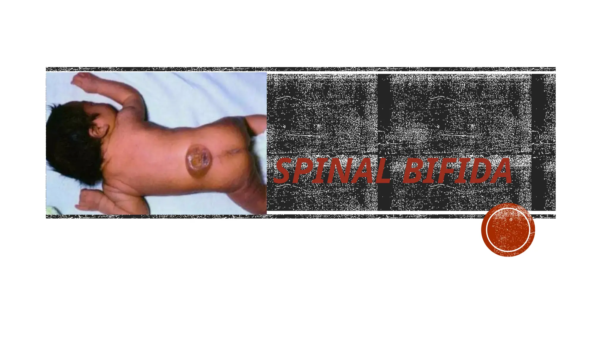
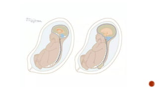
DEFINITION:
• Spina bifida(Latin: "split spine") is a developmental
congenital disorder caused by the incomplete closing of
the embryonic neural tube.
• Some vertebrae overlying the spinal cord are not fully
formed and remain unfused and open. lf the opening is
large enough, this allows a portion of the spinal cord to
protrude through the opening in the bones.
• There may or may not be a fluid-filled sac surrounding
the spinal cord.
3.
INCIDENCE:
• Spina bifidais one of the most common. birth
defects, with an average worldwide incidence of one to
two cases per 1000 births, but certain populations
have a significantly greater risk.
• Myelomeningocele is the most significant and
common form, and this leads to disability in most
affected individuals.
• This condition is more likely to appear in females; the
cause for this is unknown.
4.
CAUSES:
• Maternal diabetes
•Family history
• Obesity
• Increased body temperature from fever or external sources such as
hot tubs and electric blankets may increase the chances of delivery of a
baby with a spinal bitifa.
• Medications such as some anticonvulsants.
• Pregnant women taking Valproic acid have an increased risk of having
children with spina bifida
• Genetic basis.
• Folic acid deficiency
5.
EMBRYOLOGY:
Spina bifida iscaused by the failure of the neural tube to close
during the first month of embryonic development (often before
the mother knows she is pregnant).
Under normal circumstances, the closure of the neural tube
occurs around the 23rd (rostral closure) and 27th (caudal closure)
day after fertilization.
7.
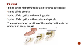
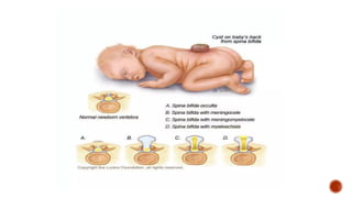
TYPES:
Spina bifida malformationsfall into three categories:
spina bifida occulta
spina bifida cystica with meningocele
spina bifida cystica with myelomeningocele.
(The most common location of the malformations is the
lumbar and sacral areas)
9.
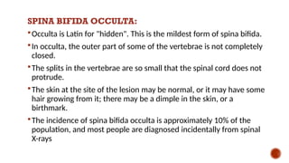
SPINA BIFIDA OCCULTA:
Occultais Latin for "hidden". This is the mildest form of spina bifida.
In occulta, the outer part of some of the vertebrae is not completely
closed.
The splits in the vertebrae are so small that the spinal cord does not
protrude.
The skin at the site of the lesion may be normal, or it may have some
hair growing from it; there may be a dimple in the skin, or a
birthmark.
The incidence of spina bifida occulta is approximately 10% of the
population, and most people are diagnosed incidentally from spinal
X-rays
10.
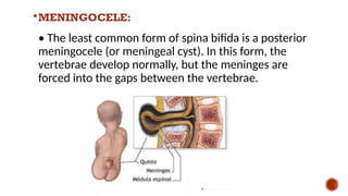
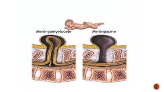
MENINGOCELE:
• The leastcommon form of spina bifida is a posterior
meningocele (or meningeal cyst). In this form, the
vertebrae develop normally, but the meninges are
forced into the gaps between the vertebrae.
12.
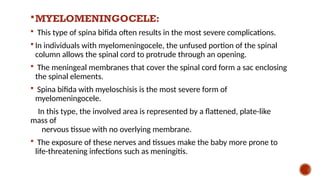
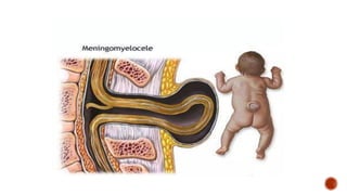
MYELOMENINGOCELE:
This typeof spina bifida often results in the most severe complications.
In individuals with myelomeningocele, the unfused portion of the spinal
column allows the spinal cord to protrude through an opening.
The meningeal membranes that cover the spinal cord form a sac enclosing
the spinal elements.
Spina bifida with myeloschisis is the most severe form of
myelomeningocele.
In this type, the involved area is represented by a flattened, plate-like
mass of
nervous tissue with no overlying membrane.
The exposure of these nerves and tissues make the baby more prone to
life-threatening infections such as meningitis.
14.
CONTD…
• The protrudedportion of the spinal cord and the nerves that
originate at that level of the cord are damaged or not properly
developed.
• As a result, there is usually some degree of paralysis and loss
of sensation below the level of the spinal cord defect.
15.
CLINICAL MANIFESTATIONS:
Physical Signs:
Orthopedic abnormalities (i.e., club foot, hip dislocation)
Bladder and bowel control problems, including
incontinence, urinary tract infections, and poor renal
function
Pressure sores and skin irritations
Abnormal eye movement
68% of children with spina bifida have an allergy to latex
Paralysis
16.
CONTD..
Scoliosis
Backpain
Partial or complete lack of sensation
Weakness of the hips, legs, or feet of a newborn
Other symptoms may include:
Hair at the back part of the pelvis called the sacral area
Dimpling of the sacral area
Difficulty swallowing, which can lead to choking.
Hoarseness.
Breath-holding and problems breathing during sleep.
Below-average intelligence.
17.
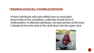
NEUROLOGICAL COMPLICATIONS
• Manyindividuals with spina bifida have an associated
abnormality of the cerebellum, called the Arnold Chiari Il
malformation. In affected individuals, the back portion of the brain
is displaced from the back of the skull down into the upper neck.
18.
EXECUTIVE FUNCTION:
• Specificareas of difficulty in some individuals include planning,
organizing, initiating, and working memory. Problem-solving,
abstraction, and visual planning may also be impaired. o Children
with spina bifida and shunted hydrocephalus have higher rates of
ADHD.
19.
ACADEMIC SKILLS:
Individualswith spina bifida may struggle academically,
especially in the subjects of mathematics and reading. In one
study, 60% of children with spina bifida were diagnosed with a
learning disability.
20.
SOCIAL COMPLICATIONS:
• Comparedto typically developing children, youths with spina
bifida may have fewer friends and spend less time with peers.
21.
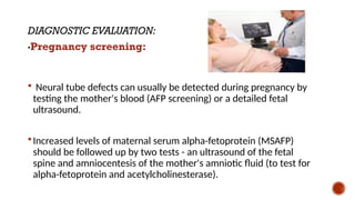
DIAGNOSTIC EVALUATION:
•Pregnancy screening:
Neural tube defects can usually be detected during pregnancy by
testing the mother's blood (AFP screening) or a detailed fetal
ultrasound.
Increased levels of maternal serum alpha-fetoprotein (MSAFP)
should be followed up by two tests - an ultrasound of the fetal
spine and amniocentesis of the mother's amniotic fluid (to test for
alpha-fetoprotein and acetylcholinesterase).
22.
PREVENTION:
Dietary supplementationwith folic acid has been shown to be
helpful in reducing the incidence of spina bifida. Sources of folic acid
include whole grains, fortified breakfast cereals, dried beans, leaf
vegetables and fruits.
It is recommended that any woman considering becoming pregnant
take 0.4 mg of folic acid a day. Pregnant women need 1 mg per day.
23.
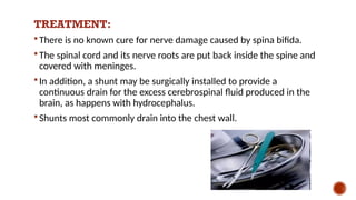
TREATMENT:
There isno known cure for nerve damage caused by spina bifida.
The spinal cord and its nerve roots are put back inside the spine and
covered with meninges.
In addition, a shunt may be surgically installed to provide a
continuous drain for the excess cerebrospinal fluid produced in the
brain, as happens with hydrocephalus.
Shunts most commonly drain into the chest wall.
24.
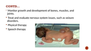
CONTD...
Monitor growthand development of bones, muscles, and
joints.
Treat and evaluate nervous system issues, such as seizure
disorders.
Physical therapy
Speech therapy
25.
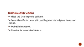
IMMEDIATE CARE:
• Placethe child in prone position.
• Cover the affected area with sterile gauze piece dipped in normal
saline.
• Maintain hydration.
• Monitor for associated defects.
26.
LIFE LONG TREATMENT:
•Catheters
• Braces
• High. fiber diet
• Antibiotics may be used to treat or prevent infections such as
meningitis or urinary tract infections.
27.
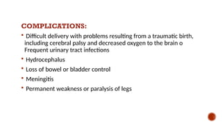
COMPLICATIONS:
Difficult deliverywith problems resulting from a traumatic birth,
including cerebral palsy and decreased oxygen to the brain o
Frequent urinary tract infections
Hydrocephalus
Loss of bowel or bladder control
Meningitis
Permanent weakness or paralysis of legs