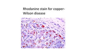
This document provides information and methods for special stains used to identify various tissues and microorganisms under the microscope. It discusses stains for amyloid, pigments, microbes and calcium. Specific stains covered include Congo red for amyloid, Perls' Prussian blue for iron, Fouchet's for bile pigments, Masson Fontana for melanin, von Kossa for calcium, rhodanine for copper, Gram stain for bacteria identification, Ziehl-Neelsen for acid-fast mycobacteria, and Warthin-Starry for spirochetes. Detailed protocols are provided for each staining method.
This document discusses special stains used in histopathology, specifically stains used to identify lipids. It describes the Oil Red O and Sudan Black B stains. Oil Red O stains neutral triglycerides and lipids red and nuclei blue. It is used to identify tumors containing fat cells and fatty emboli. Sudan Black B stains fats blue-black and nuclei red. It can identify phospholipids and is used to differentiate myeloblasts from lymphoblasts in hematological disorders. Special stains are important tools to identify tissue components not visible with routine H&E staining.
This document discusses fixatives used in histopathology. It describes the process of fixation and how fixatives preserve tissue by denaturing or precipitating proteins. The ideal properties of a fixative are described, including preventing autolysis and allowing for staining. Common fixatives are classified and their mechanisms and uses are explained. Factors that affect fixation such as temperature, size, volume ratio, time, choice of fixative, and penetration are also summarized.
This document discusses the Lupus erythematosus cell (LE cell), which is a neutrophil or monocyte that has ingested altered nuclear material from another cell. The presence of LE cells in blood smears is strongly suggestive of systemic lupus erythematosus (SLE). The document describes a method called the Rotary Method of Zinkham and Conley for inducing the formation of LE cells from a patient's blood sample through rotation and heating. It notes that a positive LE cell test is found in 75% of SLE patients but there can be false positives, so it is not a definitive diagnostic test.
Hematoxylin and eosin (H&E) staining is the most common histological staining method. Hematoxylin stains cell nuclei blue by combining with oxidized hematin and a mordant like alum. Eosin stains cytoplasm and extracellular substances pink. For H&E staining, tissue sections are stained in hematoxylin, rinsed in acid alcohol to differentiate nuclei, rinsed in water to turn nuclei blue, and then stained in eosin to color non-nuclear structures pink, allowing easy visualization of cell morphology. H&E staining provides essential structural information and is useful for pathology examinations.
This document discusses the process of decalcification, which is the removal of calcium salts from bones and calcified tissues. There are four main methods: 1) using simple dilute mineral acids like nitric acid or formic acid; 2) ion exchange resins with acid fluids; 3) electrolytic decalcification using electrodes; and 4) chelating agents like EDTA that bind calcium ions. The document provides details on procedures and advantages/disadvantages of each method.
The document discusses various histological staining techniques. It begins by explaining hematoxylin and eosin staining, which provides basic diagnostic information. It then covers special stains that highlight specific tissue components, categorized by the structures they identify such as carbohydrates, amyloid, lipids, nucleic acids, and microorganisms. Carbohydrate stains discussed include periodic acid Schiff, alcian blue, mucicarmine, and others. Amyloid identification using Congo red and methyl violet is explained. Lipid stains using Sudan dyes are also summarized. The document provides details on techniques for staining nucleic acids and identifying bacteria by Gram staining.
This document discusses various fixatives used for small biopsy specimens and their applications. It describes the ideal properties of a fixative and the mechanisms of different types of fixatives including aldehydes, mercurials, dichromates, picric acid and alcohol-containing fixatives. For each fixative, the document outlines their composition, principle of action, tissues they are suitable for, fixation time and advantages and disadvantages. It also provides recommendations for fixation of specific tissue biopsies like renal, muscle and liver biopsies. The conclusion emphasizes the importance of using an appropriate fixative to ensure optimal specimen evaluation and diagnosis.
The document discusses various carbohydrate stains including periodic acid Schiff (PAS), Alcian blue, mucicarmine, and others. It describes the principles, procedures, and results of PAS staining for glycogen and fungi. Different types of mucins are discussed along with combined staining techniques to differentiate neutral from acidic mucins. The document also covers connective tissue stains for collagen and elastic fibers, as well as special stains for pigments, minerals, and lipids.
This document discusses various staining techniques used in cytology, including both routine and special stains. It provides details on the principles, procedures, and applications of stains such as May-Grunwald Giemsa, Diff-Quik, Papanicolaou, hematoxylin and eosin, periodic acid Schiff, mucicarmine, Alcian blue, Oil red O, Congo red, Feulgen, and Ziehl-Neelsen. The stains are used to demonstrate cellular and extracellular components, identify infectious organisms, and examine DNA content. Proper staining allows visualization of structures like glycogen, mucin, lipids, amyloid, and acid-fast bacteria under the microscope.
COMPARISON OF CONVENTIONAL PAPANICOLAOU STAIN WITH MODIFIED ULTRAFAST PAPANIC...SURAMYA BABU
• Body fluid cytology is vital in diagnosis of various neoplastic and non neoplastic lesions and conventional Pap stain is the staining method of choice for the same.
• MUFP is a quick and cheap staining technique which gives good interpretation of cytological features with easily available reagents.
• Preservation of cell morphology and nuclear staining are superior with conventional Papanicolaou technique whereas cytoplasmic staining is comparable with conventional pap and MUFP techniques.
• Though background of stained smears was slightly better with conventional Pap staining; MUFP was superior in case of hemorrhagic samples.
This document provides information about Ziehl-Neelsen (ZN) staining for acid-fast bacteria. It describes the principle, reagents, procedure, interpretation and quality control for ZN staining. The principle is that acid-fast bacteria contain mycolic acid in their cell wall, allowing the primary stain to bind strongly and resist decolorization by acid. Reagents include carbol fuchsin, sulfuric acid, and methylene blue. The procedure involves staining, heating, decolorizing, counterstaining and examining under a microscope. Positive results show pink rods on a blue background. Modifications to the sulfuric acid concentration are also discussed.
This document discusses methods for decalcifying bone and teeth for histological examination. It begins with an introduction to decalcification and describes the process of removing calcium ions from bone to make it flexible for pathological investigation. It then classifies common decalcifying agents such as acids, chelating agents, and other solutions. The document discusses factors that affect the rate of decalcification and tests used to determine when decalcification is complete. It provides details on processing and staining decalcified bone and teeth. The document concludes with precautions for decalcification and references.
This document provides information on staining blood films and smears. It discusses the different types of stains used including Romanowsky stains like Leishman stain, Giemsa stain, Wright stain, and Field stain. Specimens should be collected in EDTA and smears prepared within an hour then fixed in methanol or ethanol to preserve cell morphology before staining. Romanowsky stains use methylene blue and eosin dyes to reveal subtle differences in cell structures and components.
This document provides information about phosphotungstic acid hematoxylin (PTAH) staining, including its purpose, principle, procedure, and results. PTAH staining demonstrates intercellular proteins, muscle cross-striations, and fibrin. It is used to identify diseases involving gliosis, skeletal muscle tumors, cardiac abnormalities, and fibrin deposition. The staining procedure involves fixing sections, treating with various reagents including Lugol's iodine, potassium permanganate, and oxalic acid, and overnight staining with PTAH. This results in muscle cross-striations, fibrin, and glial fibers staining blue, while collagen appears red-brown.
Haemoglobin is a protein in red blood cells that carries oxygen throughout the body. It is composed of heme and globin. There are several methods to estimate haemoglobin levels, including physical, chemical, gasometric, and colorimetric techniques. The cyanmeth haemoglobin method is recommended, as it is accurate and converts haemoglobin to stable cyanmethemoglobin that can be read photometrically. This involves mixing blood with Drabkin's reagent containing potassium cyanide and ferricyanide to form cyanmethemoglobin, which is then measured colorimetrically at 540nm and quantified against a standard curve.
This document discusses the process of decalcification, which is the removal of calcium from tissues to make them suitable for section cutting. It outlines the key steps: selection of tissue, fixation, decalcification using mineral acids, chelating agents, or electrophoresis, detection of endpoint, neutralization, and washing. Common decalcifying agents discussed include Gooding and Stewart's fluid, von Ebner's fluid, citrate-citric acid buffer, and chelating agents like EDTA. The factors that influence decalcification speed and the importance of determining the endpoint are also summarized.
The tissue section is colourless because the fixed protein has the same refractive index as that of glass. We use dyes that have specific affinity with the different tissue proteins and colour them differently.
Colour is seen by the eye as a result of the effect of certain electromagnetic waves on the rods and cones of the retina. These waves, which have a varying length, will determine the colour that is seen.
White light being composed of all the colours of the visible spectrum varies in wavelength from 4,000 Â to 8,000 Â.
If light of a specific wavelength is absorbed from white light the resultant light will then be coloured, the colour being dependent upon the particular wavelength that has been removed.
The document describes the procedure for performing an antigen-antibody reaction test. It involves collecting whole blood from patients, preparing red blood cell suspensions, and diluting the suspensions in normal saline solution. The diluted suspensions are then added to tubes containing different antisera and incubated. Initial and final grading of agglutination is observed after shaking and further incubation. Anti-human globulin is added to some tubes to differentiate naturally occurring from immune antibodies.
This document discusses various dyes and stains used in biological studies. It begins by distinguishing between dyes, which are manufactured substances, and stains, which are dye solutions. Dyes can be natural, extracted from sources like lichens, or synthetic and manufactured. Common stains discussed include Wright's stain, Leishman's stain, Giemsa stain, and Romanowsky stains. The document provides details on the composition and preparation of these stains as well as their staining properties and applications in studying blood smears, bone marrow, and malaria parasites.
Staining ( rouine and special in cytology) rajiv kumarrajusehrawat
The document discusses staining techniques used in histology and cytology. It provides details on the preparation, components, and use of common stains including Hematoxylin, Giemsa stain, Papanicolaou stain, and Periodic acid–Schiff stain. The stains are used to differentially color structures like nuclei, cytoplasm, muscles, bones, parasites and glycogen under the microscope to enable examination of tissue samples and identification of cells and microorganisms.
This document describes capsule staining and metachromatic staining techniques. It discusses the principle, reagents used, and procedures for each staining method. Capsule staining distinguishes capsular material from bacterial cells using negative stains like India ink or positive stains and a mordant. Metachromatic staining demonstrates granules in Corynebacterium diphtheriae using Albert's solutions, which cause the granules to appear a different color than the cells. Both techniques provide specific staining to identify important bacterial structures.
The document describes the oxidase test, which is used to identify bacteria. It works by detecting cytochrome c, an iron-containing protein in the respiratory chain, which causes an oxidase reagent to turn purple in oxidase-positive bacteria within 5-10 seconds. The document outlines several methods for performing the test, such as using filter paper or commercial strips soaked in reagents like tetramethyl-p-phenylenediamine. It lists both oxidase-positive and -negative bacteria and notes some factors that can cause false results. Quantitative oxidase tests have also been developed that correlate with other identification tests and can differentiate bacteria at various taxonomic levels.
Hematoxylin and eosin (H&E) staining is the most common histology stain. Hematoxylin stains cell nuclei blue by binding to DNA and RNA, while eosin stains cytoplasm and extracellular components pink. The staining process involves deparaffinizing tissue sections, staining with hematoxylin, differentiating with acid to remove excess stain, staining with eosin, and mounting for examination. Hematoxylin is extracted from logwood and oxidized to hematin, which binds tissue as a cationic dye with a mordant like alum. Eosin Y is the typical counterstain used to visualize cytoplasm. Together, H&E staining provides excellent contrast to study cell and
This document discusses peroxidase staining, which is used to differentiate between myelogenous or monocytic leukemia and acute lymphocytic leukemia. Peroxidase is present in the primary granules of neutrophils, eosinophils, and monocytes, and activity increases with maturation. The peroxidase stain principle involves myeloperoxidase catalyzing hydrogen peroxide to oxidize a substrate like benzidine or DAB, forming a black precipitate. A peroxidase stain will show red-brown staining in neutrophils, eosinophils in promyelocyte through metamyelocyte stages, and finely granular staining in monocytes.
Fetal hemoglobin and rh incompatibilityrohini sane
A comprehensive presentation on fetal hemoglobin & Rh incompatibility for undergraduate medical, dental, biotechnology & pharmacology students for self-learning .Presentation has physical & chemical properties of fetal hemoglobin along with its function. Binding affinity for O₂ of HbF and oxygen dissociation curve for HbF elucidated with suitable diagrams. Molecular constitution of Embryonic Hb ( Grover I &Grover II )with electrophoretic patterns are presented here . Importance of Kleihauer staining for detection of fetal cells is described briefly.
Diagrammatic representation of Rh- incompatibility is done for complete understanding of the concept. Signs & symptoms Kernicterus are presented diagrammatically.
Direct and indirect Coomb’s Test for Rh- incompatibility for diagnosis of Erythroblastosis Fetalis is illustrated. Biochemical aspects of Hemolytic Disease of Newborn (HDN) and Physiological /Neonatal Jaundice are presented. Comparison of Causes & biochemical findings for Hemolytic Jaundice along hepatic and obstructive jaundice is done in this presentation.
Molecular mechanism involved in biosynthesis of Hb Bart and Hb H along with their electrophoretic patterns for their detection are illustrated.
Hereditary persistent fetal Hb( HPFH ) & Point mutations causing HPFH are described in lucid manner. Google images are used for intense impact of the subject.
This document summarizes haematological changes that can occur in systemic diseases. It discusses anaemia of chronic disorders which occurs in patients with inflammatory or malignant conditions. The anaemia is caused by decreased iron availability and erythropoietin response. It also occurs in rheumatoid arthritis, lupus, chronic kidney disease, liver disease, and hypothyroidism due to various contributing factors like blood loss, bone marrow suppression, and cytokine effects. Coagulation abnormalities can also arise in cancers, liver disease, and nephrotic syndrome.
This document provides information on various histological staining techniques used to identify different types of tissues and biomolecules. It discusses connective tissue stains like Van Gieson's stain, Masson's trichrome stain, and Verhoeff's stain used to identify collagen fibers. It also describes reticulin stains, elastic stains, carbohydrate stains like periodic acid Schiff, and mucin stains like Alcian blue to identify different components of tissues under the microscope. Procedures for each stain are outlined along with the expected results.
The document provides information on the preparation, properties, assays, and uses of several inorganic compounds including sodium chloride, calcium gluconate, ammonium chloride, sodium bicarbonate, hydrogen peroxide, chlorinated lime, copper sulphate, ferrous sulphate, and sodium thiosulphate. For each compound, methods of preparation, physical and chemical properties, assay methods (often titration based), and common uses are described. The compounds discussed are commonly used in pharmaceutical, medical, industrial, and laboratory applications.
This document discusses tissue fixation techniques and histochemical staining methods. It begins by defining tissue fixation as a process that preserves tissues from decay through chemical means. It then describes various types of fixatives including simple fixatives like formalin and compound fixatives. Specific fixatives discussed include 10% neutral buffered formalin, Bouin's fluid, Zenker's fluid, and osmium tetroxide. Modes of action and uses of these fixatives are provided. The document concludes by outlining various histochemical staining techniques for identifying elements like glycogen, calcium, iron, uric acid crystals and amyloid. Specific stains discussed are PAS, Best's carmine, von Kossa's, Prussian blue, de Gal
The document discusses various histological staining techniques. It describes the principles, reagents used, and results of common stains like H&E, PAS, Alcian blue, mucicarmine, Oil red O, Verhoeff's stain, Masson's trichrome, Azan-Mallory stain, Weigert's iron hematoxylin and more specialized stains. The staining methods allow visualization and differentiation of tissues, cells, proteins, carbohydrates, lipids, fibers and other structures under the microscope.
The document summarizes a seminar presentation on histopathology staining techniques. It discusses the routine H&E stain and provides details on the principle, reagents, and procedure. It also describes special stains used to identify substances like carbohydrates, amyloid, nucleic acids, and lipids. Specific stains covered include periodic acid Schiff, Congo red, Feulgen, methyl green pyronin, and Oil red O. The document aims to inform attendees about common and special staining methods in histopathology.
Phyochemical screening of Plant Ectractsarjunaliya
This document describes methods for the preliminary phytochemical screening and qualitative analysis of plant extracts to identify various phytochemical constituents. It provides details on qualitative chemical tests to detect primary and secondary metabolites like alkaloids, saponins, tannins, flavonoids, cardiac glycosides, anthraquinone glycosides, and tests specific for detecting constituents in particular plants like aloe. The aim is to determine the presence or absence of these important phytochemicals in plant samples.
The document summarizes the PAS staining technique and Ziehl-Neelsen staining technique for acid-fast bacteria. The PAS stain involves oxidizing carbohydrates using periodic acid, then treating with Schiff's reagent to produce a magenta color. The Ziehl-Neelsen stain takes advantage of mycobacteria's lipid-rich cell wall, using a carbol fuchsin solution which is retained after differentiation in acid or alcohol, allowing visualization of acid-fast bacilli. Both techniques are described in detail, outlining the required solutions and step-by-step procedures.
This document discusses Perls stain, which is used to identify iron deposits in tissue samples. It provides background on pigments in living tissue, including endogenous pigments like hemosiderin and hematogenous pigments. The history of Prussian blue and its use as Perls stain is described. The principle of the stain is that hydrochloric acid releases ferric ions from hemosiderin, which then react with potassium ferrocyanide to form insoluble Prussian blue pigment. Staining protocols, quality control, and clinical applications for identifying iron deposits in organs are covered.
Expectorants are drugs that enhance sputum secretion from the airways, making it easier to cough up phlegm. Ipecacuanha and potassium iodide are common expectorants that work by irritating the stomach or bronchial mucosa to stimulate sputum production. Ammonium chloride and antimony potassium tartrate are also used as expectorants. Potassium iodide, ammonium chloride, and sodium potassium tartrate (Rochelle salt) can also act as emetics in small doses to induce vomiting. Activated charcoal, sodium nitrite, and sodium thiosulphate are common antidotes used to treat poisoning from substances like cyanide.
This document discusses several types of gastrointestinal agents including acidifiers, antacids, cathartics, and antimicrobials. It provides details on specific agents such as ammonium chloride, dilute hydrochloric acid, aluminum hydroxide gel, milk of magnesia, sodium bicarbonate, and their uses, preparations, and assays. Antacids are discussed in terms of their ideal properties and combinations used. Acidifiers are described as agents that increase acidity in the gastrointestinal tract for various purposes.
1. This document discusses various oxidation-reduction titration methods including those using ceric ammonium sulfate, potassium iodate, potassium bromate, and titanous chloride as titrants.
2. Preparation and standardization procedures are provided for 0.1N ceric ammonium sulfate, 0.05M potassium iodate, 0.1N potassium bromate, and titanous chloride solutions.
3. Examples of titrations discussed include assays of ferrous fumarate, acetomenaphthone, ferrous gluconate tablets using ceric ammonium sulfate; assays of benzalkonium chloride and hydralazine hydrochloride using potassium iodate; and assays
Ziehl-Neelsen stain uses carbol fuchsin to stain mycobacteria pink to red and a counterstain of methylene blue or malachite green. Fite acid fast stain combines peanut oil and xylene to retain acid fastness of mycobacteria stained red. Periodic acid Schiff stain oxidizes glycans magenta to demonstrate substances like glycogen, fungi, and mucins, using liver and intestine as controls.
Analytical Procedures in Elemental Impuritiesumaiya fatima
This document discusses analytical procedures for determining elemental impurities like arsenic, lead, and mercury. It provides details on the methodology and reagents used for arsenic detection using the molybdenum blue method and atomic absorption spectrometry. For lead, the dithizone method is described where lead ions form a brown colloid with dithizone. Mercury detection uses the mercury(II) thiocyanate method where chloride ions displace thiocyanate and form a colored complex with iron(III). Spectrophotometric techniques are used for quantification and calibration curves.
The document discusses water quality assessment and surveillance. It outlines various physical, chemical and biological parameters used to evaluate drinking water quality according to WHO guidelines. These include turbidity, total dissolved solids, colour, odor, taste, temperature, pH and presence of inorganic constituents like chloride, calcium, magnesium, iron and sodium. Methods for testing parameters like turbidity, chloride, hardness, iron and fluoride are described. The document also covers bacteriological indicators of water quality including coliforms, E. coli and presence of pathogens. It provides methods for testing coliform bacteria using membrane filtration and multiple tube techniques.
This document discusses various staining techniques used in cytology, including both routine and special stains. It provides details on the principles, procedures, and applications of common stains like Papanicolaou stain, hematoxylin and eosin stain, periodic acid Schiff stain, mucicarmine stain, oil red O stain, and Ziehl-Neelsen stain. The stains highlighted aid in the examination of cellular details, identification of keratin and malignant cells, and demonstration of substances like glycogen, lipids, acid mucins, amyloid, and mycobacteria. Proper staining is crucial in cytological evaluation and disease diagnosis.
The document discusses electrolyte replacement therapy used to treat dehydration and describes the concentrations of various electrolytes like sodium, potassium, calcium, and magnesium that must be replaced. It also provides information on common electrolyte solutions used for replacement including their compositions, properties, methods of preparation, and uses. Specific electrolytes discussed in more detail include sodium chloride, potassium chloride, and calcium gluconate.
Rasamanikya is a excellent preparation in the field of Rasashastra, it is used in various Kushtha Roga, Shwasa, Vicharchika, Bhagandara, Vatarakta, and Phiranga Roga. In this article Preparation& Comparative analytical profile for both Formulationon i.e Rasamanikya prepared by Kushmanda swarasa & Churnodhaka Shodita Haratala. The study aims to provide insights into the comparative efficacy and analytical aspects of these formulations for enhanced therapeutic outcomes.
8 Surprising Reasons To Meditate 40 Minutes A Day That Can Change Your Life.pptxHolistified Wellness
We’re talking about Vedic Meditation, a form of meditation that has been around for at least 5,000 years. Back then, the people who lived in the Indus Valley, now known as India and Pakistan, practised meditation as a fundamental part of daily life. This knowledge that has given us yoga and Ayurveda, was known as Veda, hence the name Vedic. And though there are some written records, the practice has been passed down verbally from generation to generation.
Our backs are like superheroes, holding us up and helping us move around. But sometimes, even superheroes can get hurt. That’s where slip discs come in.
Promoting Wellbeing - Applied Social Psychology - Psychology SuperNotesPsychoTech Services
A proprietary approach developed by bringing together the best of learning theories from Psychology, design principles from the world of visualization, and pedagogical methods from over a decade of training experience, that enables you to: Learn better, faster!
Does Over-Masturbation Contribute to Chronic Prostatitis.pptxwalterHu5
In some case, your chronic prostatitis may be related to over-masturbation. Generally, natural medicine Diuretic and Anti-inflammatory Pill can help mee get a cure.
Travel vaccination in Manchester offers comprehensive immunization services for individuals planning international trips. Expert healthcare providers administer vaccines tailored to your destination, ensuring you stay protected against various diseases. Conveniently located clinics and flexible appointment options make it easy to get the necessary shots before your journey. Stay healthy and travel with confidence by getting vaccinated in Manchester. Visit us: www.nxhealthcare.co.uk
Histololgy of Female Reproductive System.pptxAyeshaZaid1
Dive into an in-depth exploration of the histological structure of female reproductive system with this comprehensive lecture. Presented by Dr. Ayesha Irfan, Assistant Professor of Anatomy, this presentation covers the Gross anatomy and functional histology of the female reproductive organs. Ideal for students, educators, and anyone interested in medical science, this lecture provides clear explanations, detailed diagrams, and valuable insights into female reproductive system. Enhance your knowledge and understanding of this essential aspect of human biology.
2. SPECIAL STAINS FOR:
Amyloid
Pigments (Iron, bile, melanin, calcium, copper)
Microbes (gram positive bacteria, mycobacterium, spirochaete and
fungi )
3. AMYLOID-Congo red stain
Principle of congo red staining:
Amyloids are homogenous and eosinophilic,
proteinaceous deposits, that are extracellular and may become
sufficiently large enough to cause damage to surrounding tissues.
When stained with the congo red stain the amyloid will show
bifringence an apple green color, under the polarizing microscope.
4. Bennhold's congo red staining:
Method
(1) Bring sections to water,
(2) Stain with Ehrliçh haematoxylin for 20 minutes,
(3) Differentiate with 1% acid alcohol.
(4) Wash in running water for 1 minute to remove the acid
(5) Stain with 1% aqueous Congo red for 20-30 minutes.
5. (6) Pour off stain and flood slide with a saturated aqueous solution of lithium carbonate;
leave for 15 seconds.
(7) Differentiate in 80% alcohol until excess Congo red is removed.
(8) Wash in running water for 10 minutes.
(9) Dehydrate, clear and mount
Results
Amyloid -Pink to red
Nuclei -Blue
6. With polarized light - amyloid exhibits bright green birefringence,
‘apple-green birefringence’
This property is shared by other β-pleated sheet proteins and specific
to that conformation.
8. Congo red stained section of kidney
demonstrating amyloid deposition(red)
9. Same section viewed by polarized light showing
green birefringence against dark background
10. USES
It is used to demonstrate amyloid deposits in
Renal amyloidosis
Medullary carcinoma thyroid
Vessel wall in case of Alzheimer disease
Cardiac arrythmias
11. In upper dermis
there is deposit of eosinophilic
amorphous material (H&E)
Dermal amyloid deposits under
polarizing microscopy
(Thioflavin-T stain)
Other methods: 1)Flourescent
Microscopy Technique: Thioflavin T
13. Perls’ Prussian blue
Hemosiderin is a brown pigment that is present in reticuloendothelial
cells of bone marrow, spleen and liver. It is formed by partial
degradation of aggregates of ferritin by lysosomes.
PRINCIPLE:
Treatment of tissue with an acid ferrocyanide solution will result in
the unmasking of ferric iron in hemosiderin, in the form of the
hydroxide, Fe(OH)3. The ferric iron then reacts with a dilute
potassium ferrocyanide solution to produce an insoluble blue
compound, ferric ferrocyanide (Prussian blue).
14. PURPOSE:
• To demonstrate ferric iron in tissue sections.
• Small amounts of iron are found normally in spleen
and bone marrow.
• Excessive amount are present in hemochromatosis,
with deposits found in the liver and pancreas,
hemosiderosis, with deposits in the liver, spleen, and
lymph nodes.
15. METHOD
(1)Bring sections to water.
(2) Transfer to a fresh solution of equal parts of 2% aqueous potassium
ferrocyanide and 2% hydrochloric acid, for 30 minutes.
(3) Wash thoroughly in water.
(4) Counterstain lightly with 1% neutral red for 10-15 seconds.
(5) Wash in water, dehydrate, clear and mount.
Results
Ferric-iron-containing pigments (haemosiderin)- Blue
Nuclei -Red
18. Liver section from a patient with
hemochromatosis stained for ferric iron with Perl’s
method. (Ferric iron is stained blue).
19. Fouchet technique for bile pigments
Bile pigments are endogenous breakdown products of haemoglobin-
bilirubin and biliverdin that are excreted in bile.
Principle of fouchets:
The pigment is converted to the green color of biliverdin and
blue cholecyanin by the oxidative action of the ferric chloride in the
presence of trichloroacetic acid .
20. Solutions:
Fouchet’s solution :
25% aqueous trichloroacetic acid -36 ml
10% aqueous ferric chloride -4 ml
Van Gieson stain solution :
Dissolve 100 mg of acid fuchsin in 100 ml of saturated aqueous
picric acid
21. Method
1. Take sections to water.
2. Treat with the freshly prepared Fouchet’s solution for 10 minutes.
3. Wash well in running tap water for 1 minute.
4. Rinse in distilled water.
22. • 5. Counterstain with van Gieson solution for 2 minutes.
• 6. Dehydrate, clear and mount
Results
Bile pigments- emerald to blue-green
24. Bile in liver section stained with fouchet’s
technique. Bilirubin is stained emerald green.
25. OTHER METHODS
• 1)GLENNER METHOD- treat sections with 3% potassium dichromate
bilirubin is stained emerald green
• 2)STEIN’S METHOD- treat sections with mixture of 3 parts lugol’s
iodine and 1 part tincture iodine, decolorise with sodium sulphite and
counterstain in neutral red. Bilirubin appears green
26. MELANIN
Melanin is non lipid, non hematogenous pigment.
It is a brown black pigment present normally in hair, skin, retina, iris
and certain parts of CNS.
27. Principle:
The solutions of ammoniacal silver nitrate are reduced by
melanin to black metallic silver, basis for demonstrating melanin.
28. METHOD: Masson Fontana for melanin
1.Take sections to water.
2.Treat with the ammoniacal silver solution in a Coplin jar for 30–40
minutes at 56°C or overnight at room temperature.
3.Wash well in water.
4. Treat sections with 5% aqueous sodium thiosulfate for 1 minute.
5. Wash well in running tap water for 2–3 minutes.
29. 6.Lightly counterstain in 0.5% aqueous neutral red for 5 minutes
7. Rinse in distilled water.
8.Dehydrate, clear and mount.
Results
Melanin, argentaffin, chromaffin and some lipofuscins- black
Nuclei- red
30. Melanin pigment of skin showing black color
RESULTS:
Melanin,
argentaffin cells -black
Nuclei - red
31. USES
To identify melanin and argentaffin granules.
In diagnosis of malignant melanoma.
Argentaffin granules are found in carcinoid tumors.
34. OTHER METHODS- 1)ENZYME DOPA METHOD
Principle :
The enzyme tyrosinase is located within some cells producing melanin
will oxidize DOPA to form an insoluble brown – black pigment.
Purpose :
Cells that are capable of producing melanin can be demonstrated by
DOPA (dihydroxy- phenylalanine) method.
Result
DOPA oxidase – Brown
Nuclei - blue
35. 2)Fluoroscence methods-melanin precursors give yellow
fluorescence
3)Bleaching methods: melanin is bleached by strong
oxidising agents like hydrogen peroxide, potassium
chlorate or treatment with potassium permangate for
half hour followed by 1% oxalic acid.
4)Nile blue method: melanin stained dark blue
36. CALCIUM-VON KOSSA METHOD
Calcium is plentiful mineral found in human body in teeth, bones, blood and nerve cells. Abnormal
deposits of calcium may be found in any area of the body
Principle of von kossa method:
Tissue sections are treated with silver nitrate solution, silver is deposited by
replacing the calcium and then it is reduced by the strong light and visualized as metallic silver.
o With H&E stain, calcium appear deep blue purple.
o On von kossa method it appears black.
37. Method
1) Bring sections to water.
2) Immerse section in pH 45 citrate buffer for 20 minutes
3) Wash well in distilled water.
4) Flood slides with 5% silver nitrate.
5) Expose to bright sunlight or to a 60-watt electric bulb at a range of 4-
5 inches for 30-60 minutes.
38. (6) Wash in several changes of distilled water
(7) Treat with 5% sodium thiosulphate for 2-3 minutes.
(8) Counterstain with neutral red
(9) Dehydrate, clear and mount
Result
Calcium deposits- Black
41. OTHER METHODS
1)FLUOROSCENT METHOD: Stain with morin in 85% alcohol containing
0.5% acetic acid and view under fluorescent microscope. Greenish
white fluorescence indicates presence of calcium.
2)ALIZARIN RED S METHOD:stain in alizarin solution(alizarin red S 1g
and 50ml distilled water. Calcium deposits are stained orange red.
43. COPPER
Copper is excreted in bile and accumulates in liver in chronic biliary
diseases
Rhodanine technique for copper
Principle:
Rhodanine stain demonstrates protein to which copper binds rather
than copper itself. It is used to identify copper deposits in Wilson
disease
44. Rhodanine technique for copper
• Solutions
Rhodanine stock solution 5-p-Dimethylaminobenzylidene-
rhodanine 0.05 g
Absolute ethanol 25 ml
Working solution 5 ml of the stock rhodanine solution added to 45 ml of
2% sodium acetate trihydrate.
Borax solution
Disodium tetraborate 0.5 g
Distilled water 100 ml
45. Method
1. Take sections to water.
2. Incubate in the rhodanine working solution at 56°C for 3 hours or
overnight in a 37°C oven.
3. Rinse in several changes of distilled water for 3 minutes each.
4. Stain in acidified alum hematoxylin for 10 seconds.
5. Briefly rinse in distilled water and place immediately in borax
solution for 15 seconds.
46. • 6. Rinse well in distilled water.
• 7. Dehydrate clear and mount.
Results
Copper and copper-associated protein red to orange-red
Nuclei blue
49. GRAM STAIN
• Gram positive bacteria have thick cell wall without an outer membrane and
stain purple with gram stain.eg: cyanobacteria
• Gram negative bacteria have thin cell wall with an outer membrane and
stain red with gram stain .eg: salmonella
Principle of gram stain:
The structure of organism’s cell wall determines whether the organism is
gram positive or negative. Those bacteria which retain the primary stain by
resisting decolorization are gram positive and those which get decolorized
and then get counterstained are called gram negative.
50. Gram's method
Reagent
Lugol's iodine
Iodine 1 g
Potassium iodide 2 g
Distilled water to 100 ml
• Dissolve the potassium iodide in 4-5 ml of water; dissolve the iodine
in this. Dilute to 100 ml to make Lugol's iodine
51. Method
(1) Bring sections to water.
(2) Stain with 0.5% aqueous methyl violet 6 B for 1-3 minutes.
(3) Rinse with water.
(4) Pour on Lugol's iodine for 1-3 minutes
52. 5) Differentiate rapidly with acetone (1-2 seconds) and wash
immediately in running water.
6) Counterstain with 1% neutral red for 1 minute.
7) Wash in water.
8) Dehydrate, clear and mount.
Results
Gram-positive organisms- Blue-black
Other tissue structures -Shades of red
54. Ziehl-Neelsen (ZN) Stain
Mycobacteria are difficult to demonstrate by the Gram
technique because they possess a capsule containing a
long chain fatty acids, mycolic acid that make them
hydrophobic.
Phenolic acid or heat may be used to reduce the surface
tension and increase the porosity.
55. Ziehl-Neelsen method
Principle:
Mycobacterias (tubercle bacilli) have a lipid rich cell wall
which is capable of taking up strong phenol dye solutions (eg.carbol
fuschin solution) such that the dye is retained upon subsequent
differentiation in acid or alcohol called as acid and alcohol fast(AAFB=
acid and alcohol fast bacilli)
57. METHOD
1) Bring section to water.
2) Stain in hot carbol-fuchsin, either in a Coplin jar in a 56 °C
oven for 30 minutes, or flooding the slide with stain, heating until
the stain steams and leaving for 10 minutes.
3) Wash in water to remove excess stain
4) Differentiate in 1 % acid alcohol, 10 min.
58. 5) Wash in water.
6) Counterstain lightly in 0.1% méthylène blue for 10-15 seconds
7) Wash in water
8) Dehydrate, clear and mount.
60. OTHER METHODS
1) Modified Zeihl Neelsen method
Lepra bacillus is not as acid fast as M.
tuberculosis, so conc. of H2SO4 is less.
Decolorise with 5%-8% H2SO4 ( instead of 20%
in ZN method).
Results : lepra bacilli - red
other tissue - blue
2)Modified fite procedure:
lepra bacilli - red
other tissue - blue
61. 3)Bleach Concentration Technique (cytology)
Pus in tube
mixed with 2ml of 5% sod hypochlorite
incubated for 15min at 37c
2ml distilled water added
centrifuged at 3000rpm for 15 min
air dried smear using 1 drop sediment
stained by ZN stain
64. SPIROCHAETE
Spirochaetes are elongated motile, flexible bacteria twisted spirally along the long
axis with a periplasmic flagella.eg: Treponema pallidium(syphilis), borrelia
burgodorferi (lyme disease), leptospira (leptospirosis)
Principle of warthin-starry stain method:
Spirochaetes have ability to bind silver ions.It involves
impregnation of spirochaetes in tissues with silver ions with subsequent
reduction of these ions to metallic silver using a developer containing
hydroquinone.
65. SPIROCHAETE
Warthin-Starry method for spirochetes
Solutions
Acetate buffer, pH 3.6
• Sodium acetate 4.1 g
• Acetic acid 6.25 ml
• Distilled water 500 ml
Silver solution 1% silver nitrate in pH 3.6 acetate buffer
Developer Dissolve 0.3 g of hydroquinone in 10 ml pH 3.6 acetate buffer, and
mix 1 ml of this solution and 15 ml of warmed 5% Scotch glue or gelatin;
keep at 40°C. Take 3 ml of 2% silver nitrate in pH 3.6 buffer solution and keep
at 55°C. Mix these two solutions immediately before use.
66. Method
• 1. Bring section to water.
• 2. Celloidinize in 0.5% celloidin, drain and harden in distilled water
for 1 minute.
• 3. Impregnate in preheated 55–60°C silver solution for 90–105
minutes.
• 4. Prepare and preheat developer in a water bath.
• 5. Treat with developer for 30 minutes at 55°C. Sections should be
golden-brown at this point.
67. • 6. Remove from developer and rinse in tap water for several minutes
at 55–60°C, then in buffer at room temperature.
• 7. Tone in 0.2% gold chloride.
• 8. Dehydrate, clear, and mount.
Results
Spirochetes black
Background golden/yellow
70. SILVER METHENAMINE STAIN FOR
FUNGI
o GMS staining is a silver staining technique for demonstrating
fungi in tissue sections.
o It is primarily based on staining the polysaccharides in fungal
cell walls.
• PRINCIPLE
This method depends upon the reduction of the silver by the
aldehyde groups produced after oxidation of fungal wall
components with chromic acid.
72. 0.21% silver nitrate, stock solution A
Silver nitrate 2.1 g
Distilled water 1000 ml
Methenamine-sodium borate, stock solution B
Methenamine 27 g
Sodium borate decahydrate (borax) 3.8 g
Distilled water 1000 ml
Methenamine-silver sodium borate working solution Equal parts of
solutions A and B.
73. Method
• 1. Bring section to water.
• 2. Oxidize in 4% aqueous chromic acid (chromium trioxide) for 30
minutes.
• 3. Wash briefly in distilled water.
• 4. Dip briefly in 1% sodium bisulfite.
• 5. Wash well in distilled water
• 6. Place in preheated (56–60°C water bath) working silver solution for
15–20 minutes. If section is ‘paper bag brown’ then rinse in distilled
water.
74. • 7. Tone in 0.1% gold chloride for 5 seconds. Rinse in distilled water.
• 8. Place in 5% sodium thiosulfate for 5 seconds.
• 9. Rinse well in running tap water.
• 10. Counterstain in light green solution until a medium green
(usually 5–15 seconds).
• 11. Dehydrate, clear and mount
75. • Fungi -black
• Hyphae and yeast-form cells sharply delineated in black of fungi
• Background-pale green
77. GMS showing light green counterstain for
Histoplasma capsulatum, dimorphic fungus.
78. Periodic - acid – Schiff Stain (PAS)
Principle:
Certain tissue elements are oxidised by the periodic
acid. One of the reaction products is an aldehyde.
which combine with Schiff's reagent to form an
insoluble magenta compound
Periodic acid
Tissue element Dialdehydes
( containing 1:2 glycol groups
or 1:2 aminohydroxyl groups ) Schiff reagent
Coloured end product
79. Procedure :
Bring sections to water
Treat with periodic acid for 5 min
Wash with tap water for 5 min
Stain with Schiff’s reagent for 8-10 min
Wash with tap water for 5 min
counterstain with Haematoxylin for 3 min
Wash with tap water for 5min
Dehydrate, clear & mount