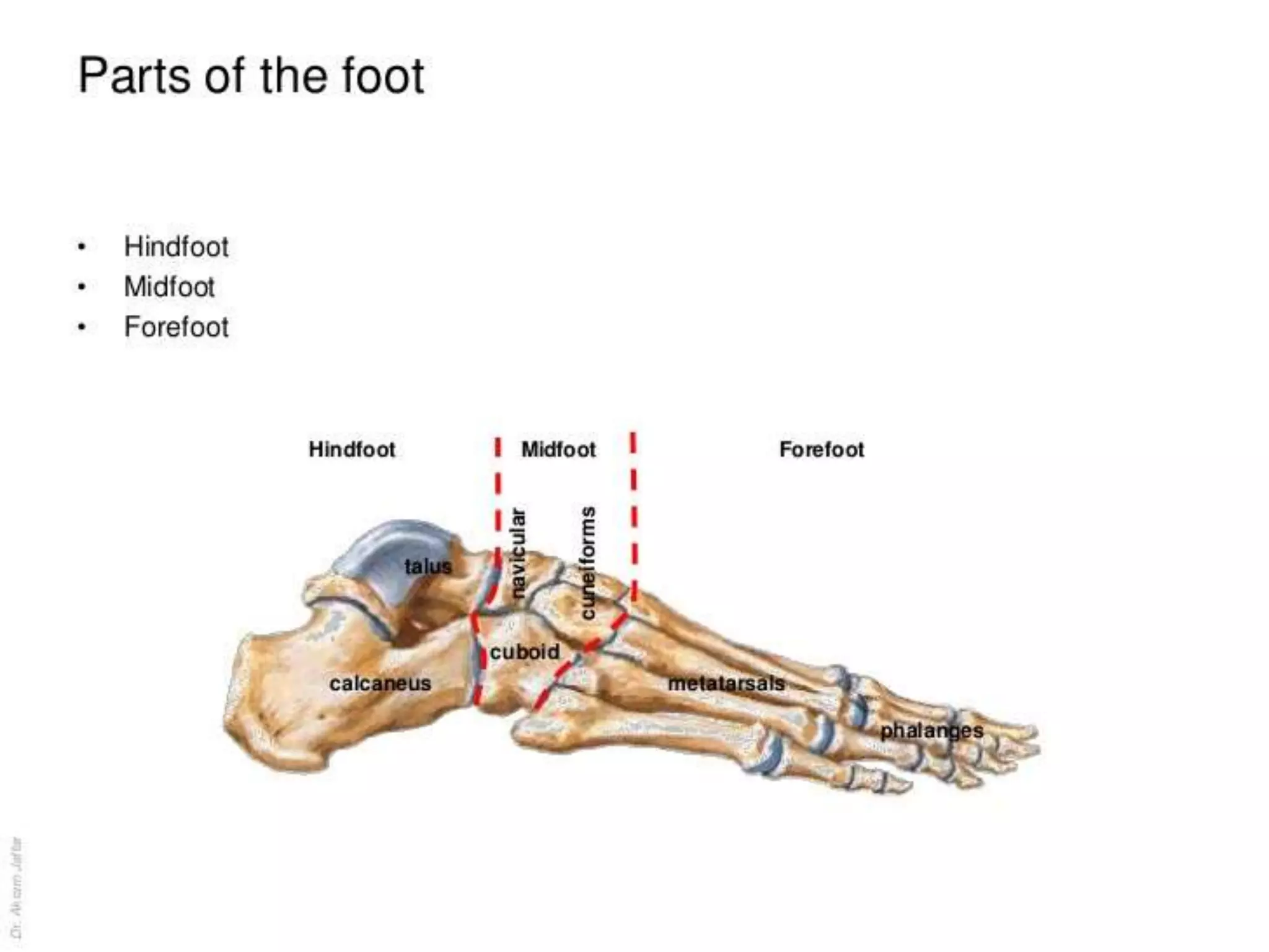
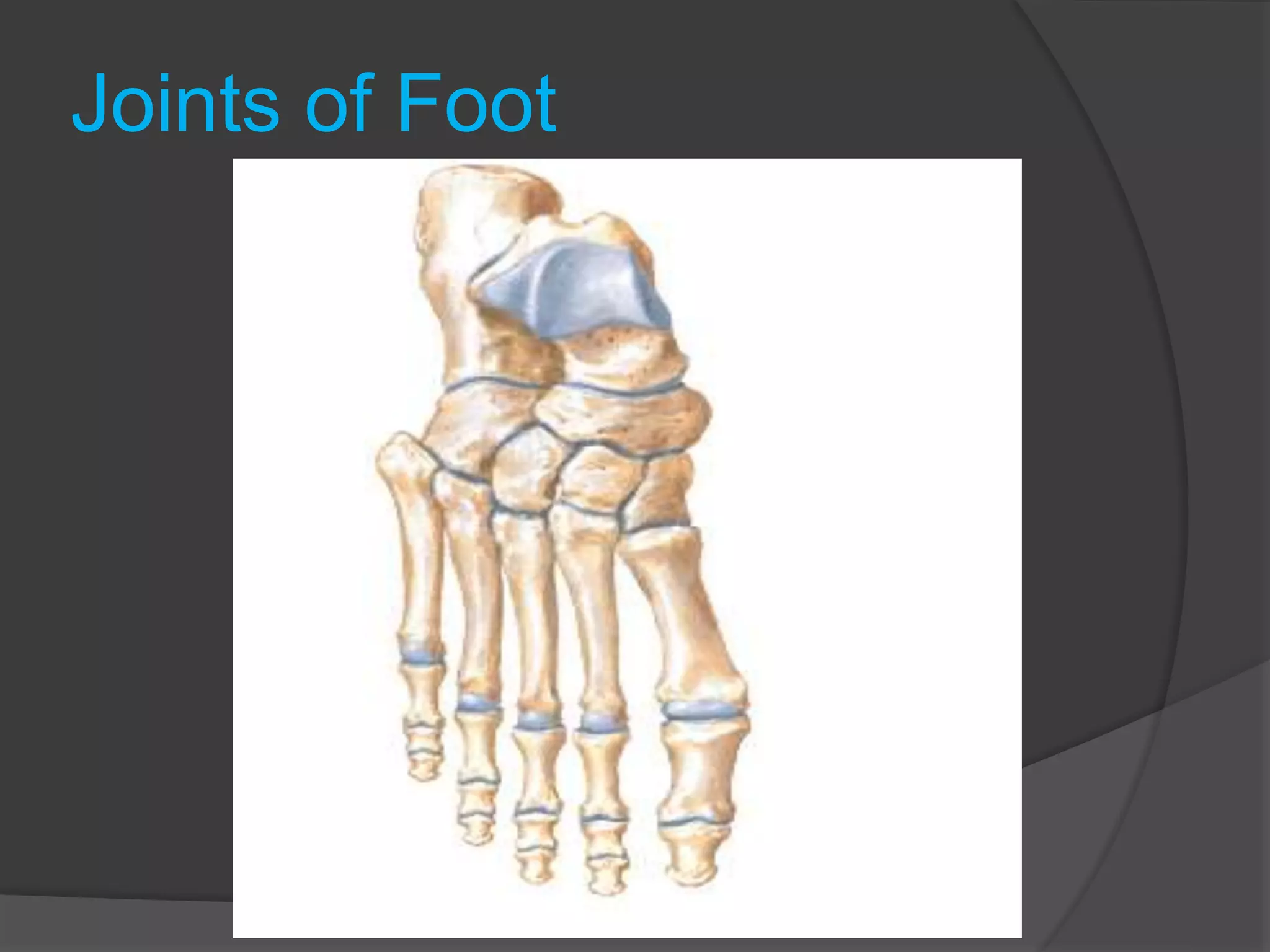
1- Calcaneus
2- Talus
3- Navicular
4- Cuboid
5- 1st Cuneiform
6- 2nd Cuneiform
7- 3rd Cuneiform
8- Metatarsals
9- Proximal phalanges
10- Middle phalanges
11- Distal phalanges
In total there are 26 bones in the foot.
In triple arthodesis
there is fusion of joints:
Calcaneo cuboid
Talo Navicular
Talo Calcaial
Talus bone
Numerous important ligaments are attached to the talus,
but no muscles are attached to this bone
The talus articulates above at the ankle joint with the tibia
and fibula, below with the calcaneum, and in front with the
navicular bone
Joints of Foot
Ankle joint
Formed by
• Talus
• Articular surface of lower tibia and medial malleolus
• Lateral malleolus of fibula
It allows movements
• Dorsi flexion
• Planter flexiom
Tibia and fibula tightly bound by anterior and posterior
tibiofibular ligaments
Ankle joint bound by multiple medial and lateral
collateral ligaments
Arches of Foot
Three arches are
• Longitudinal (Medial and lateral)
• Transverse
Together they make a tripod foot which
share the weight in
balanced position
Posterior Aspect of the
Ankle
StructuresThat Pass Behind the Medial Malleolus
Beneath the Flexor Retinaculum with synovial sheath
•Tibialis posterior tendon
•Flexor digitorum longus
•Posterior tibial artery with venae comitantes
•Tibial nerve
•Flexor hallucis longus
StructuresThat Pass Behind the Lateral Malleolus
Superficial to the Superior Peroneal Retinaculum
•The sural nerve
•Small saphenous vein
StructuresThat Pass Behind the Lateral Malleolus
Beneath the Superior and Inferior Peroneal
Retinaculuae
•The peroneus longus
•Peronius brevis tendons
They share a common synovial sheath when they
pass beneath superior peroneal retinaculum, but
behind inferior peroneal retinaculum they have
separate sheaths.
Anterior Aspect of the Ankle
StructuresThat Pass Beneath orThrough
Extensor
Retinaculae
•Tibialis anterior tendon
•Extensor hallucis longus tendon
•Anterior tibial artery with venae comitantes
•Deep peroneal nerve
•Extensor digitorum longus tendons
•Peroneus tertius
StructuresThat Pass in Front of the Medial
Malleolus
•Great saphenous vein
•Saphenous nerve
As each of the tendons passes through the
extensor
retinacula, it is surrounded by a synovial
sheath
Sole of Foot
• Skin
• Plantar aponeurosis
• Attached to
• Medial and lateral tubercles of
calcaneus
• Five slips
• Divide into two
• Fuse deep transverse ligament of
sole
• Abduction and adduction from a line
drawn through the middle of second
toe
EXTENSOR EXPANSION OF
LATERAL 4 TOES
Origin:
 Fibrous expansion on the dorsal surface of the proximal phalanx.
 Formation:
 Tendons of the extensor digitorum longus for 2nd, 3rd, 4th & 5th toes.
 Tendon of the extensor digitorum brevis for 2nd, 3rd & 4th toes.
 Tendons of lumbricals muscle for 2nd, 3rd, 4th & 5th toes.
 Tendons of dorsal interossei 2 for 2nd toe and one for each of 3rd and 4th toe.
The tendons of planter interossei for 3rd, 4th & 5th toes.
All these tendons unite on the dorsum of the proximal phalanx to form this expansion.
The expansion is divided into three slips.
1) The thick central part of the expansion is inserted into the dorsal surface of the
base of the middle phalanx.
2) The lateral and medial parts joint each other to be inserted into the base of the
terminal phalanx.
Flexor digitorum brevis insert into middle phalanx by
piercing flexor digitorum longus
Flexor digitorum longus inserts into distal phanalnges of 4toes
Tibialis anterior inserts into 1st metatarsal head and invert the
foot at subtaler and transverse taler joint
Tibialis posterior inserts into 2nd to 4th metatarsal and
cuniform and navicular bones and invert the foot
Peronius longus and brevis evert the foot
All small muscles are supplied by tibial nerve
Seasmoid Bones
• Usually 2 bones
• Larger medial in
Abductor hallucis and
medial half of flexor
hallucis brevis
• Smaller lateral in
Adductor hallucis and
ateral half of flexor
hallucis brevis
Blood Supply of the foot
Dorsalis pedis artery branch of anterior tibial
artery
which becomes
• Arcuate artery
• Dorsal metatarsal arteries
• Digital arteries
Arcuate artery also give perforating arteries to
communicate with deep talar artery
DORSALIS PEDIS ARTERY
 Origin:
 It is the continuation of the anterior tibial
artery.
 Beginning
 On the front of the ankle joint at a point
midway between the medial and lateral
malleoli
 Course:
 Runs forwards on the dorsum of the foot
accompanied by two venae comitants.
 Termination:
 At the proximal end of the first
interosseous space, the artery turns
towards the sole between the two heads
of the first dorsal interosseous muscle to
anastomose with the end of the planter
arch.
 It is superficial in position and is crossed by the inferior
extensor retinaculum and the first tendon of extensor
digitorum brevis
 Its course can be indicated by a line extends from
 Point mid way between the 2 malleoli to
Point at the proximal end of thefirst intermetatarsal space.
 Pulsations can be easily felt
lateral to the tendon of
extensor hallucis longus.
Branches.
1. Medial and lateral tarsal arteries:
 They arise just distal to the ankle joint
 Supply the joint of the tarsus
 Share in the anastomosis around the
ankle joint
2. Arcuate artery:
 Origin: arises opposite the bases of
metatarsal bones
 Course: runs laterally with an arched
course
 Branches:
 It gives off the 2nd, 3rd and 4th dorsal
metatarsal arteries. Each dorsal
metatarsal divides into two dorsal digitalarteries to the sides of the adjoining
toes.
 The 4th dorsal metatarsal artery gives off
a branch, which supplies the lateral side
of the little toe.
3. 1st dorsal metatarsal artery:
 Arises from the termination of the
dorsalis pedis immediately before it
pierces the 1st interosseous space.
 Runs forwards on the 1st dorsal
interosseus muscle & divided into two
branches:
 Medial branch: to the medial border
of the big toe.
 Lateral branch: divides into two
dorsal digital branches, which supply
the adjoining sides of the big &
second toes.
4. 1st planter metatarsal artery:
 Arises from the ends of the
dorsalis pedis in the sole.
 Branches:
 Planter digital branch to the
medial side of the big toe.
 Common planter digital
branch, which divided into
two planter digital branches
supplies the adjoining sides
of the big & 2nd toe.
MEDIAL PLANTAR
ARTERY
 Origin:
 One of the two terminal branches of
the posterior tibial nerve.
 Course:
 Enter the foot midway between the
medial malleolus and the medial
tubercle of the calcaneus, under
cover the flexor retinaculum.
 Passes forwards deep to the
abductor hallucis muscle.
 Passes between the abductor
hallucis and flexor digitorum brevis.
 It is accompanied by two venae
comitantes.
 The medial planter nerve runs along
its lateral side.
 Termination:
 By anastmosing with the 1st planter
metatarsal artery.
 Branches:
 Muscular:
 to the surrounding muscles.
 Digital:
 These are 3 superficial digital
branches these branches end by
anastmosing with the first, second
and third planter metatarsal
arteries.
LATERAL PLANTAR
ARTERY
 Origin:
 One of the two terminal branches of
the posterior tibial nerve.
 Course:
 At first between the 1st and 2nd
layers, then curves medially
between the 3rd and 4th layers of
the sole.
 The lateral planter nerve lies along
its medial side.
 Termination:
 Turns medially with the deep
branch of the lateral planter nerve
with slight forward convexity to
from the plantar arch between
the 3rd & 4th layers of muscles.
 Branches:
 Muscular: to the surrounding muscles.
 Anastomotic branches:
 Anastomosis with branches of arcuate & lateral
tarsal arteries of the dorsalis pedis artery.
 Posterior perforating arteries:
 3 branches which ascend through the proximal
ends of the 2nd, 3rd & 4th interosseous spaces to
anastomose with the dorsal metatarsal arteries.
 Planter digital artery: to the lateral side of
the little toe. From the beginning of the arch.
 Three planter metatarsal arteries:
 Each divides into two planter digital arteries
which supply the adjacent sides of two toes.
These planter metatarsal arteries supply the
adjacent sides between the 2nd, 3rd, 4th toes.
Before it division into planter digital arteries,
each planter metatarsal artery sends an anterior
perforating artery to joint the corresponding
dorsal metatarsal artery.
Nerve Supply of the Foot
• Medial plantar nerve
Three and half toes
• Lateral plantar nerves
One and half toes
Nerve Supply of Dorsum
• Superficial peroneal
nerve
• Deep peroneal nerve,
first cleft
• Saphenous nerve,
medial border to base
of big toe
• Sural nerve, lateral
border
Cutaneous nerve supply
• Dorsum by Superficial peroneal nerve by
medial and
lateral branches
• 1st web by deep peroneal nerve
• Lateral side by sural nerve
• Medial side by saphanous nerve
MEDIAL PLANTAR NERVE
 Origin:
 The larger of the two terminal
branches of the posterior tibial
nerve.
 Course:
 Enter the foot midway between the
medial malleolus and the medial
tubercle of the calcaneus, under
cover the flexor retinaculum.
 Passes forwards deep to the
abductor hallucis muscle.
 Pass between the abductor hallucis
and flexor digitorum brevis muscle.
 The medial planter vessels run
along its medial side
 Termination:
 At the bases of the metatarsal
bones by dividing into 3 planter
digital nerves.
 Branches:
 Muscular (to four muscles) to:
1) Abductor hallucis.
2) Flexor digitorum brevis.
3) Flexor hallucis brevis
4) First lumbrical muscle
 Cutaneous:
Planter cutaneous branches:
1) To the skin of the medial 2/3 of the
sole of the foot.
2) Planter digital nerves
 Articular branches: To intertarsal
and tarso-metatarsal joints.
LATERAL PLANTAR
NERVE
 Origin:
 The smaller of the two terminal
branches of the posterior tibial
nerve.
 Course:
 Enters the foot midway between
the medial malleolus and the medial
tubercle of the calcaneus under
cover the flexor retinaculum.
 Passes forwards and laterally deep
to abductor hallusis.
 Passes between flexor digitorum
brevis & flexor digitoum
accessorius.
 The lateral planter vessels rum
along its lateral side.
 Termination:
 At the base of the 5th metatarsal
bone, by dividing into a superficial
and a deep branches.
 Branches:
 Muscular :
1) Flexor digitoum accessorius muscle
2) Abductor digiti minimi
3) Flexor digiti minimi brevis
4) Adductor halucis muscle.
5) Interossei
6) 2nd, 3rd & 4th lumbricals.
 Cutaneous:
1) Skin of the lateral 1/3 of the sole
2) Skin on the lateral side of the
planter surface of the little toe and
the adjoining sides of the 4th & 5th
toes.
3) The planter digital branches, also,
supply the skin on the dorsum of
the terminal phalanges of the lateral
one and half toes.
Motor nerve supply
Foot Compartments
- Anatomy:
- the 9 compartments of the foot can be placed
into 4 groups;
- Intrinsic Compartment:
- 4 intrinsic muscles between the 1st and 5th
metatarsals;
- Medial Compartment:
- abductor hallucis;
- flexor hallucis brevis;
- Central Compartment: (Calcaneal
Compartment)
- flexor digitorum brevis;
- quadratus plantae;
- adductor hallucis;
- Lateral Compartment:
- flexor digiti minimi brevis;
- abductor digiti minimi;
- medial approach:
- this is usually the approach of choice;
- can be used to decompress the medial and central compartments as well as
the remaining foot compartments;
- extends from a point below the medial malleolus (3 cm from the sole) to
proximal aspect of first metatarsal;
- once the neurovascular bundle has been retracted out of the way, the
fascia overlying the abduction hallucis and FDB is released;
- medial intermuscular septum is opened longitudinally;
- the lateral plantar neurovascular bundle is found coursing over
the quadratus plantae (central compartment) as they course laterally;
- the remaining compartments (central, lateral, intrinsic) are entered thru blunt
dissection w/ a clamp;3
- lateral compartment is found by retracting the FDB out of the way;
- dorsal approach:
- often the dorsal approach is not necessary unless there is
concomitant metatarsal or Lisfranc fractures;
- accomplished through 2 dorsal incisions centered just medial to the 2nd
metatarsal and just lateral to the 4th metatarsals (to maximize skin bridge);
- avoid injury to sensory nerves and extensor tendons;
- superficial fascia is divided and interosseous are elevated off the
metatarsals to further decompress the compartments;
- clamp is used to bluntly dissect thru the central, medial, and lateral
compartments;
- separate medial incision may be needed to release the abductor;
- fasciotomy incisions may be used for fracture fixation
Flaps of Foot
Abductor Digiti Minimi Flap
• Muscle or musculocutaneus based on branch
of lateral planter artery and nerve.
Abductor Hallucis Flap
• Muscle or musculocutaneus and based on
branch of medial planter artery and nerve.
Dorsalis Pedis Flap
Fasciocutaneous. Artery may have variations
in 17% people
Flexor Digitorum Brevis Flap
• Muscular. Based on proximal branch of
medial planter artery.
Lateral Planter Artery Flap
•.Faschicutaneous.
Medial Planter Artery Flap
• Faschicutaneous. Medial planter artery
based. From navicular tubercle to head of
first metatarsal.
Levels of amputations in the
foot
Biomechanics
Inversion and Eversion
• Inversion is raising the medial
border of the foot
• Sole of the foot looks medially
• Close pack
• Eversion is raising the lateral
border of the foot
• Sole of the foot looks laterally
• Least pack
Inversion and Eversion
• Initiated at the transverse
tarsal joint
• Calcaneocuboid
• Anterior portion of the
talocalcaneonavicular
• Main movement takes place
at the clinical sub-talar joint
i.e.
• Talocalcaneal
• Inferior portion of the
talocalcaneonavicular
• The pivot is the ligament of
the neck of the talus
Plantar Fasciitis
Plantar fasciitis, which occurs in individuals who do
a great
deal of standing or walking, causes pain and
tenderness
of the sole of the foot. It is believed to be caused by
repeated minor trauma. Repeated attacks of this
condition induce ossification in the posterior
attachment
of the aponeurosis, forming a calcaneal spur
Thank you

Foot clinical and applied anatomy

  • 2.
    1- Calcaneus 2- Talus 3-Navicular 4- Cuboid 5- 1st Cuneiform 6- 2nd Cuneiform 7- 3rd Cuneiform 8- Metatarsals 9- Proximal phalanges 10- Middle phalanges 11- Distal phalanges In total there are 26 bones in the foot.
  • 5.
    In triple arthodesis thereis fusion of joints: Calcaneo cuboid Talo Navicular Talo Calcaial
  • 6.
    Talus bone Numerous importantligaments are attached to the talus, but no muscles are attached to this bone The talus articulates above at the ankle joint with the tibia and fibula, below with the calcaneum, and in front with the navicular bone
  • 8.
  • 9.
    Ankle joint Formed by •Talus • Articular surface of lower tibia and medial malleolus • Lateral malleolus of fibula It allows movements • Dorsi flexion • Planter flexiom Tibia and fibula tightly bound by anterior and posterior tibiofibular ligaments Ankle joint bound by multiple medial and lateral collateral ligaments
  • 14.
    Arches of Foot Threearches are • Longitudinal (Medial and lateral) • Transverse Together they make a tripod foot which share the weight in balanced position
  • 27.
    Posterior Aspect ofthe Ankle StructuresThat Pass Behind the Medial Malleolus Beneath the Flexor Retinaculum with synovial sheath •Tibialis posterior tendon •Flexor digitorum longus •Posterior tibial artery with venae comitantes •Tibial nerve •Flexor hallucis longus StructuresThat Pass Behind the Lateral Malleolus Superficial to the Superior Peroneal Retinaculum •The sural nerve •Small saphenous vein
  • 30.
    StructuresThat Pass Behindthe Lateral Malleolus Beneath the Superior and Inferior Peroneal Retinaculuae •The peroneus longus •Peronius brevis tendons They share a common synovial sheath when they pass beneath superior peroneal retinaculum, but behind inferior peroneal retinaculum they have separate sheaths.
  • 32.
    Anterior Aspect ofthe Ankle StructuresThat Pass Beneath orThrough Extensor Retinaculae •Tibialis anterior tendon •Extensor hallucis longus tendon •Anterior tibial artery with venae comitantes •Deep peroneal nerve •Extensor digitorum longus tendons •Peroneus tertius
  • 33.
    StructuresThat Pass inFront of the Medial Malleolus •Great saphenous vein •Saphenous nerve As each of the tendons passes through the extensor retinacula, it is surrounded by a synovial sheath
  • 35.
    Sole of Foot •Skin • Plantar aponeurosis • Attached to • Medial and lateral tubercles of calcaneus • Five slips • Divide into two • Fuse deep transverse ligament of sole • Abduction and adduction from a line drawn through the middle of second toe
  • 42.
    EXTENSOR EXPANSION OF LATERAL4 TOES Origin:  Fibrous expansion on the dorsal surface of the proximal phalanx.  Formation:  Tendons of the extensor digitorum longus for 2nd, 3rd, 4th & 5th toes.  Tendon of the extensor digitorum brevis for 2nd, 3rd & 4th toes.  Tendons of lumbricals muscle for 2nd, 3rd, 4th & 5th toes.  Tendons of dorsal interossei 2 for 2nd toe and one for each of 3rd and 4th toe. The tendons of planter interossei for 3rd, 4th & 5th toes. All these tendons unite on the dorsum of the proximal phalanx to form this expansion. The expansion is divided into three slips. 1) The thick central part of the expansion is inserted into the dorsal surface of the base of the middle phalanx. 2) The lateral and medial parts joint each other to be inserted into the base of the terminal phalanx.
  • 43.
    Flexor digitorum brevisinsert into middle phalanx by piercing flexor digitorum longus Flexor digitorum longus inserts into distal phanalnges of 4toes Tibialis anterior inserts into 1st metatarsal head and invert the foot at subtaler and transverse taler joint Tibialis posterior inserts into 2nd to 4th metatarsal and cuniform and navicular bones and invert the foot Peronius longus and brevis evert the foot All small muscles are supplied by tibial nerve
  • 45.
    Seasmoid Bones • Usually2 bones • Larger medial in Abductor hallucis and medial half of flexor hallucis brevis • Smaller lateral in Adductor hallucis and ateral half of flexor hallucis brevis
  • 46.
    Blood Supply ofthe foot Dorsalis pedis artery branch of anterior tibial artery which becomes • Arcuate artery • Dorsal metatarsal arteries • Digital arteries Arcuate artery also give perforating arteries to communicate with deep talar artery
  • 47.
    DORSALIS PEDIS ARTERY Origin:  It is the continuation of the anterior tibial artery.  Beginning  On the front of the ankle joint at a point midway between the medial and lateral malleoli  Course:  Runs forwards on the dorsum of the foot accompanied by two venae comitants.  Termination:  At the proximal end of the first interosseous space, the artery turns towards the sole between the two heads of the first dorsal interosseous muscle to anastomose with the end of the planter arch.
  • 48.
     It issuperficial in position and is crossed by the inferior extensor retinaculum and the first tendon of extensor digitorum brevis  Its course can be indicated by a line extends from  Point mid way between the 2 malleoli to Point at the proximal end of thefirst intermetatarsal space.  Pulsations can be easily felt lateral to the tendon of extensor hallucis longus.
  • 49.
    Branches. 1. Medial andlateral tarsal arteries:  They arise just distal to the ankle joint  Supply the joint of the tarsus  Share in the anastomosis around the ankle joint 2. Arcuate artery:  Origin: arises opposite the bases of metatarsal bones  Course: runs laterally with an arched course  Branches:  It gives off the 2nd, 3rd and 4th dorsal metatarsal arteries. Each dorsal metatarsal divides into two dorsal digitalarteries to the sides of the adjoining toes.  The 4th dorsal metatarsal artery gives off a branch, which supplies the lateral side of the little toe.
  • 50.
    3. 1st dorsalmetatarsal artery:  Arises from the termination of the dorsalis pedis immediately before it pierces the 1st interosseous space.  Runs forwards on the 1st dorsal interosseus muscle & divided into two branches:  Medial branch: to the medial border of the big toe.  Lateral branch: divides into two dorsal digital branches, which supply the adjoining sides of the big & second toes.
  • 51.
    4. 1st plantermetatarsal artery:  Arises from the ends of the dorsalis pedis in the sole.  Branches:  Planter digital branch to the medial side of the big toe.  Common planter digital branch, which divided into two planter digital branches supplies the adjoining sides of the big & 2nd toe.
  • 52.
    MEDIAL PLANTAR ARTERY  Origin: One of the two terminal branches of the posterior tibial nerve.  Course:  Enter the foot midway between the medial malleolus and the medial tubercle of the calcaneus, under cover the flexor retinaculum.  Passes forwards deep to the abductor hallucis muscle.  Passes between the abductor hallucis and flexor digitorum brevis.  It is accompanied by two venae comitantes.  The medial planter nerve runs along its lateral side.  Termination:  By anastmosing with the 1st planter metatarsal artery.
  • 53.
     Branches:  Muscular: to the surrounding muscles.  Digital:  These are 3 superficial digital branches these branches end by anastmosing with the first, second and third planter metatarsal arteries.
  • 54.
    LATERAL PLANTAR ARTERY  Origin: One of the two terminal branches of the posterior tibial nerve.  Course:  At first between the 1st and 2nd layers, then curves medially between the 3rd and 4th layers of the sole.  The lateral planter nerve lies along its medial side.  Termination:  Turns medially with the deep branch of the lateral planter nerve with slight forward convexity to from the plantar arch between the 3rd & 4th layers of muscles.
  • 55.
     Branches:  Muscular:to the surrounding muscles.  Anastomotic branches:  Anastomosis with branches of arcuate & lateral tarsal arteries of the dorsalis pedis artery.  Posterior perforating arteries:  3 branches which ascend through the proximal ends of the 2nd, 3rd & 4th interosseous spaces to anastomose with the dorsal metatarsal arteries.  Planter digital artery: to the lateral side of the little toe. From the beginning of the arch.  Three planter metatarsal arteries:  Each divides into two planter digital arteries which supply the adjacent sides of two toes. These planter metatarsal arteries supply the adjacent sides between the 2nd, 3rd, 4th toes. Before it division into planter digital arteries, each planter metatarsal artery sends an anterior perforating artery to joint the corresponding dorsal metatarsal artery.
  • 56.
    Nerve Supply ofthe Foot • Medial plantar nerve Three and half toes • Lateral plantar nerves One and half toes
  • 57.
    Nerve Supply ofDorsum • Superficial peroneal nerve • Deep peroneal nerve, first cleft • Saphenous nerve, medial border to base of big toe • Sural nerve, lateral border
  • 58.
    Cutaneous nerve supply •Dorsum by Superficial peroneal nerve by medial and lateral branches • 1st web by deep peroneal nerve • Lateral side by sural nerve • Medial side by saphanous nerve
  • 60.
    MEDIAL PLANTAR NERVE Origin:  The larger of the two terminal branches of the posterior tibial nerve.  Course:  Enter the foot midway between the medial malleolus and the medial tubercle of the calcaneus, under cover the flexor retinaculum.  Passes forwards deep to the abductor hallucis muscle.  Pass between the abductor hallucis and flexor digitorum brevis muscle.  The medial planter vessels run along its medial side  Termination:  At the bases of the metatarsal bones by dividing into 3 planter digital nerves.
  • 61.
     Branches:  Muscular(to four muscles) to: 1) Abductor hallucis. 2) Flexor digitorum brevis. 3) Flexor hallucis brevis 4) First lumbrical muscle  Cutaneous: Planter cutaneous branches: 1) To the skin of the medial 2/3 of the sole of the foot. 2) Planter digital nerves  Articular branches: To intertarsal and tarso-metatarsal joints.
  • 62.
    LATERAL PLANTAR NERVE  Origin: The smaller of the two terminal branches of the posterior tibial nerve.  Course:  Enters the foot midway between the medial malleolus and the medial tubercle of the calcaneus under cover the flexor retinaculum.  Passes forwards and laterally deep to abductor hallusis.  Passes between flexor digitorum brevis & flexor digitoum accessorius.  The lateral planter vessels rum along its lateral side.  Termination:  At the base of the 5th metatarsal bone, by dividing into a superficial and a deep branches.
  • 63.
     Branches:  Muscular: 1) Flexor digitoum accessorius muscle 2) Abductor digiti minimi 3) Flexor digiti minimi brevis 4) Adductor halucis muscle. 5) Interossei 6) 2nd, 3rd & 4th lumbricals.  Cutaneous: 1) Skin of the lateral 1/3 of the sole 2) Skin on the lateral side of the planter surface of the little toe and the adjoining sides of the 4th & 5th toes. 3) The planter digital branches, also, supply the skin on the dorsum of the terminal phalanges of the lateral one and half toes.
  • 64.
  • 65.
  • 67.
    - Anatomy: - the9 compartments of the foot can be placed into 4 groups; - Intrinsic Compartment: - 4 intrinsic muscles between the 1st and 5th metatarsals; - Medial Compartment: - abductor hallucis; - flexor hallucis brevis; - Central Compartment: (Calcaneal Compartment) - flexor digitorum brevis; - quadratus plantae; - adductor hallucis; - Lateral Compartment: - flexor digiti minimi brevis; - abductor digiti minimi;
  • 68.
    - medial approach: -this is usually the approach of choice; - can be used to decompress the medial and central compartments as well as the remaining foot compartments; - extends from a point below the medial malleolus (3 cm from the sole) to proximal aspect of first metatarsal; - once the neurovascular bundle has been retracted out of the way, the fascia overlying the abduction hallucis and FDB is released; - medial intermuscular septum is opened longitudinally; - the lateral plantar neurovascular bundle is found coursing over the quadratus plantae (central compartment) as they course laterally; - the remaining compartments (central, lateral, intrinsic) are entered thru blunt dissection w/ a clamp;3 - lateral compartment is found by retracting the FDB out of the way; - dorsal approach: - often the dorsal approach is not necessary unless there is concomitant metatarsal or Lisfranc fractures; - accomplished through 2 dorsal incisions centered just medial to the 2nd metatarsal and just lateral to the 4th metatarsals (to maximize skin bridge); - avoid injury to sensory nerves and extensor tendons; - superficial fascia is divided and interosseous are elevated off the metatarsals to further decompress the compartments; - clamp is used to bluntly dissect thru the central, medial, and lateral compartments; - separate medial incision may be needed to release the abductor; - fasciotomy incisions may be used for fracture fixation
  • 69.
    Flaps of Foot AbductorDigiti Minimi Flap • Muscle or musculocutaneus based on branch of lateral planter artery and nerve. Abductor Hallucis Flap • Muscle or musculocutaneus and based on branch of medial planter artery and nerve. Dorsalis Pedis Flap Fasciocutaneous. Artery may have variations in 17% people
  • 70.
    Flexor Digitorum BrevisFlap • Muscular. Based on proximal branch of medial planter artery. Lateral Planter Artery Flap •.Faschicutaneous. Medial Planter Artery Flap • Faschicutaneous. Medial planter artery based. From navicular tubercle to head of first metatarsal.
  • 71.
  • 72.
  • 75.
    Inversion and Eversion •Inversion is raising the medial border of the foot • Sole of the foot looks medially • Close pack • Eversion is raising the lateral border of the foot • Sole of the foot looks laterally • Least pack
  • 76.
    Inversion and Eversion •Initiated at the transverse tarsal joint • Calcaneocuboid • Anterior portion of the talocalcaneonavicular • Main movement takes place at the clinical sub-talar joint i.e. • Talocalcaneal • Inferior portion of the talocalcaneonavicular • The pivot is the ligament of the neck of the talus
  • 82.
    Plantar Fasciitis Plantar fasciitis,which occurs in individuals who do a great deal of standing or walking, causes pain and tenderness of the sole of the foot. It is believed to be caused by repeated minor trauma. Repeated attacks of this condition induce ossification in the posterior attachment of the aponeurosis, forming a calcaneal spur
  • 86.