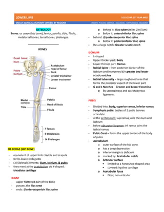
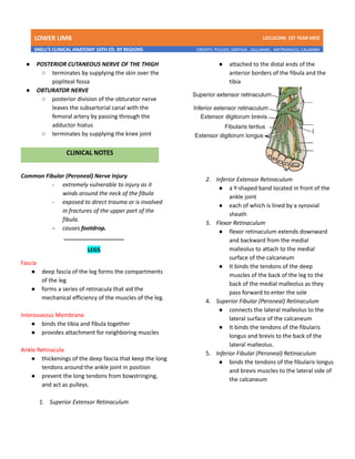
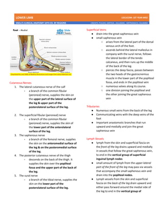
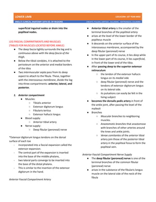
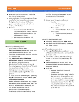
LOWER LIMB LDCUCOM: 1ST YEAR MED
SNELL’S CLINICAL ANATOMY 10TH ED. BY REGIONS CREDITS: PULIDO, ZARTIGA , GILLAMAC, ARITRANGCO, CALAMBA
OSTEOLOGY
Bones: os coxae (hip bone), femur, patella, tibia, fibula,
metatarsal bones, tarsal bones, phalanges.
_____________________
BONES
OS COXAE (HIP BONE)
- equivalent of upper limb clavicle and scapula.
- forms lower limb girdle
- (3) Skeletal Elements: ilium, ischium, & pubis
- they meet at the acetabulum via Y-shaped:
triradiate cartilage
ILIUM
- upper flattened part of the bone
- possess the iliac crest
- ends: @anterosuperioir iliac spine
o Behind it: Iliac tubercle lies 2in (5cm)
o Below it: anteroinferior iliac spine
- behind: @posterosuperior iliac spine
o Below it: posteroinferior iliac spine
- Has a large notch: Greater sciatic notch
ISCHIUM
- L-shaped
- Upper thicker part: Body
- Lower thinner part: Ramus
- Ischial Spine - from posterior border of the
ischium and intervenes b/n greater and lesser
sciatic notches
- Ischial tuberosity – large roughened area that
forms the posterior aspect of the lower part
- G and L Notches 🡪 Greater and Lesser Foramina
o By sacrospinous and sacrotuberous
ligaments
PUBIS
- Divided into: body, superior ramus, inferior ramus
- Symphysis pubis: bodies of 2 pubic bonnes
articulate
- at the acetabulum: sup ramus joins the ilium and
ischium
- below obturator foramen: inf ramus joins the
ischial ramus
- Pubic Crest – forms the upper border of the body
of pubis
- Acetabulum
o outer surface of the hip bone
o has a deep depression
o inferior margin is deficient
o marked by: Acetabular notch
o Articular surface
▪ limited to a horseshoe-shaped area
▪ covered: hyaline cartilage
o Acetabular fossa
▪ Floor, non-articular
LOWER LIMB LDCUCOM: 1ST YEAR MED
SNELL’S CLINICAL ANATOMY 10TH ED. BY REGIONS CREDITS: PULIDO, ZARTIGA , GILLAMAC, ARITRANGCO, CALAMBA
FEMUR
- Articulates:
o above with acetabulum: hip joint
o below with tibia and patella: knee joint
- Upper end: head, neck, greater and lesser
trochanters
o Head
▪ 2/3rds of a sphere & articulate
with os coaxae -> hip joint
▪ Fovea capitis – small depression,
center of the head
▪ Blood supply from the obturator
artery
o Neck
▪ Connects the head to the shaft
▪ angle 125 degree
o Greater & lesser trachanters
▪ Large eminences at the junction of
neck and shaft
▪ Intertrochanteric line – connects
anteriorly
▪ Intertrochanteric crest – connect
posteriorly
- Shaft
o Smooth and rounded on anterior surface
o Posteriorly has a ridge, linea aspera
▪ Where muscles and intermuscular
septa attached
o Medial margin: cont below the medial
supracondylar ridge to the adductor
tubercle on the medial condyle
o Lateral margin: cont below lateral
supracondylar ridge
o Gluteal tuberosity – posterior surface
below greater trochanter
o Popliteal surface – shaft becomes broader
towards distal end, flat, triangular are
- Lateral & medial condyles
o lower end
o separated by: intercondylar notch
o articular surface for the patella
o medial & lateral EPIcondyles – above the
condyles
adductor tubercle – cont with medial
epicondyle
1. Tenderness over the head of the femur
- Indicates presence of hip joint arthritis
2. Blood supply to femoral head and neck fractures
- Avascular necrosis
- Obturator artery – small branch that supply the
epiphysis of the head
- Medial femoral circumflex artery – supply the
upper part of the neck
3. Neck of the Femur and Coxa Valga and Vara
- Young: 160°; Adult: 125°
- Coxa valga – an increase in the angle
i. Congenital dislocation of hip
ii. Adduction is limited
- Coxa vara – a decrease in the angle
i. Fracture of the neck
ii. Abduction is limited
- Shelton’s Line – useful in assessing the angles
4. Femur Fractures
- Subcapital fracture
i. Elderly
ii. By minor trip or stumble
iii. Common in women after menopause (SF
femoral neck) – thinning of the cortical and
trabecular bone caused by estrogen
deficiency.
- Trochanteric fractures
i. Young and middle-aged
ii. Result of direct trauma
iii. Fracture line is extracapsular
- Fractures:
i. upper third of the shaft
● proximal fragment
o flexed: iliopsoas;
o abducted: gluteus medius and minimus;
LOWER LIMB LDCUCOM: 1ST YEAR MED
SNELL’S CLINICAL ANATOMY 10TH ED. BY REGIONS CREDITS: PULIDO, ZARTIGA , GILLAMAC, ARITRANGCO, CALAMBA
o laterally rotated: gluteus maximus, the
piriformis, the obturator internus, the
gemelli, and the quadratus femoris
● lower fragment
- adducted: adductor muscles,
- pulled upward: hamstrings and quadriceps
- laterally rotated: adductors and the weight
of the foot
ii. middle third of the shaft
● distal fragment
o pulled upward: hamstrings and the
quadriceps -> considerable shortening
o rotated backward: pull of the two heads of
the gastrocnemius
PATELLA (KNEECAP)
- largest sesamoid bone
- triangular, and its apex lies inferiorly
- Apex: connected to tuberosity of tibia by
ligamentum patellae (patellar ligament)
- Posterior surface – articulates with condyles of
femur
- Prepatellar bursa: separated from the skin
TIBIA
- large weight-bearing medial bone of the leg
- Articulates:
i. the condyles of the femur and
ii. the head of the fibula above and with the talus
and
iii. the distal end of the fibula below
- has an expanded upper end, a smaller lower end,
and a shaft.
i. Upper end
● lateral and medial condyles (lateral and
medial tibial plateaus)
o articulate with the lateral and medial
condyles of the femur and intervening
lateral and medial menisci
● Anterior and posterior intercondylar areas
- separate the upper articular surfaces of
the tibial condyles.
o intercondylar eminence lies between
these areas
● Lateral condyle
o possesses a small circular articular
facet for the head of the fibula on its
lateral aspect.
ii. Shaft
● triangular in cross section
● presenting three borders and three
surfaces.
o anterior and medial borders, with the
medial surface b/n - subcutaneous.
o anterior border
▪ prominent and forms the shin.
▪ tuberosity of the tibia - at the
junction
▪ rounded below,
▪ cont. with the medial malleolus.
o lateral (interosseous) border
▪ gives attachment to the
interosseous membrane
o posterior surface - shows an oblique
line, the soleal line
▪ for the attachment of the soleus
muscle.
iii. lower end
● slightly expanded
● shows a saddle-shaped articular surface
for the talus on its inferior aspect
● prolonged downward medially to form
the large medial malleolus.
FIBULA
- slender lateral bone of the leg
- no articulation at the knee joint; but participates
in the ankle joint below
- no transmission of body weigh; but provides
attachment for muscles
- has expanded upper end, a shaft, and a lower end
i. upper end (head)
● possesses a styloid process
● an articular surface for articulation with
the lateral condyle of the tibia
LOWER LIMB LDCUCOM: 1ST YEAR MED
SNELL’S CLINICAL ANATOMY 10TH ED. BY REGIONS CREDITS: PULIDO, ZARTIGA , GILLAMAC, ARITRANGCO, CALAMBA
ii. shaft
● long and slender
● four borders and four surfaces
o medial (interosseous) border gives
attachment to the interosseous
membrane
iii. lower end
● forms the triangular lateral malleolus,
which is subcutaneous
● triangular articular facet - articulation
with the lateral aspect of the talus is on
the medial surface of the lateral
malleolus
o malleolar fossa - depression lies
below and behind the articular facet
1. Patellar Dislocations
a. lower horizontal fibers of the vastus
medialis muscle and the large size of
the lateral condyle of the femur
i. prevents dislocation during
quadriceps muscle action
b. Congenital recurrent dislocations
i. caused by underdevelopment
of the lateral femoral condyle
c. Traumatic dislocation
i. results from direct trauma to
the quadriceps attachments
2. Patellar Fractures
a. result of direct violence
i. bone lies within the quadriceps
femoris tendon
b. result of indirect violence
i. caused by the sudden
contraction of the quadriceps
snapping
3. Tibia and Fibula Fracture
a. tibia and fibula
i. common
ii. only one bone is fractured, the
other acts as a splint
b. shaft (tibia)
i. often open because the entire
length of the medial surface is
covered only by skin and
superficial fascia
c. distal third of the shaft (tibia)
i. prone to delayed union or
nonunion
ii. bc the nutrient artery is torn at
the fracture line
d. proximal end (tibia), at tibial condyles
(tibial plateau)
i. middle-aged and elderly
ii. result from direct violence
4. Intraosseous Tibia Infusion in Infants
FOOT BONES
- Bones: the tarsal bones, the metatarsals, and the
phalanges
TARSAL BONES
- Bones: the calcaneum, the talus, the navicular,
the cuboid, and the three cuneiform bones
- ONLY talus – articulates w/ tibia & fibula at the
ankle joint
- Starts to ossify before birth
- 5th
year – ossification takes place in all
CALCANEUM
- the largest bone of the foot
- forms the prominence of the heel
- articulates: above with the talus and in front
with the cuboid
- has six surfaces:
1. anterior surface
● small and forms the articular facet that
articulates with the cuboid bone.
2. posterior surface
● forms the prominence of the heel
● gives attachment to: tendo calcaneus
(Achilles tendon)
LOWER LIMB LDCUCOM: 1ST YEAR MED
SNELL’S CLINICAL ANATOMY 10TH ED. BY REGIONS CREDITS: PULIDO, ZARTIGA , GILLAMAC, ARITRANGCO, CALAMBA
3. superior surface
● Two articular facets for the talus, separated
by a roughened groove, the sulcus calcanei
4. inferior surface
● has an anterior tubercle in the midline
● and a large medial and a smaller lateral
tubercle at the junction of the inferior and
posterior surfaces
5. medial surface
● has sustentaculum tali - a large, shelflike
process, w/c assists in the support of the
talus.
6. lateral surface
● almost flat
● peroneal tubercle - anterior part has a small
elevation, w/c separates the tendons of the
fibularis longus and brevis muscles.
TALUS
- articulates:
o above at the ankle joint with the
tibia and fibula
o below with the calcaneum
o in front with the navicular bone
- possesses a head, a neck, and a body
- NO muscles attach to this bone
- Head
o directed distally
o has an oval convex articular surface
for articulation with the navicular
bone.
▪ That continued on its inferior
surface,
▪ where it rests on the
sustentaculum tali behind
▪ and the calcaneonavicular
ligament in front.
- Neck
o lies posterior to the head
o slightly narrowed
o upper surface - roughened and
gives attachment to ligaments
o lower surface - shows a deep
groove, the sulcus tali.
o sinus tarsi - a tunnel formed the
sulcus tali and the sulcus calcanei in
the articulated foot
▪ which is occupied by the strong
interosseous talocalcaneal
ligament
- Body
o cuboidal
o superior surface - articulates with
the distal end of the tibia.
o lateral surface - presents a
triangular articular facet
▪ for articulation with the lateral
malleolus of the fibula
o medial surface - has a small,
comma-shaped articular facet
▪ for articulation with the medial
malleolus of the tibia
o posterior surface - marked by two
small tubercles
▪ separated by a groove for the
flexor hallucis longus tendon.
NAVICULAR BONE
- tuberosity: seen and felt on the medial
border of the foot 1 in. (2.5 cm) in front of
and below the medial malleolus
- gives attachment to the main part of the
tibialis posterior tendon
CUBOISAL BONE
- deep groove on the inferior aspect of the
cuboid bone lodges the tendon of the
fibularis longus muscle
CUNEIFORM BONE
- three small, wedge-shaped cuneiform
bones
- articulate:
LOWER LIMB LDCUCOM: 1ST YEAR MED
SNELL’S CLINICAL ANATOMY 10TH ED. BY REGIONS CREDITS: PULIDO, ZARTIGA , GILLAMAC, ARITRANGCO, CALAMBA
o proximally with the navicular bone
and
o distally with the first three
metatarsal bones. T
- wedge shape - contributes formation and
maintenance of the transverse arch of the
foot
METATARSAL BONES AND PHALENGES
- each possesses a head distally, a shaft, and a base
proximally
- five metatarsals - numbered from the medial to the
lateral side
- first (1st
) metatarsal
o aligns with the big toe (hallux)
o large and strong
o important In supporting wt of body
o - is grooved on its inferior aspect by the
medial and lateral sesamoid bones in the
tendons of the flexor hallucis brevis
- fifth (5th
) metatarsal
o prominent tubercle on its base
o fibularis brevis tendon attaches to the
tubercle
- each toe - three phalanges
- except the big head toe, which possesses only
two
1. Talus Fractures
- occur at the neck or body
- Neck fractures
i. occur during violent dorsiflexion of the
ankle joint
- Body fracture
i. Jumping from a height
2. Calcaneum Fractures
- from falls from a height
- drives the talus downward into the
calcaneum
3. Metatarsal Fractures
- base of the fifth metatarsal
i. during forced inversion of the foot
- Stress fracture
i. common in runners and after long
periods of walking
ii. most frequently in the distal third of the
2nd
, 3rd
, or 4th
metatarsal bone
_____________________
POPLITEAL FOSSA
● diamond-shaped intermuscular space situated
at the back of the knee
● most prominent when the knee joint is flexed.
● It contains:
○ small saphenous vein
○ common fibular (peroneal) and tibial nerves
○ posterior cutaneous nerve of the thigh
○ genicular branch of the obturator nerve
○ connective tissue
○ lymph nodes
● Boundaries
○ Laterally:
■ Biceps femoris above
■ Lateral head of the gastrocnemius and
plantaris below
○ Medially:
■ Semimembranosus and
semitendinosus above
■ medial head of the gastrocnemius
below
○ Anterior wall (floor):
■ Popliteal surface of the femur
■ Capsule of the knee joint,
■ Popliteus muscle
○ Roof:
■ Skin
■ Superficial fascia
■ Deep fascia of the thigh
LOWER LIMB LDCUCOM: 1ST YEAR MED
SNELL’S CLINICAL ANATOMY 10TH ED. BY REGIONS CREDITS: PULIDO, ZARTIGA , GILLAMAC, ARITRANGCO, CALAMBA
● MUSCLES
○ Back of the thigh:
■ Biceps femoris
■ Semimembranosus
■ Semitendinosus muscles.
○ Popliteus muscle (deep posterior leg)
- a key role in the movements of the
knee joint.
- its tendon separates the lateral
meniscus from the lateral ligament of
the joint
- ACTION:
-flexes the knee
-produce medial rotation of the tibia on
the femur or,
-lateral rotation of the femur on the
tibia (occurs at the commencement of
flexion of the extended knee, and its
rotatory action slackens the ligaments
of the knee joint - unlocking the knee
joint.)
● POPLITEAL ARTERY
○ continuation of the femoral artery
○ deeply placed and enters the popliteal
fossa through the adductor hiatus
○ ends at the level of the lower border of the
popliteus muscle by dividing into anterior
and posterior tibial arteries
○ Relations
■ Anteriorly:
● popliteal surface of the femur
● knee joint
● popliteus muscle
■ Posteriorly:
● popliteal vein
● tibial nerve
● Fascia
● Skin
○ Branches:
LOWER LIMB LDCUCOM: 1ST YEAR MED
SNELL’S CLINICAL ANATOMY 10TH ED. BY REGIONS CREDITS: PULIDO, ZARTIGA , GILLAMAC, ARITRANGCO, CALAMBA
● ARTERIAL ANASTOMOSIS AROUND KNEE JOINT -
to compensate for the narrowing of the
popliteal artery that occurs during extreme
flexion of the knee
○ small branches of the femoral artery
○ muscular and articular branches of the
popliteal artery
○ branches of the anterior and posterior tibial
arteries
1. Popliteal Aneurysm
- Caused by pulsations of the wall of the
femoral artery against the tendon of the
adductor magnus at the opening of the
adductor hiatus.
2. Semimembranosus Bursa Swelling
- most common swelling found in the popliteal
space
- extending the knee joint and becomes flaccid
when the joint is flexed
- should be distinguished from a Baker’s cyst,
which is centrally located and arises as a
pathologic (osteoarthritis) diverticulum of
the synovial membrane through a hole in the
back of the capsule of the knee joint
● POPLITEAL VEIN
○ formed by the junction of the venae
comitantes of the anterior and posterior
tibial arteries at the lower border of the
popliteus muscle on the medial side of the
popliteal artery
○ as it ascends through the fossa, it crosses
behind the popliteal artery so that it comes
to lie on its lateral side
○ passes through the adductor hiatus to
become the femoral vein.
○ Tributaries - veins that correspond to
branches given off by the popliteal artery
■ Small saphenous vein, which perforates
the deep fascia and passes between the
two heads of the gastrocnemius
muscle to end in the popliteal vein
● POPLITEAL LYMPH NODES
six lymph nodes are embedded
○ superficial lymph vessels
● lateral side of the foot and leg
● accompany the small saphenous vein
into the popliteal fossa.
○ deep lymph vessels
● accompanying the anterior and
posterior tibial arteries.
LOWER LIMB LDCUCOM: 1ST YEAR MED
SNELL’S CLINICAL ANATOMY 10TH ED. BY REGIONS CREDITS: PULIDO, ZARTIGA , GILLAMAC, ARITRANGCO, CALAMBA
● NERVES
●
SCIATIC NERVE
○ Divides into tibial and common fibular
components above the popliteal fossa
● Tibial Nerve
○ larger terminal branch of the sciatic nerve
○ runs downward through the popliteal fossa,
lying first on the lateral side of the popliteal
artery, then posterior to it, and finally
medial to it
○ enters the posterior compartment of the leg
by passing beneath the soleus muscle.
○ Branches
■ Cutaneous: Sural nerve
● descends between the two heads of
the gastrocnemius muscle
● joined by the sural communicating
branch of the common fibular nerve.
● supply the skin of the calf and the
back of the leg.
● accompanies the small saphenous
vein behind the lateral malleolus and
distributes to the skin along the
lateral border of the foot and the
lateral side of the little toe.
■ Muscular branches
● supply both heads of the
gastrocnemius and the plantaris,
soleus, and popliteus
■ Articular branches
● supply the knee joint
● Common Fibular (Peroneal) Nerve
○ smaller terminal branch of the sciatic nerve
○ runs downward through the popliteal fossa,
closely following the medial border of the
biceps femoris muscle
○ leaves the fossa by crossing superficial to the
lateral head of the gastrocnemius muscle
○ pierces the fibularis (peroneus) longus
muscle
○ it is subcutaneous and can easily be rolled
against the bone.
○ divides into two terminal branches:
■ superficial fibular (peroneal) nerve
■ deep fibular (peroneal) nerve
○ Branches
■ Cutaneous:
● Sural communicating branch
○ runs downward and joins the
sural nerve.
● Lateral cutaneous nerve of the calf
○ supplies the skin on the lateral
side of the back of the leg
■ Muscular branch
● to the short head of the biceps
femoris muscle, which arises high up
in the popliteal fossa
■ Articular branches to the knee joint.
LOWER LIMB LDCUCOM: 1ST YEAR MED
SNELL’S CLINICAL ANATOMY 10TH ED. BY REGIONS CREDITS: PULIDO, ZARTIGA , GILLAMAC, ARITRANGCO, CALAMBA
● POSTERIOR CUTANEOUS NERVE OF THE THIGH
○ terminates by supplying the skin over the
popliteal fossa
● OBTURATOR NERVE
○ posterior division of the obturator nerve
leaves the subsartorial canal with the
femoral artery by passing through the
adductor hiatus
○ terminates by supplying the knee joint
Common Fibular (Peroneal) Nerve Injury
- extremely vulnerable to injury as it
winds around the neck of the fibula
- exposed to direct trauma or is involved
in fractures of the upper part of the
fibula.
- causes footdrop.
___________________
LEGS
Fascia
● deep fascia of the leg forms the compartments
of the leg
● forms a series of retinacula that aid the
mechanical efficiency of the muscles of the leg.
Interosseous Membrane
● binds the tibia and fibula together
● provides attachment for neighboring muscles
Ankle Retinacula
● thickenings of the deep fascia that keep the long
tendons around the ankle joint in position
● prevent the long tendons from bowstringing,
and act as pulleys.
1. Superior Extensor Retinaculum
● attached to the distal ends of the
anterior borders of the fibula and the
tibia
2. Inferior Extensor Retinaculum
● a Y-shaped band located in front of the
ankle joint
● each of which is lined by a synovial
sheath
3. Flexor Retinaculum
● flexor retinaculum extends downward
and backward from the medial
malleolus to attach to the medial
surface of the calcaneum
● It binds the tendons of the deep
muscles of the back of the leg to the
back of the medial malleolus as they
pass forward to enter the sole
4. Superior Fibular (Peroneal) Retinaculum
● connects the lateral malleolus to the
lateral surface of the calcaneum
● It binds the tendons of the fibularis
longus and brevis to the back of the
lateral malleolus.
5. Inferior Fibular (Peroneal) Retinaculum
● binds the tendons of the fibularis longus
and brevis muscles to the lateral side of
the calcaneum
LOWER LIMB LDCUCOM: 1ST YEAR MED
SNELL’S CLINICAL ANATOMY 10TH ED. BY REGIONS CREDITS: PULIDO, ZARTIGA , GILLAMAC, ARITRANGCO, CALAMBA
Cutaneous Nerves
1. The lateral cutaneous nerve of the calf
- a branch of the common fibular
(peroneal) nerve, supplies the skin on
the upper part of the lateral surface of
the leg & upper part of the
posterolateral surface of the leg.
2. The superficial fibular (peroneal) nerve
- a branch of the common fibular
(peroneal) nerve, supplies the skin of
the lower part of the anterolateral
surface of the leg.
3. The saphenous nerve
- a branch of the femoral nerve, supplies
the skin on the anteromedial surface of
the leg & on the posteromedial surface
of the leg.
4. The posterior cutaneous nerve of the thigh
- descends on the back of the thigh. It
supplies the skin over the popliteal
fossa and the upper part of the back of
the leg.
5. The sural nerve
- a branch of the tibial nerve, supplies the
skin on the lower part of the
posterolateral surface of the leg.
Superficial Veins
● drain into the great saphenous vein
● small saphenous vein
○ arises from the lateral part of the dorsal
venous arch of the foot.
○ ascends behind the lateral malleolus in
company with the sural nerve, follows
the lateral border of the tendo
calcaneus, and then runs up the middle
of the back of the leg.
○ pierces the deep fascia, passes between
the two heads of the gastrocnemius
muscle in the lower part of the popliteal
fossa, and ends in the popliteal vein
○ numerous valves along its course.
○ one division joining the popliteal and
the other joining the great saphenous
vein
Tributaries
● Numerous small veins from the back of the leg
● Communicating veins with the deep veins of the
foot
● Important anastomotic branches that run
upward and medially and join the great
saphenous vein
Lymph Vessels
● lymph from the skin and superficial fascia on
the front of the leg drains upward and medially
in vessels that follow the great saphenous vein,
to end in the vertical group of superficial
inguinal lymph nodes
● small amount of lymph from the upper lateral
part of the front of the leg may pass via vessels
that accompany the small saphenous vein and
drain into the popliteal nodes.
● Lymph vessels from the skin and superficial
fascia on the back of the leg drain upward and
either pass forward around the medial side of
the leg to end in the vertical group of
LOWER LIMB LDCUCOM: 1ST YEAR MED
SNELL’S CLINICAL ANATOMY 10TH ED. BY REGIONS CREDITS: PULIDO, ZARTIGA , GILLAMAC, ARITRANGCO, CALAMBA
superficial inguinal nodes or drain into the
popliteal nodes.
LEG FASCIAL COMPARTMENTS AND MUSCLES
(TABLES FOR MUSCLES LOCATED BEFORE ANKLE)
● The deep fascia tightly surrounds the leg and is
continuous above with the deep fascia of the
thigh.
● Below the tibial condyles, it is attached to the
periosteum on the anterior and medial borders
of the tibia
● Two intermuscular septa pass from its deep
aspect to attach to the fibula. These, together
with the interosseous membrane, divide the leg
into three compartments: anterior, lateral, and
posterior.
1. Anterior compartment
● Muscles:
○ Tibialis anterior
○ Extensor digitorum longus
○ Fibularis tertius
○ Extensor hallucis longus
● Blood supply:
○ Anterior tibial artery
● Nerve supply:
○ Deep fibular (peroneal) nerve
*Extensor digitorum longus tendons on the dorsal
surface of each toe
- incorporated into a fascial expansion called the
extensor expansion.
- The central part of the expansion is inserted
into the base of the middle phalanx,
- two lateral parts converge to be inserted into
the base of the distal phalanx.
- This is similar to the insertion of the extensor
digitorum in the hand.
Anterior Fascial Compartment Artery
● Anterior tibial artery is the smaller of the
terminal branches of the popliteal artery
● arises at the level of the lower border of the
popliteus muscle
● It descends on the anterior surface of the
interosseous membrane, accompanied by the
deep fibular (peroneal) nerve
● In the upper part of its course, it lies deep while
in the lower part of its course, it lies superficial,
in front of the lower end of the tibia.
● After passing deep to the superior extensor
retinaculum
○ the tendon of the extensor hallucis
longus on its medial side
○ deep fibular (peroneal) nerve and the
tendons of extensor digitorum longus
on its lateral side.
○ its pulsations can easily be felt in the
living subject
● becomes the dorsalis pedis artery in front of
the ankle joint, after passing the level of the
malleoli
● Branches
○ Muscular branches to neighboring
muscles.
○ Anastomotic branches that anastomose
with branches of other arteries around
the knee and ankle joints.
○ Venae comitantes of the anterior tibial
artery join those of the posterior tibial
artery in the popliteal fossa to form the
popliteal vein.
Anterior Fascial Compartment Nerve Supply
● The deep fibular (peroneal) nerve is one of the
terminal branches of the common fibular
(peroneal) nerve
● arises in the substance of the fibularis longus
muscle on the lateral side of the neck of the
fibula
LOWER LIMB LDCUCOM: 1ST YEAR MED
SNELL’S CLINICAL ANATOMY 10TH ED. BY REGIONS CREDITS: PULIDO, ZARTIGA , GILLAMAC, ARITRANGCO, CALAMBA
● enters the anterior compartment by piercing
the anterior fascial septum.
● descends deep to the extensor digitorum longus
muscle, first lying lateral, then anterior, and
finally lateral to the anterior tibial artery.
● passes deep to the extensor retinacula and
continues into the foot.
● Branches
○ Muscular branches to the anterior
compartment (tibialis anterior, extensor
digitorum longus, fibularis tertius, and
extensor hallucis longus)
○ Articular branch to the ankle joint
Anterior Compartment Syndrome
- produced by an increase in the
intracompartmental pressure that results from
an increased production of tissue fluid.
- Soft tissue injury associated with bone fractures
is a common cause
- The deep, aching pain in the anterior
compartment of the leg that is characteristic of
this syndrome can become severe.
- Dorsiflexion of the foot at the ankle joint &
Stretching by passive plantar flexion of the ankle
increases the severity of the pain.
- As the pressure rises, the venous return is
diminished, thus producing a further rise in
pressure.
- In severe cases, the arterial supply is eventually
cut off by compression, and the dorsalis pedis
arterial pulse disappears.
- Loss of sensation is limited to the area supplied
by the deep fibular (peroneal) nerve, that is the
skin cleft between the first and second toes.
- The surgeon can open the anterior
compartment of the leg by making a
longitudinal incision through the deep fascia
and thus decompress the area and prevent
anoxic necrosis of the muscles.
2. Lateral Fascial Compartment Contents
● Muscles:
○ Fibularis longus
○ Fibularis brevis
● Blood supply:
○ Branches from the fibular artery
● Nerve supply:
○ Superficial fibular (peroneal)
nerve
Lateral Fascial Compartment Artery
● Numerous branches from the fibular artery
● pierce the posterior fascial septum and supply
the fibular muscles.
Lateral Fascial Compartment Nerve
● The superficial fibular (peroneal) nerve is one
of the terminal branches of the common fibular
(peroneal) nerve
● arises in the substance of the fibularis longus
muscle on the lateral side of the neck of the
fibula
● descends between the fibularis longus and
brevis muscles and becomes cutaneous in the
lower part of the leg
● Branches
○ Muscular branches to the lateral
compartment
○ Cutaneous: Medial and lateral branches
distribute to the skin on the lower part
of the front of the leg and the dorsum
of the foot. In addition, branches supply
the dorsal surfaces of the skin of all the
toes, except the adjacent sides of the
first and second toes and the lateral side
of the little toe.
LOWER LIMB LDCUCOM: 1ST YEAR MED
SNELL’S CLINICAL ANATOMY 10TH ED. BY REGIONS CREDITS: PULIDO, ZARTIGA , GILLAMAC, ARITRANGCO, CALAMBA
Tenosynovitis and Dislocation of Fibularis Longus and
Brevis Tendons
● Tenosynovitis (inflammation of the synovial
sheaths)
● Treatment consists of immobilization, heat, and
physiotherapy.
● Tendon dislocation can occur when the tendons
of fibularis longus and brevis dislocate forward
from behind the lateral malleolus.
● superior fibular retinaculum must be torn.
● usually occurs in older children and is caused by
trauma.
Posterior Fascial Compartment Contents
● Deep transverse fascia of the leg is a septum
that divides the muscles of the posterior
compartment into superficial and deep groups
● Superficial group of muscles:
○ Gastrocnemius
○ Plantaris
○ soleus
● Deep group of muscles:
○ Popliteus
○ flexor digitorum longus
○ flexor hallucis longus
○ tibialis posterior
● Blood supply:
○ Posterior tibial artery
● Nerve supply:
○ Tibial nerve
Triceps surae muscles:
Soleus, gastrocnemius, and plantaris
- act as powerful plantar flexors of the ankle joint
- provide the main forward propulsive force in
locomotion by using the foot as a lever and
raising the heel off the ground.
- The gastrocnemius and soleus insert into the
calcaneum together via the common tendo
calcaneus (Achilles tendon)
- three-headed muscle in the calf
Gastrocnemius and Soleus Muscle Tears
- produces severe localized pain, swelling over
the damaged muscle.
Ruptured Tendo Calcaneus
- common sport-related injury
- rupture occurs at its narrowest part, about 2 in.
(5 cm) above its insertion.
- A sudden, sharp pain is felt, with immediate
disability.
- The gastrocnemius and soleus muscles retract
proximally, leaving a palpable gap in the
tendon.
- It is impossible for the patient to actively plantar
flex the foot.
- sutured as soon as possible and the leg
immobilized with the ankle joint plantar flexed
and the knee joint flexed.
Plantaris Tendon Rupture
- rare, although tearing of the fibers of the soleus
or partial tearing of the tendo calcaneus is
frequently diagnosed as such a rupture.
Plantaris Tendon and Autografts
- plantaris muscle, which is often missing, can be
used for tendon autografts in repairing severed
flexor tendons to the fingers.
- The tendon of the palmaris longus muscle can
also be used for this purpose.
Posterior Fascial Compartment Artery
● The posterior tibial artery is one of the terminal
branches of the popliteal artery
LOWER LIMB LDCUCOM: 1ST YEAR MED
SNELL’S CLINICAL ANATOMY 10TH ED. BY REGIONS CREDITS: PULIDO, ZARTIGA , GILLAMAC, ARITRANGCO, CALAMBA
● It lies on the posterior surface of the tibialis
posterior muscle above and on the posterior
surface of the tibia below.
● In the lower part of the leg, the artery is
covered only by skin and fascia.
● The artery passes behind the medial malleolus
deep to the flexor retinaculum and terminates
by dividing into medial and lateral plantar
arteries.
● Branches
○ Fibular artery, which is a large artery
that arises close to the origin of the
posterior tibial artery
○ Muscular branches
○ Nutrient artery to the tibia.
○ Anastomotic branches
○ Medial and lateral plantar arteries
○ Venae comitantes
Deep Vein Thrombosis and Long-Distance Air Travel
- Passengers who sit immobile for hours on
long-distance travels -
- veins of the soleus muscle gives rise to mild
pain or tightness in the calf and calf muscle
tenderness.
- thrombus become dislodged, it passes rapidly to
the heart and lungs, causing pulmonary
embolism, which is often fatal.
- stretching the legs every hour to improve the
venous circulation.
Posterior Fascial Compartment Nerve
● The tibial nerve is the larger terminal branch of
the sciatic nerve
● nerve accompanies the posterior tibial artery
and lies at first on its medial side, then crosses
posterior to it, and finally lies on its lateral side.
● The nerve, with the artery, passes behind the
medial malleolus, between the tendons of the
flexor digitorum longus and the flexor hallucis
longus
● It is covered here by the flexor retinaculum and
divides into the medial and lateral plantar
nerves.
● Leg Branches (below Popliteal Fossa)
○ Muscular branches
○ Cutaneous branch:
■ The medial calcaneal branch
supplies the skin over the
medial surface of the heel
○ Articular branch to the ankle joint.
○ Medial and lateral plantar nerves:
■ The terminal branches of the
tibial nerve
LOWER LIMB LDCUCOM: 1ST YEAR MED
SNELL’S CLINICAL ANATOMY 10TH ED. BY REGIONS CREDITS: PULIDO, ZARTIGA , GILLAMAC, ARITRANGCO, CALAMBA
LOWER LIMB LDCUCOM: 1ST YEAR MED
SNELL’S CLINICAL ANATOMY 10TH ED. BY REGIONS CREDITS: PULIDO, ZARTIGA , GILLAMAC, ARITRANGCO, CALAMBA
LOWER LIMB LDCUCOM: 1ST YEAR MED
SNELL’S CLINICAL ANATOMY 10TH ED. BY REGIONS CREDITS: PULIDO, ZARTIGA , GILLAMAC, ARITRANGCO, CALAMBA
LOWER LIMB LDCUCOM: 1ST YEAR MED
SNELL’S CLINICAL ANATOMY 10TH ED. BY REGIONS CREDITS: PULIDO, ZARTIGA , GILLAMAC, ARITRANGCO, CALAMBA
_____________________
ANKLE
- transition zone between the leg and the foot
- it organized pattern that ensures optimal function
Anterior Aspect
● Structures crossing the anterior aspect of the ankle
do so anterior to the malleoli and In relation to the
extensor retinacula.
Structures That Pass Superficial to Extensor Retinacula
from Medial to lateral
● Saphenous nerve and great saphenous vein (in
front of the medial malleolus)
● Superficial fibular (peroneal) nerve (medial and
lateral branches)
Structures That Pass Deep to or through Extensor
Retinacula from Medial to Lateral
● Tibialis anterior tendon
● Extensor hallucis longus tendon
● Anterior tibial artery with venae comitantes
● Deep fibular (peroneal) nerve
● Extensor digitorum longus tendons
● Fibularis tertius
Structures That Pass Immediately Anterior to Medial
Malleolus
● Great saphenous vein
● Saphenous nerve
Posterior Aspect
● Structures crossing the posterior aspect of the
ankle do so posterior to the malleoli and in
relation to the flexor retinacula.
Structures That Pass Behind Medial Malleolus Deep to
Flexor Retinaculum from Medial to Lateral
● Tibialis posterior tendon
● flexor digitorum longus tendon
● Posterior tibial artery with vena comitantes
● Tibial nerve
● Flexor hallucis longus tendon
Structures That Pass behind Lateral Malleolus Superficial
to Superior Fibular Retinaculum
● The sural nerve
● Small saphenous vein
Structures That Pass behind Lateral Malleolus Deep to
Superior Fibular Retinaculum
● The fibularis longus and brevis tendons share a
common synovial sheath.
● Lower down, beneath the inferior fibular
retinaculum, they have separate sheaths
Structures That Lie Directly behind the Ankle
● Fat and the large tendo calcaneus lie behind the
ankle
_____________________
FOOT
- supports the body weight and provides leverage
for walking and running
- constructed in the form of arches, which enable
it to adapt its shape to uneven surfaces.
- serves as a resilient spring to absorb shocks,
such as In jumping.
- Pes - anatomical term of the foot.
- dorsum of the foot (or superior aspect) - the
top of the foot
- Sole/ plantar side/ ventral side (or inferior
aspect) - the bottom of the foot
- The great toe (big toe) - digit #1 (hallux).
SOLE
● thick and hairless
● firmly bound down to the underlying deep fascia
by numerous fibrous bands.
● The skin shows a few flexure creases at the sites
of skin movement.
● Sweat glands are present in large numbers.
LOWER LIMB LDCUCOM: 1ST YEAR MED
SNELL’S CLINICAL ANATOMY 10TH ED. BY REGIONS CREDITS: PULIDO, ZARTIGA , GILLAMAC, ARITRANGCO, CALAMBA
Cutaneous Nerves
● Medial calcaneal branch of the tibial nerve,
which innervates the medial side of the heel
● Branches
○ from the medial plantar nerve, which
innervates the medial two-thirds of the sole
○ from the lateral plantar nerve, which
innervate the lateral third of the sole
Deep Fascia
● plantar aponeurosis - a triangular thickening of
the deep fascia that protects the underlying
nerves, blood vessels, and muscles
● apex is attached to the medial and lateral
tubercles of the calcaneum.
● base of the aponeurosis divides into five slips
that pass into the toes.
Sole Muscles
● four layers from superficial to deep.
○ First layer:
■ Abductor hallucis, flexor digitorum
brevis, abductor digiti minimi
○ Second layer:
■ Quadratus plantae, lumbricals, flexor
digitorum longus tendon, flexor
hallucis longus tendon
○ Third layer:
■ Flexor hallucis brevis, adductor
hallucis, flexor digiti minimi brevis
○ Fourth layer:
■ Interossei, fibularis longus tendon,
tibialis posterior tendon
Long Tendons of Sole:
1. Flexor Digitorum Longus Tendon
● enters the sole:
○ by passing behind the medial malleolus
deep to the flexor retinaculum
● passes forward across the medial surface of
the sustentaculum tali
● crosses the tendon of flexor hallucis longus
○ which it receives a strong slip.
○ receives the insertion of the quadratus
plantae muscle on its lateral border
● The tendon next divides into its four tendons
of insertion, which pass forward: giving origin
to the lumbrical muscles
● then enter the fibrous sheaths of the lateral
four toes
● flexor digitorum brevis and passes on to insert
into the base of the distal phalanx
2. Flexor Hallucis Longus Tendon
● enters the sole
○ by passing behind the medial malleolus
deep to the flexor retinaculum
● runs forward under the sustentaculum tali
● crosses deep to the flexor digitorum longus
tendon, w/c gives a strong slip
● enters the fibrous sheath of the big toe and
inserts into the base of the distal phalanx.
FIBROUS FLEXOR SHEATHS
● the inferior surface of each toe, from the head
of the metatarsal bone to the base of the distal
phalanx,
● strong fibrous sheath, which is attached to the
sides of the phalanges
● fibrous sheath, together with the inferior
surfaces of the phalanges and the
interphalangeal joints,
○ forms a blind tunnel - w/c lie the flexor
tendons of the toe
SYNOVIAL FLEXOR SHEATHS
● surround the tendons:
○ flexor hallucis longus
○ flexor digitorum longus
3. Fibularis Longus Tendon
● enters the foo:
○ from behind the lateral malleolus
LOWER LIMB LDCUCOM: 1ST YEAR MED
SNELL’S CLINICAL ANATOMY 10TH ED. BY REGIONS CREDITS: PULIDO, ZARTIGA , GILLAMAC, ARITRANGCO, CALAMBA
● runs obliquely across the sole to insert into the
base of the first metatarsal bone and the
adjacent part of the medial cuneiform
● long plantar ligament
○ held the tendon grooves the inferior surface
of the cuboid
○ surrounded by a synovial sheath
4. Tibialis Posterior Tendon
● enters the foot:
○ from behind the medial malleolus.
● passes deep to the flexor retinaculum and runs
downward and forward above the
sustentaculum tali to insert mainly into the
tuberosity of the navicular.
● Small tendinous slips pass to the cuboid and the
cuneiforms and to the bases of the second,
third, and fourth metatarsals.
● surrounded by a synovial sheath
Arteries of Sole:
- posterior tibial artery passes behind the medial
malleolus, deep to the flexor retinaculum
- terminates by dividing into the medial and
lateral plantar arteries
1. Medial Plantar Artery
- smaller of the terminal branches of the
posterior tibial artery.
- arises deep to the flexor retinaculum and passes
forward deep to the abductor hallucis muscle
- ends by supplying the medial side of the big toe
- gives off numerous muscular, cutaneous, and
articular branches along its course.
2. Lateral Plantar Artery
- larger of the terminal branches of the posterior
tibial artery.
- arises deep to the flexor retinaculum and passes
forward deep to the abductor hallucis and the
flexor digitorum brevis
- On reaching the base of the fifth metatarsal
bone, the artery curves medially to form the
plantar arch
- the proximal end of the first intermetatarsal
space, it anastomoses with the deep plantar
artery, which is a branch of the dorsalis pedis
artery
- gives off numerous muscular, cutaneous, and
articular branches along its course.
- The plantar arch gives off plantar metalanal
arteries to the toes
Veins of Sole
- Medial and lateral plantar veins accompany the
corresponding arteries. They unite behind the
medial malleolus to form the posterior tibial
venae comitantes.
Nerves of Sole
- tibial nerve - passes behind the medial
malleolus, deep to the flexor retinaculum, and
terminates by dividIng into the medial and
lateral plantar nerves
1. Medial Plantar Nerve
- terminal branch of the tibial nerve
- arises deep to the flexor retinaculum and runs
forward deep to the abductor hallucis, with the
medial plantar artery
- lie In the interval between the abductor hallucis
and the flexor digitorum brevis.
BRANCHES
- Muscular branches: to the abductor
hallucis, the flexor digitorum brevis, the
flexor hallucis brevis, and the first lumbrical
muscle
- Cutaneous branches: Plantar digital nerves
run to the sides of the medial three and a
half toes
LOWER LIMB LDCUCOM: 1ST YEAR MED
SNELL’S CLINICAL ANATOMY 10TH ED. BY REGIONS CREDITS: PULIDO, ZARTIGA , GILLAMAC, ARITRANGCO, CALAMBA
- The nerves extend onto the dorsum and
supply the nail beds and the tips of the toes.
2. Lateral Plantar Nerve
- terminal branch of the tibial nerve
- arises deep to the flexor retinaculum and runs
forward deep to the abductor hallucis and the
flexor digitorum brevis, in company with the
lateral plantar artery
- On reaching the base of the fifth metatarsal
bone, It divides into superficial and deep
branches.
BRANCHES
- From the main trunk:
- Muscular branches - to the quadratus
plantae and abductor digiti minimi;
- cutaneous branches - to the skin of the
lateral part of the sole.
- From the superficial terminal branch:
- Muscular branches - to the nexor digiti
minimi and the interosseous muscles of
the fourth intermetatarsal space.
- Plantar digital branches pass to the
sides of the lateral one and a half toes.
The nerve extends onto the dorsum and
supplies the nail beds and tips of the
toes.
- From the deep terminal branch:
- curves medially with the lateral plantar
artery and supplies the adductor
hallucis; the second, third. and fourth
Lumbricals; and all the interossei.
except those in the fourth
intermetatarsal space
DORSUM
● thin, hairy, and freely mobile on the underlying
tendons and bones.
● This much like the condition in the dorsum of the
hand.
Cutaneous Nerves
sensory nerve supply to the skin on the dorsum of the
foot is derived from the superficial fibular (peroneal)
nerve, assisted mainly by the deep fibular (peroneal),
saphenous, and sural nerves
● superficial fibular (peroneal) nerve
○ emerges from between the fibularis brevis
and the extensor digitorum longus muscle in
the lower part of the leg.
○ divides into medial and lateral cutaneous
branches that supply the skin on the dorsum,
the medial side of the big toe, and the
adjacent sides of the second, third, fourth,
and fifth toes.
● deep fibular (peroneal) nerve
○ supplies the skin of the adjacent sides of the
big and second toes.
● saphenous nerve
○ passes onto the dorsum of the foot in front of
the medial malleolus
○ supplies the skin along the medial side of the
foot as far forward as the head of the first
metatarsal bone.
● sural nerve
○ enters the foot behind the lateral malleolus
○ supplies the skin along the lateral margin of
the foot and the lateral side of the little toe.
● medial and lateral plantar nerves
○ supply the nail beds and the skin covering the
dorsal surfaces of the terminal phalanges
Dorsal Venous Arch (Network)
● lies in the subcutaneous tissue over the heads
of the metatarsal bones
● drains:
○ on the medial side into the great saphenous
vein and
○ on the lateral side Into the small saphenous
vein.
● great saphenous vein leaves the dorsum of the
foot
LOWER LIMB LDCUCOM: 1ST YEAR MED
SNELL’S CLINICAL ANATOMY 10TH ED. BY REGIONS CREDITS: PULIDO, ZARTIGA , GILLAMAC, ARITRANGCO, CALAMBA
○ by ascending into the leg in front of the
medial malleolus
● small saphenous vein ascends into the leg
behind the lateral malleolus.
Dorsum Muscles
● extensor digitorum brevis is the sole intrinsic
muscle on the dorsum of the foot
● muscle sends a long tendon to the big toe (
effectively an extensor hallucis brevis) but does
not run to the fifth digit.
Long Extensor Tendon Insertion
❖ tendon of extensor digitorum longus passes deep
to the superior extensor retinaculum and through
the inferior extensor retinaculum, in company
with the fibularis tertius muscle
❖ The tendon divides into four, which fan out over
the dorsum of the foot and pass to the lateral four
toes
❖ dorsal surface of each toe, the extensor tendon
Joins the fascial expansion called the extensor(
dorsal) expansion.
Synovial Sheath of Tendon of Extensor
Digitorum Longus
❖ common synovial sheath surrounds the extensor
digitorum longus and fibularis tertius tendons as
they pass beneath the extensor retinaculum
Dorsum Arterial Supply
- dorsalis pedis artery ( dorsal artery of the foot)
Is the continuation of the anterior tibial artery
and supplies the dorsum of the foot
- superficial in position and is crossed by the
inferior extensor retinaculum and the first
tendon of extensor digitorum brevis
- lateral side lie the terminal part of the deep
fibular (peroneal) nerve and the extensor
digitorum longus tendons.
- medial side lies the tendon of extensor hallucis
longus. Its pulsations can easily be felt in this
location.
Branches
- Lateral tarsal artery:
- Arcuate artery:
- First dorsal metatarsal artery: supplies
both sides of the big toe.
- Deep plantar artery: It anastomoses with
the end of the lateral plantar artery to
complete the plantar arterial arch.
Dorsum Nerve Supply
- deep fibular (peroneal) nerve enters the dorsum
of the foot by passing deep to the extensor
retinacula on the lateral side of the dorsalis
pedis artery
- divides into terminal, medial, and lateral
branches.
- The medial branch supplies the skin of the
adjacent sides of the big and second toes
- The lateral branch supplies the extensor
digitorum brevis muscle
LOWER LIMB LDCUCOM: 1ST YEAR MED
SNELL’S CLINICAL ANATOMY 10TH ED. BY REGIONS CREDITS: PULIDO, ZARTIGA , GILLAMAC, ARITRANGCO, CALAMBA
_____________________
JOINTS
HIP JOINTS
Joints - are the areas where 2 or more bones meet.
The major joints in the lower limb are the hip, knee, and
ankle.
Joints consist of the following:
● Cartilage. This is a type of tissue that covers the
surface of a bone at a joint. Cartilage helps
reduce the friction of movement within a joint.
● Synovial membrane. A tissue called the synovial
membrane lines the joint and seals it into a joint
capsule. The synovial membrane secretes a
clear, sticky fluid (synovial fluid) around the joint
to lubricate it.
● Ligaments. Strong ligaments (tough, elastic
bands of connective tissue) surround the joint
to give support and limit the joint's movement.
Ligaments connect bones together.
● Tendons. Tendons (another type of tough
connective tissue) on each side of a joint attach
to muscles that control movement of the joint.
Tendons connect muscles to bones.
● Bursas. Fluid-filled sacs, called bursas, between
bones, ligaments, or other nearby structures.
They help cushion the friction in a joint.
● Synovial fluid. A clear, sticky fluid secreted by
the synovial membrane.
● Meniscus. This is a curved part of cartilage in
the knees and other joints.
Different types of joints:
There are many types of joints, including joints that
don’t move in adults, such as the suture joints in the
skull. Joints that don’t move are called fixed. Other
joints may move a little, such as the vertebrae.
Examples of mobile joints include the following:
● Ball-and-socket joints. Ball-and-socket joints,
such as the shoulder and hip joints, allow
backward, forward, sideways, and rotating
movements. The hip joint is the articulation
between the hemispherical head of the femur
and the cup-shaped acetabulum of the os
coxae. The articular surface of the acetabulum
LOWER LIMB LDCUCOM: 1ST YEAR MED
SNELL’S CLINICAL ANATOMY 10TH ED. BY REGIONS CREDITS: PULIDO, ZARTIGA , GILLAMAC, ARITRANGCO, CALAMBA
is horseshoe shaped and is deficient inferiorly at
the acetabular notch. The cavity of the
acetabulum is deepened by the presence of a
fibrocartilaginous rim called the acetabular
labrum. The labrum bridges across the
acetabular notch and is here called the
transverse acetabular ligament. The articular
surfaces are covered with hyaline cartilage.
● Hinge joints. Hinge joints, such as in the fingers,
knees, elbows, and toes, allow only bending and
straightening movements.
● Pivot joints. Pivot joints, such as the neck joints,
allow limited rotating movements.
● Ellipsoidal joints. Ellipsoidal joints, such as the
wrist joint, allow all types of movement except
pivotal movements.
CAPSULE OF THE HIP JOINT
● The capsule encloses the joint and attaches to
the acetabular labrum medially.
● Laterally it attaches to the intertrochanteric line
of the femur in front and halfway along the
posterior aspect of the neck of the bone behind.
● Attached to the intertrochanteric line in front,
some of its fibers, accompanied by blood
vessels, reflect upward along the neck as bands
called retinacula.
● These blood vessels supply the head and neck
of the femur.
LIGAMENTS OF THE HIP JOINT
● ligaments (tough, elastic bands of connective
tissue) surround the joint to give support and
limit the joint's movement.
● Ligaments connect bones together.
Anterior aspect (A) and posterior aspect (B) of the right hip joint.
Iliofemoral ligament is a strong, inverted Y-shaped
ligament.
● Its base attaches to the anteroinferior iliac
spine above. Below, the two limbs of the Y
attach to the upper and lower parts of the
intertrochanteric line of the femur.
LOWER LIMB LDCUCOM: 1ST YEAR MED
SNELL’S CLINICAL ANATOMY 10TH ED. BY REGIONS CREDITS: PULIDO, ZARTIGA , GILLAMAC, ARITRANGCO, CALAMBA
● This strong ligament prevents overextension
during standing.
Pubofemoral ligament is triangular.
● The base of the ligament attaches to the
superior ramus of the pubis, and the apex
attaches below to the lower part of the
intertrochanteric line.
● This ligament limits extension and abduction.
Ischiofemoral ligament is spiral shaped and is attached
to the body of the ischium near the acetabular margin.
● The fibers pass upward and laterally and attach
to the greater trochanter.
● This ligament limits extension.
Transverse acetabular ligament is formed by the
acetabular labrum as it bridges the acetabular notch.
● The ligament converts the notch into a tunnel
through which the blood vessels and nerves
enter the joint.
Ligament of the head of the femur is flat and triangular.
● It attaches by its apex to the pit on the head of
the femur (fovea capitis) and by its base to the
transverse ligament and the margins of the
acetabular notch.
● It lies within the joint and is ensheathed by
synovial membrane.
SYNOVIAL MEMBRANE OF THE HIP JOINT
The synovial membrane lines the capsule and attaches
to the margins of the articular surfaces.
● It covers the portion of the neck of the femur
that lies within the joint capsule.
● It ensheaths the ligament of the head of the
femur and covers the pad of fat contained in the
acetabular fossa.
● A pouch of synovial membrane frequently
protrudes through a gap in the anterior wall of
the capsule, between the pubofemoral and
iliofemoral ligaments, and forms the psoas
bursa deep to the psoas tendon.
BLOOD SUPPLY
The arteries supplying the hip joint include the
following:
Retinacular branches of the medial and lateral
circumflex femoral arteries .
● The retinacular arteries, especially those from
the medial circumflex femoral artery, are the
major supply to the head and neck of the
femur and the hip joint.
Artery to the head of the femur (acetabular branch of
the obturator artery).
● variably sized branch of the obturator artery
● It traverses the ligament of the head of the
femur and supplies the head of the femur
● It may form anastomoses with the retinacular
arteries.
NERVE SUPPLY
Femoral, obturator, and sciatic nerves and the nerve to
the quadratus femoris supply the area.
MOVEMENTS
The hip joint has a wide range of movements. The
strength of the joint depends largely on the shape of the
bones taking part in the articulation and on the strong
ligaments.
● Flexion is performed by the iliopsoas, rectus
femoris, and sartorius and also by the adductor
muscles.
● Extension (a backward movement of the flexed
thigh) is performed by the gluteus maximus and
the hamstring muscles.
LOWER LIMB LDCUCOM: 1ST YEAR MED
SNELL’S CLINICAL ANATOMY 10TH ED. BY REGIONS CREDITS: PULIDO, ZARTIGA , GILLAMAC, ARITRANGCO, CALAMBA
● Abduction is performed by the gluteus medius
and minimus, assisted by the sartorius, tensor
fasciae latae, and piriformis.
● Adduction is performed by the adductor longus
and brevis and the adductor fibers of the
adductor magnus. These muscles are assisted by
the pectineus and the gracilis.
● Lateral rotation is performed by the piriformis,
obturator internus and externus, superior and
inferior gemelli, and quadratus femoris, assisted
by the gluteus maximus.
● Medial rotation is performed by the anterior
fibers of the gluteus medius and gluteus
minimus and the tensor fasciae latae.
● Circumduction is a combination of the previous
movements
The extensor group of muscles is more powerful than
the flexor group, and the lateral rotators are more
powerful than the medial rotators
Important Relations:
Anteriorly: Iliopsoas, pectineus, and rectus femoris
muscles. The iliopsoas and pectineus separate the
femoral vessels and nerve from the joint.
Posteriorly: The obturator internus, the gemelli, and the
quadratus femoris muscles separate the joint from the
sciatic nerve.
Superiorly: Piriformis and gluteus minimus.
Inferiorly: Obturator externus tendon.
Referred Hip Joint Pain
● The femoral nerve supplies the hip joint and
also the skin of the front and medial sides of the
thigh. Thus, pain originating in the hip joint may
refer to the front and medial side of the thigh.
● The posterior division of the obturator nerve
supplies both the hip and knee joints. This
explains why hip joint disease sometimes gives
rise to pain in the knee joint.
Congenital Hip Dislocation
● The stability of the hip joint depends on the
ball-and-socket arrangement of the articular
surfaces and the strong ligaments.
● In congenital dislocation of the hip, the upper
lip of the acetabulum fails to develop
adequately, and the head of the femur, having
no stable platform under which it can lodge,
rides up out of the acetabulum onto the gluteal
surface of the ilium.
Traumatic Hip Dislocation
● Traumatic dislocation of the hip is rare because
of its strength; it is usually caused by motor
vehicle accidents. However, should it occur, it
usually does so when the joint is flexed and
adducted.
● The head of the femur is displaced posteriorly
out of the acetabulum, and it comes to rest on
the gluteal surface of the ilium (posterior
dislocation).
● he close relation of the sciatic nerve to the
posterior surface of the joint makes it prone to
injury in posterior dislocations.
Hip Joint Stability and Trendelenburg Sign
The stability of the hip joint when a person stands on
one leg with the foot of the opposite leg raised above
the ground depends on three factors:
● The gluteus medius and minimus must be
functioning normally.
● The head of the femur must be located normally
within the acetabulum.
● The neck of the femur must be intact and must
have a normal angle with the shaft of the femur.
If any one of these factors is defective, then the pelvis
will sink downward on the opposite, unsupported side.
LOWER LIMB LDCUCOM: 1ST YEAR MED
SNELL’S CLINICAL ANATOMY 10TH ED. BY REGIONS CREDITS: PULIDO, ZARTIGA , GILLAMAC, ARITRANGCO, CALAMBA
The patient is then said to exhibit a positive
Trendelenburg sign.
Hip Joint Arthritis
● A patient with an inflamed hip joint will place
the femur in the position that gives minimum
discomfort—that is, the position in which the
joint cavity has the greatest capacity to contain
the increased amount of synovial fluid secreted.
● The hip joint is partially flexed, abducted, and
externally rotated.
Osteoarthritis
● Most common disease of the hip joint in the
adult, causes pain, stiffness, and deformity.
● The pain may be in the hip joint itself or
referred to the knee (the obturator nerve
supplies both joints).
● The stiffness is caused by the pain and reflex
spasm of the surrounding muscles.
● The deformity is flexion, adduction, and
external rotation and is produced initially by
muscle spasm and later by muscle contracture.
Femoral Head Avascular Necrosis
● Fractures of the neck of the femur occur
commonly in older adults, especially women,
because of osteoporosis-related degeneration in
bone structure.
● Such fractures often include tearing of the
retinacular branches of the medial circumflex
femoral artery, resulting in compromised blood
flow to the head of the femur.
● In some such cases, the artery to the head of
the femur may be the sole remaining source of
blood to the femoral head.
● However, if the artery to the head is absent or
inadequate for sufficient blood flow (common
situations), the femoral head may undergo
avascular necrosis.
● In children, traumatic dislocations of the hip or
fractures that disrupt the femoral epiphysis
between the head and neck may result in
damage to the artery of the head or the
retinacular arteries. Again, the outcome may be
avascular necrosis of the femoral head.
LOWER LIMB LDCUCOM: 1ST YEAR MED
SNELL’S CLINICAL ANATOMY 10TH ED. BY REGIONS CREDITS: PULIDO, ZARTIGA , GILLAMAC, ARITRANGCO, CALAMBA
KNEE JOINT
The knee joint is the largest and most complicated joint
in the body.
It consists of two main parts:
(1) paired condylar joints between the rounded medial
and lateral condyles of the femur above and the
corresponding condyles of the tibia and their
cartilaginous menisci below.
(2) a gliding joint between the patella and the patellar
surface of the femur.
● Note that the fibula is not directly involved in
the joint.
● The articular surfaces of the femur, tibia, and
patella are covered with hyaline cartilage.
● The articular surfaces of the medial and lateral
condyles of the tibia are often referred to
clinically as the medial and lateral tibial
plateaus.
● The joint between the femur and tibia is a
synovial joint of the hinge variety, but some
degree of rotatory movement is possible.
● The joint between the patella and femur is a
synovial joint of the plane gliding variety.
CAPSULE OF THE KNEE JOINT
● The capsule is attached to the margins of the
articular surfaces and surrounds the sides and
posterior aspect of the joint.
● On the front of the joint, the capsule is absent,
permitting the synovial membrane to pouch
upward beneath the quadriceps tendon,
forming the suprapatellar bursa.
● On each side of the patella, the capsule is
strengthened by expansions from the tendons
of vastus lateralis and medialis.
● Behind the joint, the capsule is strengthened by
an expansion of the semimembranosus muscle
called the oblique popliteal ligament.
● An opening in the capsule behind the lateral
tibial condyle permits the tendon of the
popliteus to emerge.
LIGAMENTS OF THE KNEE JOINT
The ligaments are divided into those that lie outside the
joint capsule (extracapsular ligaments) and those that
lie within the capsule (intracapsular ligaments).
Extracapsular Ligaments:
Ligamentum patellae
● attaches above to the lower border of the
patella and below to the tuberosity of the tibia.
● It is, in fact, a continuation of the central
portion of the common tendon of the
quadriceps femoris muscle.
Lateral collateral ligament
● is cordlike and attaches above to the lateral
condyle of the femur and below to the head of
the fibula.).
● The tendon of the popliteus muscle intervenes
between the ligament and the lateral meniscus.
Medial collateral ligament
● is a flat band and attaches above to the medial
condyle of the femur and below to the medial
surface of the shaft of the tibia.
● It is firmly attached to the edge of the medial
meniscus.
Oblique popliteal ligament
● is a tendinous expansion derived from the
semimembranosus muscle.
● It strengthens the posterior aspect of the
capsule.
LOWER LIMB LDCUCOM: 1ST YEAR MED
SNELL’S CLINICAL ANATOMY 10TH ED. BY REGIONS CREDITS: PULIDO, ZARTIGA , GILLAMAC, ARITRANGCO, CALAMBA
Intracapsular Ligaments:
Anterior Cruciate Ligament (ACL)
● attaches to the anterior intercondylar area of
the tibia and passes upward, backward, and
laterally to attach to the posterior part of the
medial surface of the lateral femoral condyle.
● the ACL prevents posterior displacement of the
femur on the tibia. Conversely, with the knee
joint flexed, the ACL prevents the tibia from
being pulled anteriorly relative to the femur.
Posterior Cruciate Ligament (PCL)
● attaches to the posterior intercondylar area of
the tibia and passes upward, forward, and
medially to attach to the anterior part of the
lateral surface of the medial femoral condyle.
● the PCL prevents anterior displacement of the
femur on the tibia. With the knee joint flexed,
the PCL prevents the tibia from being pulled
posteriorly relative to the femur.
Menisci
● are C-shaped sheets of fibrocartilage. The
peripheral border is thick and attached to the
capsule, and the inner border is thin and
concave and forms a free edge.
● The upper surfaces are in contact with the
femoral condyles. The lower surfaces are in
contact with the tibial condyles.
● Their function is to deepen the articular
surfaces of the tibial condyles to receive the
convex femoral condyles; they also serve as
cushions between the two bones and distribute
forces transmitted through the joint.
● Each meniscus is attached to the upper surface
of the tibia by anterior and posterior horns.
Because the medial meniscus also attaches to
the medial collateral ligament, it is relatively
immobile.
SYNOVIAL MEMBRANE OF THE KNEE JOINT
The synovial membrane lines the capsule and attaches
to the margins of the articular surfaces.
● On the front and above the joint, it forms a
pouch, which extends up beneath the
quadriceps femoris muscle for three
fingerbreadths above the patella, forming the
suprapatellar bursa.
● This is held in position by the attachment of a
small portion of the vastus intermedius muscle,
called the articularis genus muscle.
● At the back of the joint, the synovial membrane
is prolonged downward on the deep surface of
the tendon of the popliteus, forming the
popliteal bursa.
● A bursa is interposed between the medial head
of the gastrocnemius and the medial femoral
condyle and the semimembranosus tendon; this
is termed the semimembranosus bursa, and it
frequently communicates with the synovial
cavity of the joint.
● The synovial membrane is reflected forward
from the posterior part of the capsule around
the front of the cruciate ligaments. As a result,
the cruciate ligaments lie behind the synovial
cavity and are not bathed in synovial fluid.
● In the anterior part of the joint, the synovial
membrane is reflected backward from the
posterior surface of the ligamentum patellae to
form the infrapatellar fold; the free borders of
the fold are termed the alar folds.
BURSAE RELATED TO KNEE JOINT
● Found wherever skin, muscle, or tendon rubs
against bone.
● Four are situated in front of the joint
● Six are found behind the joint.
● The suprapatellar bursa and the popliteal bursa
always communicate with the joint.
● The semimembranosus bursa may communicate
with the joint.
LOWER LIMB LDCUCOM: 1ST YEAR MED
SNELL’S CLINICAL ANATOMY 10TH ED. BY REGIONS CREDITS: PULIDO, ZARTIGA , GILLAMAC, ARITRANGCO, CALAMBA
Anterior Bursae:
● Suprapatellar bursa lies beneath the quadriceps
muscle and communicates with the joint cavity.
● Prepatellar bursa lies in the subcutaneous
tissue between the skin and the front of the
lower half of the patella and the upper part of
the ligamentum patellae.
● Superficial infrapatellar bursa lies in the
subcutaneous tissue between the skin and the
front of the lower part of the ligamentum
patellae.
● Deep infrapatellar bursa lies between the
ligamentum patellae and the tibia.
Posterior Bursae:
● Popliteal bursa is found in association with the
tendon of the popliteus and communicates with
the joint cavity. It was described previously.
● Semimembranosus bursa is found related to
the insertion of the semimembranosus muscle
and may communicate with the joint cavity. It
was described previously.
● The remaining four posterior bursae are found
related to the tendon of insertion of the biceps
femoris; related to the tendons of the sartorius,
gracilis, and semitendinosus muscles as they
pass to their insertion on the tibia; beneath the
lateral head of origin of the gastrocnemius
muscle; and beneath the medial head of origin
of the gastrocnemius muscle.
NERVE SUPPLY
The femoral, obturator, common fibular, and tibial
nerves supply the knee joint
MOVEMENTS
● The knee joint can flex, extend, and rotate.
● When the foot is planted on the ground and the
knee joint assumes the position of full
extension, the femur medially rotates on the
tibia resulting in a twisting and tightening of all
the major ligaments of the joint, and the knee
becomes a mechanically rigid structure.
● If the foot is off the ground, the tibia may
laterally rotate on the femur. In both instances,
the cartilaginous menisci are compressed like
rubber cushions between the femoral and tibial
condyles.
● The fully extended knee is said to be in the
locked and stabilized position.
● Before flexion of the knee joint can occur, it is
essential that the major ligaments be untwisted
and slackened to permit movements between
the joint surfaces.
● This unlocking or untwisting process is
accomplished by the popliteus muscle, which
laterally rotates the femur on the tibia.
● Menisci have to adapt their shape to the
changing contour of the femoral condyles. The
attachment of the popliteus to the lateral
meniscus results in that structure being pulled
backward also.
● When the knee joint is flexed to a right angle, a
considerable range of rotation is possible.
● In the flexed position, the tibia can also be
moved passively forward and backward on the
femur. This is possible because the major
ligaments, especially the cruciate ligaments, are
slack in this position. The stability of the knee
joint depends on the tone of the strong muscles
acting on the joint and the strength of the
ligaments.
● Of these factors, the tone of the muscles is the
most important, and it is the job of the
physiotherapist to build up the strength of these
muscles, especially the quadriceps femoris,
after injury to the knee joint.
LOWER LIMB LDCUCOM: 1ST YEAR MED
SNELL’S CLINICAL ANATOMY 10TH ED. BY REGIONS CREDITS: PULIDO, ZARTIGA , GILLAMAC, ARITRANGCO, CALAMBA
The following muscles produce movements of the knee
joint:
● Flexion The biceps femoris, semitendinosus,
and semimembranosus muscles, assisted by the
gracilis, sartorius, and popliteus muscles,
produce flexion. Flexion is limited by the contact
of the back of the leg with the thigh.
● Extension The quadriceps femoris produces
extension. Extension is limited by the tension of
all the major ligaments of the joint.
● Medial Rotation The sartorius, gracilis, and
semitendinosus produce medial rotation.
● Lateral Rotation The biceps femoris produces
lateral rotation.
Important Relations
● Anteriorly: The prepatellar bursa
● Posteriorly: The popliteal vessels; tibial and
common fibular nerves; lymph nodes; and the
muscles that form the boundaries of the
popliteal fossa, namely, the semimembranosus,
the semitendinosus, the biceps femoris, the two
heads of the gastrocnemius, and the plantaris
● Medially: Sartorius, gracilis, and semitendinosus
muscles
● Laterally: Biceps femoris and common fibular
nerve
Knee Joint Strength
● The strength of the knee joint depends on the
strength of the ligaments that bind the femur to
the tibia and on the tone of the muscles acting
on the joint.
● The most important muscle group is the
quadriceps femoris. Provided that this is well
developed, it is capable of stabilizing the knee in
the presence of torn ligaments.
Knee Injury and Synovial Membrane
● The synovial membrane of the knee joint is
extensive, and if the articular surfaces, menisci,
or ligaments of the joint are damaged, the large
synovial cavity becomes distended with fluid.
● The wide communication between the
suprapatellar bursa and the joint cavity results
in this structure becoming distended also.
● The swelling of the knee extends three or four
fingerbreadths above the patella and laterally
and medially beneath the aponeuroses of
insertion of the vastus lateralis and medialis
Ligamentous Injury of Knee Joint
● Four ligaments—the medial collateral ligament,
the lateral collateral ligament, the ACL, and the
PCL—are commonly injured in the knee. Sprains
or tears occur depending on the degree of force
applied.
Medial Collateral Ligament
● Forced abduction of the tibia on the femur can
result in partial tearing of the medial collateral
ligament, which can occur at its femoral or tibial
attachments.
● It is useful to remember that tears of the
menisci result in localized tenderness on the
joint line, whereas sprains of the medial
collateral ligament result in tenderness over the
femoral or tibial attachments of the ligament.
Lateral Collateral Ligament
● Forced adduction of the tibia on the femur can
result in injury to the lateral collateral ligament.
● This occurs less commonly than medial ligament
injury.
LOWER LIMB LDCUCOM: 1ST YEAR MED
SNELL’S CLINICAL ANATOMY 10TH ED. BY REGIONS CREDITS: PULIDO, ZARTIGA , GILLAMAC, ARITRANGCO, CALAMBA
Cruciate Ligaments
● Injury to the cruciate ligaments can occur when
excessive force is applied to the knee joint.
● Tears of the ACL are common. It is the most
frequently injured ligament in the body, for
which surgery is performed.
● The condition is more common in women, and
this may be explained by the different alignment
of the thigh on the leg in women associated
with the wider pelvis.
● There is also an increased risk in women during
the preovulatory phase of the menstrual cycle,
possibly due to the influence of the female sex
hormones.
● Tears of the PCL are less common.
● Injury to the cruciate ligaments is always
accompanied by damage to other knee
structures; the collateral ligaments are
commonly torn, or the capsule may be
damaged.
● The joint cavity quickly fills with blood
(hemarthrosis) so that the joint is swollen.
● Examination of patients with a ruptured ACL
shows that the tibia can be pulled excessively
forward on the femur; with rupture of the
posterior cruciate ligament, the tibia can be
made to move excessively backward on the
femur.
● Such excessive movement is referred to as a
positive drawer sign because the tibial motion
is similar to that of opening or closing a drawer.
Because the stability of the knee joint depends
largely on the tone of the quadriceps femoris
muscle and the integrity of the collateral
ligaments, operative repair of isolated torn
cruciate ligaments is not always attempted.
Meniscal Injury
● Injuries of the menisci are common.
● The medial meniscus is damaged much more
frequently than the lateral, and this is probably
because of its strong attachment to the medial
collateral ligament of the knee joint, which
restricts its mobility.
● The injury occurs when the femur is rotated on
the tibia, or the tibia is rotated on the femur,
with the knee joint partially flexed and taking
the weight of the body.
● The tibia is usually abducted on the femur, and
the medial meniscus is pulled into an abnormal
position between the femoral and tibial
condyles.
● A sudden movement between the condyles
results in the meniscus being subjected to a
severe grinding force, and it splits along its
length.
● When the torn part of the meniscus becomes
wedged between the articular surfaces, further
movement is impossible, and the joint is said to
“lock”.
● Injury to the lateral meniscus is less common,
probably because it is not attached to the lateral
collateral ligament of the knee joint and is
consequently more mobile.
● The popliteus muscle sends a few of its fibers
into the lateral meniscus, and these can pull the
meniscus into a more favorable position during
sudden movements of the knee joint.
Pneumoarthrography
● Air can be injected into the synovial cavity of
the knee joint so that soft tissues can be
studied.
● This technique is based on the fact that air is
less radiopaque than structures such as the
medial and lateral menisci, so their outline can
be visualized on a radiograph.
LOWER LIMB LDCUCOM: 1ST YEAR MED
SNELL’S CLINICAL ANATOMY 10TH ED. BY REGIONS CREDITS: PULIDO, ZARTIGA , GILLAMAC, ARITRANGCO, CALAMBA
Arthroscopy
● involves the introduction of a lighted instrument
into the synovial cavity of the knee joint through
a small incision.
● This technique permits the direct visualization
of structures, such as the cruciate ligaments and
the menisci, for diagnostic purposes.
DISTAL TUBULAR JOINT
● Articulation is between the fibular notch at the
lower end of the tibia and the lower end of the
fibula.
● The opposed bony surfaces are roughened.
● The distal tibiofibular joint is a fibrous joint.
● There is no capsule
LIGAMENTS DISTAL TUBULAR JOINT
● Interosseous ligament is a strong, thick band of
fibrous tissue that binds the two bones
together.
● Interosseous membrane, which connects the
shafts of the tibia and fibula together, also
greatly strengthens the joint.
● The anterior and posterior ligaments are flat
bands of fibrous tissue connecting the two
bones together in front and behind the
interosseous ligament.
● The inferior transverse ligament runs from the
medial surface of the upper part of the lateral
malleolus to the posterior border of the lower
end of the tibia.
NERVE SUPPLY
Deep fibular and tibial nerves supply the joint.
MOVEMENTS
A small amount of movement takes place during
movements at the ankle joint.
ANKLE JOINT
● The ankle joint is between the lower end of the
tibia, the two malleoli, and the body of the
talus.
● It consists of a deep socket formed by the lower
ends of the tibia and fibula, into which is fitted
the upper part of the body of the talus.
● The inferior transverse tibiofibular ligament,
which runs between the lateral malleolus and
the posterior border of the lower end of the
tibia, deepens the socket into which the body of
the talus fits snugly.
● The articular surfaces are covered with hyaline
cartilage. The shape of the bones and the
strength of the ligaments and the surrounding
tendons make this joint strong and stable.
LOWER LIMB LDCUCOM: 1ST YEAR MED
SNELL’S CLINICAL ANATOMY 10TH ED. BY REGIONS CREDITS: PULIDO, ZARTIGA , GILLAMAC, ARITRANGCO, CALAMBA
TYPE
● The ankle is a synovial joint.
● The talus moves on a transverse axis in a
hingelike manner.
CAPSULE
● The capsule encloses the joint and attaches to
the bones near their articular margins.
LIGAMENTS
Medial (deltoid) ligament
● is a strong triangular unit composed of three
parts.
● its apex attaches to the tip of the medial
malleolus.
● The deep fibers attach to the nonarticular area
on the medial surface of the body of the talus.
● The superficial fibers attach to the medial side
of the talus, the sustentaculum tali, the plantar
calcaneonavicular ligament, and the tuberosity
of the navicular bone.
Lateral ligament
● is weaker than the medial ligament and consists
of three bands.
Anterior talofibular ligament
● runs from the lateral malleolus to the lateral
surface of the talus.
Calcaneofibular ligament
● runs from the tip of the lateral malleolus
downward and backward to the lateral surface
of the calcaneum.
Posterior talofibular ligament
● runs from the lateral malleolus to the posterior
tubercle of the talus.
SYNOVIAL MEMBRANE
● The synovial membrane lines the capsule.
NERVE SUPPLY
● Deep fibular and tibial nerves supply the ankle
joint.
MOVEMENTS
● Dorsiflexion (toes pointing upward) and plantar
flexion (toes pointing downward) are the
primary movements.
● The movements of inversion and eversion take
place at the tarsal joints and not at the ankle
joint.
Dorsiflexion
● is performed by the tibialis anterior, extensor
hallucis longus, extensor digitorum longus, and
fibularis tertius. It is limited by the tension of
the tendo calcaneus, the posterior fibers of the
medial ligament, and the calcaneofibular
ligament.
● During dorsiflexion of the ankle joint, the wider
anterior part of the articular surface of the talus
is forced between the medial and lateral
malleoli, causing them to separate slightly and
tighten the ligaments of the distal tibiofibular
joint.
● This arrangement greatly increases the stability
of the ankle joint when the foot is in the initial
position for major thrusting movements in
walking, running, and jumping.
Plantar flexion
● is performed by the gastrocnemius, soleus,
plantaris, fibularis longus, fibularis brevis,
tibialis posterior, flexor digitorum longus, and
flexor hallucis longus. It is limited by the tension
of the opposing muscles, the anterior fibers of
the medial ligament, and the anterior talofibular
ligament.
LOWER LIMB LDCUCOM: 1ST YEAR MED
SNELL’S CLINICAL ANATOMY 10TH ED. BY REGIONS CREDITS: PULIDO, ZARTIGA , GILLAMAC, ARITRANGCO, CALAMBA
● when the ankle joint is fully plantar flexed, the
ligaments of the distal tibiofibular joint are less
taut and small amounts of rotation, abduction,
and adduction are possible.
Important Relations
● Anteriorly: The tibialis anterior, the extensor
hallucis longus, the anterior tibial vessels, the
deep fibular nerve, the extensor digitorum
longus, and the fibularis tertius.
● Posteriorly: Tendo calcaneus and plantaris
● Posterolaterally (behind the lateral malleolus):
Fibularis longus and brevis
● Posteromedially (behind the medial malleolus):
The tibialis posterior, the flexor digitorum
longus, the posterior tibial vessels, the tibial
nerve, and the flexor hallucis longus.
Ankle Joint Stability
● The ankle joint is a hinge joint possessing great
stability. The deep mortise formed by the lower
end of the tibia and the medial and lateral
malleoli securely holds the talus in position.
“Lateral Ankle” Acute Sprains
● Acute sprains of the lateral ankle are usually
caused by excessive inversion of the foot with
plantar flexion of the ankle. The anterior
talofibular ligament and the calcaneofibular
ligament are partially torn, giving rise to great
pain and local swelling.
“Medial Ankle” Acute Sprains
● Acute sprains of the medial ankle are similar to
but less common than those of the lateral ankle.
They may occur to the medial (deltoid) ligament
as a result of excessive eversion. The great
strength of the medial ligament usually results
in the ligament pulling off the tip of the medial
malleolus.
Ankle Joint Fracture Dislocations
● Fracture dislocations of the ankle are common
and are caused by forced external rotation and
overeversion of the foot. The talus is externally
rotated forcibly against the lateral malleolus of
the fibula. The torsion effect on the lateral
malleolus causes it to fracture spirally. If the
force continues, the talus moves laterally, and
the medial ligament of the ankle joint becomes
taut and pulls off the tip of the medial
malleolus. If the talus is forced to move still
farther, its rotary movement results in its violent
contact with the posteroinferior margin of the
tibia, which shears off.
Other less common types of fracture dislocation are
caused by forced overeversion
● (without rotation), in which the talus presses
the lateral malleolus laterally and causes it to
fracture transversely. Overinversion (without
rotation), in which the talus presses against the
medial malleolus, produces a vertical fracture
through the base of the medial malleolus.
TARSAL JOINTS
● The tarsal joints are those between the tarsal
bones. They are all synovial joints. Multiple
joints and numerous ligaments occupy this area.
SUBTALAR JOINTS
● The subtalar joint is the posterior joint between
the talus and the calcaneum.
● Articulation is between the inferior surface of
the body of the talus and the facet on the
middle of the upper surface of the calcaneum.
The articular surfaces are covered with hyaline
cartilage.
LOWER LIMB LDCUCOM: 1ST YEAR MED
SNELL’S CLINICAL ANATOMY 10TH ED. BY REGIONS CREDITS: PULIDO, ZARTIGA , GILLAMAC, ARITRANGCO, CALAMBA
TYPE
● This is a plane joint.
CAPSULE
● The capsule encloses the joint and is attached to
the margins of the articular areas of the two
bones.
LIGAMENTS
● Medial and lateral (talocalcaneal) ligaments
strengthen the capsule.
● The interosseous (talocalcaneal) ligament is
strong and is the main bond of union between
the two bones. It attaches above to the sulcus
tali and below to the sulcus calcanei.
SYNOVIAL MEMBRANE
● The synovial membrane lines the capsule.
MOVEMENTS
● Gliding and rotatory movements are possible.
TALOCALCANEONAVICULAR JOINT
● The talocalcaneonavicular joint is the anterior
joint between the talus and the calcaneum and
also involves the navicular bone.
● Articulation is between the rounded head of the
talus, the upper surface of the sustentaculum
tali, and the posterior concave surface of the
navicular bone.
● The articular surfaces are covered with hyaline
cartilage.
TYPE
● The joint is a plane type.
CAPSULE
● The capsule incompletely encloses the joint.
LIGAMENTS
● The plantar calcaneonavicular ligament is
strong and runs from the anterior margin of the
sustentaculum tali to the inferior surface and
tuberosity of the navicular bone.
● The superior surface of the ligament is covered
with fibrocartilage and supports the head of the
talus.
SYNOVIAL MEMBRANE
● The synovial membrane lines the capsule.
MOVEMENTS
● Gliding and rotatory movements are possible.
CALCANEOCUBOID JOINT
● Articulation is between the anterior end of the
calcaneum and the posterior surface of the
cuboid.
● The articular surfaces are covered with hyaline
cartilage.
● The calcaneocuboid joint is a plane type.
● The capsule encloses the joint.
● The synovial membrane lines the capsule.
LIGAMENTS
Bifurcated ligament
● is a strong ligament on the upper surface of the
joint. It is Y shaped, and the stem attaches to
the upper surface of the anterior part of the
calcaneum.
● the lateral limb attaches to the upper surface of
the cuboid and the medial limb to the upper
surface of the navicular bone.
Long plantar ligament
● is a strong ligament on the lower surface of the
joint.
LOWER LIMB LDCUCOM: 1ST YEAR MED
SNELL’S CLINICAL ANATOMY 10TH ED. BY REGIONS CREDITS: PULIDO, ZARTIGA , GILLAMAC, ARITRANGCO, CALAMBA
● It attaches to the undersurface of the
calcaneum behind and to the undersurface of
the cuboid and the bases of the third, fourth,
and fifth metatarsal bones in front.
● It bridges over the groove for the fibularis
longus tendon, converting it into a tunnel.
Short plantar ligament
● is a wide, strong ligament that attaches to the
anterior tubercle on the undersurface of the
calcaneum and to the adjoining part of the
cuboid bone.
MOVEMENTS
● The talocalcaneonavicular and the
calcaneocuboid joints are together referred to
as the midtarsal or transverse tarsal joints.
Inversion is the movement of the foot so that the sole
faces medially.
Eversion is the opposite movement of the foot so that
the sole faces in the lateral direction. The movement of
inversion is more extensive than eversion.
CUNEONAVICULAR JOINT
● The cuneonavicular joint is the articulation
between the navicular bone and the three
cuneiform bones.
● It is a gliding-type joint.
● Dorsal and plantar ligaments strengthen the
capsule.
● The joint cavity is continuous with those of the
intercuneiform and cuneocuboid joints and also
with the cuneometatarsal and intermetatarsal
joints, between the bases of the second and
third and the third and fourth metatarsal bones.
CUBOIDEONAVICULAR JOINT
● The cuboideonavicular joint is usually a fibrous
joint, with the two bones connected by dorsal,
plantar, and interosseous ligaments.
INTERCUNEIFORM AND CUNEOCUBOID JOINTS
● The intercuneiform and cuneocuboid joints are
plane types.
● Their joint cavities are continuous with that of
the cuneonavicular joint. Dorsal, plantar, and
interosseous ligaments connect the bones.
TARSOMETATARSAL AND INTERMETATARSAL JOINTS
● The tarsometatarsal and intermetatarsal joints
are synovial joints of the plane variety.
● Dorsal, plantar, and interosseous ligaments
connect the bones.
● The tarsometatarsal joint of the big toe has a
separate joint cavity.
METATARSOPHALANGEAL AND INTERPHALANGEAL
JOINTS
● The metatarsophalangeal and interphalangeal
joints closely resemble those of the hand.
● The deep transverse ligaments connect the
joints of the five toes.
● The movements of abduction and adduction of
the toes, performed by the interossei muscles,
are minimal and take place from the midline of
the second digit and not the third, as in the
hand.
Metatarsophalangeal Joint of Big Toe
● Hallux valgus, which is a lateral deviation of the
great toe at the metatarsophalangeal joint, is a
common condition.
● Its incidence is greater in women than in men
and is associated with badly fitting shoes.
LOWER LIMB LDCUCOM: 1ST YEAR MED
SNELL’S CLINICAL ANATOMY 10TH ED. BY REGIONS CREDITS: PULIDO, ZARTIGA , GILLAMAC, ARITRANGCO, CALAMBA
● It is often accompanied by the presence of a
short first metatarsal bone.
● Once the deformity is established, it is
progressively worsened by the pull of the flexor
hallucis longus and extensor hallucis longus
muscles.
● Later, osteoarthritic changes occur in the
metatarsophalangeal joint, which then becomes
stiff and painful; the condition is then known as
hallux rigidus.
-END-
JOINTS AND LIGAMENTS OF THE PELVIS
JOINT OR LIGAMENT DESCRIPTION SIGNIFICANCE
pubic symphysis symphysis midline joint uniting the bodies of the pubic bones
iliolumbar ligament syndesmosis connects transverse process of L5 to iliac crest
sacrospinous ligament syndesmosis connects ischial spine to lateral surface of sacrum & coccyx;
together with sacrotuberous ligament, converts greater &
lesser ischiadic (sciatic) notches into greater & lesser
ischiadic (sciatic) foramina
sacrotuberous ligament syndesmosis connects ischial tuberosity to lateral surface of sacrum &
coccyx; together with sacrospinous ligament, converts
greater & lesser ischiadic (sciatic) notches into greater &
lesser ischiadic (sciatic) foramina
sacroiliac joint synovial, plane connects ala of sacrum to auricular surface of ilium; fibrous
capsule reinforced by dorsal & ventral sacroiliac ligaments
acetabular labrum fibrocartilage fibrocartilage forming rim of acetabulum; deepens the
acetabulum
ankle joint synovial, hinge articulation between distal end of tibia, medial malleolus,
lateral malleolus and talus; reinforced by deltoid ligament
(anterior tibiotalar, tibionavicular, tibiocalcaneal, posterior
tibiotalar), anterior talofibular, calcaneofibular & posterior
talofibular ligaments
anterior cruciate ligament intracapsular ligament connecting anterior aspect of
intercondylar eminence of tibia with medial surface of
lateral femoral condyle
anterior talofibular ligament connects lateral malleolus with talus anterolaterally
anterior tibiofibular ligament connects distal ends of tibia & fibula anteriorly
anterior tibiotalar ligament part of deltoid ligament connecting medial malleolus with
talus
calcaneofibular ligament connects lateral malleolus with calcaneus
deltoid ligament connects medial malleolus with talus, navicular & calcaneus;
its four parts arranged from anterior to posterior: anterior
tibiotalar, tibionavicular, tibiocalcaneal, posterior tibiotalar;
also known as: medial ligament of ankle
fibular collateral ligament extracapsular ligament, not part of the fibrous capsule of
the knee; connects lateral epicondyle of femur with fibular
head
hip joint synovial acetabulum of os coxae articulates with femoral head;
reinforced by capsular ligaments (iliofemoral, pubofemoral,
ischiofemoral, zona orbicularis) and containing acetabular
labrum & ligamentum capitis femoris
iliofemoral ligament capsular ligament of the hip joint connecting anterior
inferior iliac spine to intertrochanteric line
infrapatellar bursa, deep bursa bursa lying between patellar tendon & tibia
infrapatellar bursa, superficial bursa subcutaneous bursa overlying patellar tendon
interosseous ligaments of foot intracapsular ligaments connecting adjacent tarsal bones
interosseous membrane of leg syndesmosis connects shafts of tibia & fibula
ischiofemoral ligament capsular ligament of the hip joint connecting body of
ischium to posterior femoral neck
knee joint synovial femoral condyles articulate with tibial condyles; reinforced
by intracapsular ligaments (anterior cruciate & posterior
cruciate), a capsular ligament (tibial collateral ligament), and
an extracapsular ligament (fibular collateral ligament);
contains medial & lateral menisci
lateral meniscus fibrocartilage intra-articular disc within knee joint between lateral femoral
condyle & lateral tibial condyle; attached to coronary
ligament & intercondylar eminence
ligament of the femoral head intracapsular ligament connecting transverse acetabular
ligament with fovea capitis femoris; also known as:
ligamentum capitis femoris
ligamentum capitis femoris intracapsular ligament connecting transverse acetabular
ligament with fovea capitis femoris; also known as: ligament
of the femoral head
long plantar ligament syndesmosis connects calcaneus with cuboid & bases of lateral 3
metatarsals
longitudinal arch of the foot combination of synovial joints
& syndesmoses
passes from calcaneus to talus to navicular to cuneiforms to
metatarsals; supported by plantar calcaneonavicular
ligament & many other ligaments; supported by tendons of
tibialis anterior & tibialis posterior mm.
medial meniscus fibrocartilage intra-articular disc within knee joint between medial
femoral condyle & medial tibial condyle; attached to tibial
collateral ligament, coronary ligament, & intercondylar
eminence
metatarsophalangeal joints (5) synovial chondyloid joint connecting head of metatarsal with
proximal phalanx; reinforced by collateral ligaments
patellar ligament tendon connects quadriceps femoris muscle group to the tibial
tuberosity; patella is a sesamoid bone within the quadriceps
tendon
plantar calcaneocuboid ligament syndesmosis connects calcaneus & cuboid inferiorly
plantar calcaneonavicular
ligament
syndesmosis connects sustentaculum tali with inferior surface of
navicular; also known as: spring ligament
posterior cruciate ligament intracapsular ligament connecting posterior aspect of
intercondylar eminence of tibia with lateral surface of
medial femoral condyle
posterior meniscofemoral
ligament
intracapsular ligament connects posterior part of lateral
meniscus with lateral surface of medial femoral condyle
posterior talofibular ligament connects lateral malleolus with talus posterolaterally
posterior tibiofibular ligament connects distal ends of tibia & fibula posteriorly
posterior tibiotalar ligament part of deltoid ligament connecting medial malleolus with
talus posteriorly
prepatellar bursa bursa subcutaneous bursa overlying patella
pubofemoral ligament capsular ligament of hip joint connecting superior pubic
ramus to medial surface of femoral neck
suprapatellar bursa bursa superior extension of the synovial membrane of the knee
joint; passes deep to the quadriceps tendon
tarsometatarsal joints synovial reinforced by dorsal & plantar ligaments
tibial collateral ligament capsular ligament of the knee joint connecting medial
epicondyle of femur with medial surface of medial tibial
condyle; its attachment to the medial meniscus is clinically
relevant
tibiocalcaneal ligament part of deltoid ligament connecting medial malleolus with
sustentaculum tali
tibionavicular ligament part of deltoid ligament connecting medial malleolus with
navicular
transverse acetabular ligament bridges the acetabular notch; forms a bridge over the artery
in the ligament of the femoral head (ligamentum capitis
femoris)
transverse arch of the foot combination of synovial joints
& syndesmoses
passes through a distal row of tarsal bones; supported by
shape of bones and many ligaments; supported by tendons
of fibularis longus & tibialis anterior & tibialis posterior mm.
zona orbicularis fibers of hip joint capsule encircling femoral neck

Lower limb-snells

  • 1.
    LOWER LIMB LDCUCOM:1ST YEAR MED SNELL’S CLINICAL ANATOMY 10TH ED. BY REGIONS CREDITS: PULIDO, ZARTIGA , GILLAMAC, ARITRANGCO, CALAMBA OSTEOLOGY Bones: os coxae (hip bone), femur, patella, tibia, fibula, metatarsal bones, tarsal bones, phalanges. _____________________ BONES OS COXAE (HIP BONE) - equivalent of upper limb clavicle and scapula. - forms lower limb girdle - (3) Skeletal Elements: ilium, ischium, & pubis - they meet at the acetabulum via Y-shaped: triradiate cartilage ILIUM - upper flattened part of the bone - possess the iliac crest - ends: @anterosuperioir iliac spine o Behind it: Iliac tubercle lies 2in (5cm) o Below it: anteroinferior iliac spine - behind: @posterosuperior iliac spine o Below it: posteroinferior iliac spine - Has a large notch: Greater sciatic notch ISCHIUM - L-shaped - Upper thicker part: Body - Lower thinner part: Ramus - Ischial Spine - from posterior border of the ischium and intervenes b/n greater and lesser sciatic notches - Ischial tuberosity – large roughened area that forms the posterior aspect of the lower part - G and L Notches 🡪 Greater and Lesser Foramina o By sacrospinous and sacrotuberous ligaments PUBIS - Divided into: body, superior ramus, inferior ramus - Symphysis pubis: bodies of 2 pubic bonnes articulate - at the acetabulum: sup ramus joins the ilium and ischium - below obturator foramen: inf ramus joins the ischial ramus - Pubic Crest – forms the upper border of the body of pubis - Acetabulum o outer surface of the hip bone o has a deep depression o inferior margin is deficient o marked by: Acetabular notch o Articular surface ▪ limited to a horseshoe-shaped area ▪ covered: hyaline cartilage o Acetabular fossa ▪ Floor, non-articular
  • 2.
    LOWER LIMB LDCUCOM:1ST YEAR MED SNELL’S CLINICAL ANATOMY 10TH ED. BY REGIONS CREDITS: PULIDO, ZARTIGA , GILLAMAC, ARITRANGCO, CALAMBA FEMUR - Articulates: o above with acetabulum: hip joint o below with tibia and patella: knee joint - Upper end: head, neck, greater and lesser trochanters o Head ▪ 2/3rds of a sphere & articulate with os coaxae -> hip joint ▪ Fovea capitis – small depression, center of the head ▪ Blood supply from the obturator artery o Neck ▪ Connects the head to the shaft ▪ angle 125 degree o Greater & lesser trachanters ▪ Large eminences at the junction of neck and shaft ▪ Intertrochanteric line – connects anteriorly ▪ Intertrochanteric crest – connect posteriorly - Shaft o Smooth and rounded on anterior surface o Posteriorly has a ridge, linea aspera ▪ Where muscles and intermuscular septa attached o Medial margin: cont below the medial supracondylar ridge to the adductor tubercle on the medial condyle o Lateral margin: cont below lateral supracondylar ridge o Gluteal tuberosity – posterior surface below greater trochanter o Popliteal surface – shaft becomes broader towards distal end, flat, triangular are - Lateral & medial condyles o lower end o separated by: intercondylar notch o articular surface for the patella o medial & lateral EPIcondyles – above the condyles adductor tubercle – cont with medial epicondyle 1. Tenderness over the head of the femur - Indicates presence of hip joint arthritis 2. Blood supply to femoral head and neck fractures - Avascular necrosis - Obturator artery – small branch that supply the epiphysis of the head - Medial femoral circumflex artery – supply the upper part of the neck 3. Neck of the Femur and Coxa Valga and Vara - Young: 160°; Adult: 125° - Coxa valga – an increase in the angle i. Congenital dislocation of hip ii. Adduction is limited - Coxa vara – a decrease in the angle i. Fracture of the neck ii. Abduction is limited - Shelton’s Line – useful in assessing the angles 4. Femur Fractures - Subcapital fracture i. Elderly ii. By minor trip or stumble iii. Common in women after menopause (SF femoral neck) – thinning of the cortical and trabecular bone caused by estrogen deficiency. - Trochanteric fractures i. Young and middle-aged ii. Result of direct trauma iii. Fracture line is extracapsular - Fractures: i. upper third of the shaft ● proximal fragment o flexed: iliopsoas; o abducted: gluteus medius and minimus;
  • 3.
    LOWER LIMB LDCUCOM:1ST YEAR MED SNELL’S CLINICAL ANATOMY 10TH ED. BY REGIONS CREDITS: PULIDO, ZARTIGA , GILLAMAC, ARITRANGCO, CALAMBA o laterally rotated: gluteus maximus, the piriformis, the obturator internus, the gemelli, and the quadratus femoris ● lower fragment - adducted: adductor muscles, - pulled upward: hamstrings and quadriceps - laterally rotated: adductors and the weight of the foot ii. middle third of the shaft ● distal fragment o pulled upward: hamstrings and the quadriceps -> considerable shortening o rotated backward: pull of the two heads of the gastrocnemius PATELLA (KNEECAP) - largest sesamoid bone - triangular, and its apex lies inferiorly - Apex: connected to tuberosity of tibia by ligamentum patellae (patellar ligament) - Posterior surface – articulates with condyles of femur - Prepatellar bursa: separated from the skin TIBIA - large weight-bearing medial bone of the leg - Articulates: i. the condyles of the femur and ii. the head of the fibula above and with the talus and iii. the distal end of the fibula below - has an expanded upper end, a smaller lower end, and a shaft. i. Upper end ● lateral and medial condyles (lateral and medial tibial plateaus) o articulate with the lateral and medial condyles of the femur and intervening lateral and medial menisci ● Anterior and posterior intercondylar areas - separate the upper articular surfaces of the tibial condyles. o intercondylar eminence lies between these areas ● Lateral condyle o possesses a small circular articular facet for the head of the fibula on its lateral aspect. ii. Shaft ● triangular in cross section ● presenting three borders and three surfaces. o anterior and medial borders, with the medial surface b/n - subcutaneous. o anterior border ▪ prominent and forms the shin. ▪ tuberosity of the tibia - at the junction ▪ rounded below, ▪ cont. with the medial malleolus. o lateral (interosseous) border ▪ gives attachment to the interosseous membrane o posterior surface - shows an oblique line, the soleal line ▪ for the attachment of the soleus muscle. iii. lower end ● slightly expanded ● shows a saddle-shaped articular surface for the talus on its inferior aspect ● prolonged downward medially to form the large medial malleolus. FIBULA - slender lateral bone of the leg - no articulation at the knee joint; but participates in the ankle joint below - no transmission of body weigh; but provides attachment for muscles - has expanded upper end, a shaft, and a lower end i. upper end (head) ● possesses a styloid process ● an articular surface for articulation with the lateral condyle of the tibia
  • 4.
    LOWER LIMB LDCUCOM:1ST YEAR MED SNELL’S CLINICAL ANATOMY 10TH ED. BY REGIONS CREDITS: PULIDO, ZARTIGA , GILLAMAC, ARITRANGCO, CALAMBA ii. shaft ● long and slender ● four borders and four surfaces o medial (interosseous) border gives attachment to the interosseous membrane iii. lower end ● forms the triangular lateral malleolus, which is subcutaneous ● triangular articular facet - articulation with the lateral aspect of the talus is on the medial surface of the lateral malleolus o malleolar fossa - depression lies below and behind the articular facet 1. Patellar Dislocations a. lower horizontal fibers of the vastus medialis muscle and the large size of the lateral condyle of the femur i. prevents dislocation during quadriceps muscle action b. Congenital recurrent dislocations i. caused by underdevelopment of the lateral femoral condyle c. Traumatic dislocation i. results from direct trauma to the quadriceps attachments 2. Patellar Fractures a. result of direct violence i. bone lies within the quadriceps femoris tendon b. result of indirect violence i. caused by the sudden contraction of the quadriceps snapping 3. Tibia and Fibula Fracture a. tibia and fibula i. common ii. only one bone is fractured, the other acts as a splint b. shaft (tibia) i. often open because the entire length of the medial surface is covered only by skin and superficial fascia c. distal third of the shaft (tibia) i. prone to delayed union or nonunion ii. bc the nutrient artery is torn at the fracture line d. proximal end (tibia), at tibial condyles (tibial plateau) i. middle-aged and elderly ii. result from direct violence 4. Intraosseous Tibia Infusion in Infants FOOT BONES - Bones: the tarsal bones, the metatarsals, and the phalanges TARSAL BONES - Bones: the calcaneum, the talus, the navicular, the cuboid, and the three cuneiform bones - ONLY talus – articulates w/ tibia & fibula at the ankle joint - Starts to ossify before birth - 5th year – ossification takes place in all CALCANEUM - the largest bone of the foot - forms the prominence of the heel - articulates: above with the talus and in front with the cuboid - has six surfaces: 1. anterior surface ● small and forms the articular facet that articulates with the cuboid bone. 2. posterior surface ● forms the prominence of the heel ● gives attachment to: tendo calcaneus (Achilles tendon)
  • 5.
    LOWER LIMB LDCUCOM:1ST YEAR MED SNELL’S CLINICAL ANATOMY 10TH ED. BY REGIONS CREDITS: PULIDO, ZARTIGA , GILLAMAC, ARITRANGCO, CALAMBA 3. superior surface ● Two articular facets for the talus, separated by a roughened groove, the sulcus calcanei 4. inferior surface ● has an anterior tubercle in the midline ● and a large medial and a smaller lateral tubercle at the junction of the inferior and posterior surfaces 5. medial surface ● has sustentaculum tali - a large, shelflike process, w/c assists in the support of the talus. 6. lateral surface ● almost flat ● peroneal tubercle - anterior part has a small elevation, w/c separates the tendons of the fibularis longus and brevis muscles. TALUS - articulates: o above at the ankle joint with the tibia and fibula o below with the calcaneum o in front with the navicular bone - possesses a head, a neck, and a body - NO muscles attach to this bone - Head o directed distally o has an oval convex articular surface for articulation with the navicular bone. ▪ That continued on its inferior surface, ▪ where it rests on the sustentaculum tali behind ▪ and the calcaneonavicular ligament in front. - Neck o lies posterior to the head o slightly narrowed o upper surface - roughened and gives attachment to ligaments o lower surface - shows a deep groove, the sulcus tali. o sinus tarsi - a tunnel formed the sulcus tali and the sulcus calcanei in the articulated foot ▪ which is occupied by the strong interosseous talocalcaneal ligament - Body o cuboidal o superior surface - articulates with the distal end of the tibia. o lateral surface - presents a triangular articular facet ▪ for articulation with the lateral malleolus of the fibula o medial surface - has a small, comma-shaped articular facet ▪ for articulation with the medial malleolus of the tibia o posterior surface - marked by two small tubercles ▪ separated by a groove for the flexor hallucis longus tendon. NAVICULAR BONE - tuberosity: seen and felt on the medial border of the foot 1 in. (2.5 cm) in front of and below the medial malleolus - gives attachment to the main part of the tibialis posterior tendon CUBOISAL BONE - deep groove on the inferior aspect of the cuboid bone lodges the tendon of the fibularis longus muscle CUNEIFORM BONE - three small, wedge-shaped cuneiform bones - articulate:
  • 6.
    LOWER LIMB LDCUCOM:1ST YEAR MED SNELL’S CLINICAL ANATOMY 10TH ED. BY REGIONS CREDITS: PULIDO, ZARTIGA , GILLAMAC, ARITRANGCO, CALAMBA o proximally with the navicular bone and o distally with the first three metatarsal bones. T - wedge shape - contributes formation and maintenance of the transverse arch of the foot METATARSAL BONES AND PHALENGES - each possesses a head distally, a shaft, and a base proximally - five metatarsals - numbered from the medial to the lateral side - first (1st ) metatarsal o aligns with the big toe (hallux) o large and strong o important In supporting wt of body o - is grooved on its inferior aspect by the medial and lateral sesamoid bones in the tendons of the flexor hallucis brevis - fifth (5th ) metatarsal o prominent tubercle on its base o fibularis brevis tendon attaches to the tubercle - each toe - three phalanges - except the big head toe, which possesses only two 1. Talus Fractures - occur at the neck or body - Neck fractures i. occur during violent dorsiflexion of the ankle joint - Body fracture i. Jumping from a height 2. Calcaneum Fractures - from falls from a height - drives the talus downward into the calcaneum 3. Metatarsal Fractures - base of the fifth metatarsal i. during forced inversion of the foot - Stress fracture i. common in runners and after long periods of walking ii. most frequently in the distal third of the 2nd , 3rd , or 4th metatarsal bone _____________________ POPLITEAL FOSSA ● diamond-shaped intermuscular space situated at the back of the knee ● most prominent when the knee joint is flexed. ● It contains: ○ small saphenous vein ○ common fibular (peroneal) and tibial nerves ○ posterior cutaneous nerve of the thigh ○ genicular branch of the obturator nerve ○ connective tissue ○ lymph nodes ● Boundaries ○ Laterally: ■ Biceps femoris above ■ Lateral head of the gastrocnemius and plantaris below ○ Medially: ■ Semimembranosus and semitendinosus above ■ medial head of the gastrocnemius below ○ Anterior wall (floor): ■ Popliteal surface of the femur ■ Capsule of the knee joint, ■ Popliteus muscle ○ Roof: ■ Skin ■ Superficial fascia ■ Deep fascia of the thigh
  • 7.
    LOWER LIMB LDCUCOM:1ST YEAR MED SNELL’S CLINICAL ANATOMY 10TH ED. BY REGIONS CREDITS: PULIDO, ZARTIGA , GILLAMAC, ARITRANGCO, CALAMBA ● MUSCLES ○ Back of the thigh: ■ Biceps femoris ■ Semimembranosus ■ Semitendinosus muscles. ○ Popliteus muscle (deep posterior leg) - a key role in the movements of the knee joint. - its tendon separates the lateral meniscus from the lateral ligament of the joint - ACTION: -flexes the knee -produce medial rotation of the tibia on the femur or, -lateral rotation of the femur on the tibia (occurs at the commencement of flexion of the extended knee, and its rotatory action slackens the ligaments of the knee joint - unlocking the knee joint.) ● POPLITEAL ARTERY ○ continuation of the femoral artery ○ deeply placed and enters the popliteal fossa through the adductor hiatus ○ ends at the level of the lower border of the popliteus muscle by dividing into anterior and posterior tibial arteries ○ Relations ■ Anteriorly: ● popliteal surface of the femur ● knee joint ● popliteus muscle ■ Posteriorly: ● popliteal vein ● tibial nerve ● Fascia ● Skin ○ Branches:
  • 8.
    LOWER LIMB LDCUCOM:1ST YEAR MED SNELL’S CLINICAL ANATOMY 10TH ED. BY REGIONS CREDITS: PULIDO, ZARTIGA , GILLAMAC, ARITRANGCO, CALAMBA ● ARTERIAL ANASTOMOSIS AROUND KNEE JOINT - to compensate for the narrowing of the popliteal artery that occurs during extreme flexion of the knee ○ small branches of the femoral artery ○ muscular and articular branches of the popliteal artery ○ branches of the anterior and posterior tibial arteries 1. Popliteal Aneurysm - Caused by pulsations of the wall of the femoral artery against the tendon of the adductor magnus at the opening of the adductor hiatus. 2. Semimembranosus Bursa Swelling - most common swelling found in the popliteal space - extending the knee joint and becomes flaccid when the joint is flexed - should be distinguished from a Baker’s cyst, which is centrally located and arises as a pathologic (osteoarthritis) diverticulum of the synovial membrane through a hole in the back of the capsule of the knee joint ● POPLITEAL VEIN ○ formed by the junction of the venae comitantes of the anterior and posterior tibial arteries at the lower border of the popliteus muscle on the medial side of the popliteal artery ○ as it ascends through the fossa, it crosses behind the popliteal artery so that it comes to lie on its lateral side ○ passes through the adductor hiatus to become the femoral vein. ○ Tributaries - veins that correspond to branches given off by the popliteal artery ■ Small saphenous vein, which perforates the deep fascia and passes between the two heads of the gastrocnemius muscle to end in the popliteal vein ● POPLITEAL LYMPH NODES six lymph nodes are embedded ○ superficial lymph vessels ● lateral side of the foot and leg ● accompany the small saphenous vein into the popliteal fossa. ○ deep lymph vessels ● accompanying the anterior and posterior tibial arteries.
  • 9.
    LOWER LIMB LDCUCOM:1ST YEAR MED SNELL’S CLINICAL ANATOMY 10TH ED. BY REGIONS CREDITS: PULIDO, ZARTIGA , GILLAMAC, ARITRANGCO, CALAMBA ● NERVES ● SCIATIC NERVE ○ Divides into tibial and common fibular components above the popliteal fossa ● Tibial Nerve ○ larger terminal branch of the sciatic nerve ○ runs downward through the popliteal fossa, lying first on the lateral side of the popliteal artery, then posterior to it, and finally medial to it ○ enters the posterior compartment of the leg by passing beneath the soleus muscle. ○ Branches ■ Cutaneous: Sural nerve ● descends between the two heads of the gastrocnemius muscle ● joined by the sural communicating branch of the common fibular nerve. ● supply the skin of the calf and the back of the leg. ● accompanies the small saphenous vein behind the lateral malleolus and distributes to the skin along the lateral border of the foot and the lateral side of the little toe. ■ Muscular branches ● supply both heads of the gastrocnemius and the plantaris, soleus, and popliteus ■ Articular branches ● supply the knee joint ● Common Fibular (Peroneal) Nerve ○ smaller terminal branch of the sciatic nerve ○ runs downward through the popliteal fossa, closely following the medial border of the biceps femoris muscle ○ leaves the fossa by crossing superficial to the lateral head of the gastrocnemius muscle ○ pierces the fibularis (peroneus) longus muscle ○ it is subcutaneous and can easily be rolled against the bone. ○ divides into two terminal branches: ■ superficial fibular (peroneal) nerve ■ deep fibular (peroneal) nerve ○ Branches ■ Cutaneous: ● Sural communicating branch ○ runs downward and joins the sural nerve. ● Lateral cutaneous nerve of the calf ○ supplies the skin on the lateral side of the back of the leg ■ Muscular branch ● to the short head of the biceps femoris muscle, which arises high up in the popliteal fossa ■ Articular branches to the knee joint.
  • 10.
    LOWER LIMB LDCUCOM:1ST YEAR MED SNELL’S CLINICAL ANATOMY 10TH ED. BY REGIONS CREDITS: PULIDO, ZARTIGA , GILLAMAC, ARITRANGCO, CALAMBA ● POSTERIOR CUTANEOUS NERVE OF THE THIGH ○ terminates by supplying the skin over the popliteal fossa ● OBTURATOR NERVE ○ posterior division of the obturator nerve leaves the subsartorial canal with the femoral artery by passing through the adductor hiatus ○ terminates by supplying the knee joint Common Fibular (Peroneal) Nerve Injury - extremely vulnerable to injury as it winds around the neck of the fibula - exposed to direct trauma or is involved in fractures of the upper part of the fibula. - causes footdrop. ___________________ LEGS Fascia ● deep fascia of the leg forms the compartments of the leg ● forms a series of retinacula that aid the mechanical efficiency of the muscles of the leg. Interosseous Membrane ● binds the tibia and fibula together ● provides attachment for neighboring muscles Ankle Retinacula ● thickenings of the deep fascia that keep the long tendons around the ankle joint in position ● prevent the long tendons from bowstringing, and act as pulleys. 1. Superior Extensor Retinaculum ● attached to the distal ends of the anterior borders of the fibula and the tibia 2. Inferior Extensor Retinaculum ● a Y-shaped band located in front of the ankle joint ● each of which is lined by a synovial sheath 3. Flexor Retinaculum ● flexor retinaculum extends downward and backward from the medial malleolus to attach to the medial surface of the calcaneum ● It binds the tendons of the deep muscles of the back of the leg to the back of the medial malleolus as they pass forward to enter the sole 4. Superior Fibular (Peroneal) Retinaculum ● connects the lateral malleolus to the lateral surface of the calcaneum ● It binds the tendons of the fibularis longus and brevis to the back of the lateral malleolus. 5. Inferior Fibular (Peroneal) Retinaculum ● binds the tendons of the fibularis longus and brevis muscles to the lateral side of the calcaneum
  • 11.
    LOWER LIMB LDCUCOM:1ST YEAR MED SNELL’S CLINICAL ANATOMY 10TH ED. BY REGIONS CREDITS: PULIDO, ZARTIGA , GILLAMAC, ARITRANGCO, CALAMBA Cutaneous Nerves 1. The lateral cutaneous nerve of the calf - a branch of the common fibular (peroneal) nerve, supplies the skin on the upper part of the lateral surface of the leg & upper part of the posterolateral surface of the leg. 2. The superficial fibular (peroneal) nerve - a branch of the common fibular (peroneal) nerve, supplies the skin of the lower part of the anterolateral surface of the leg. 3. The saphenous nerve - a branch of the femoral nerve, supplies the skin on the anteromedial surface of the leg & on the posteromedial surface of the leg. 4. The posterior cutaneous nerve of the thigh - descends on the back of the thigh. It supplies the skin over the popliteal fossa and the upper part of the back of the leg. 5. The sural nerve - a branch of the tibial nerve, supplies the skin on the lower part of the posterolateral surface of the leg. Superficial Veins ● drain into the great saphenous vein ● small saphenous vein ○ arises from the lateral part of the dorsal venous arch of the foot. ○ ascends behind the lateral malleolus in company with the sural nerve, follows the lateral border of the tendo calcaneus, and then runs up the middle of the back of the leg. ○ pierces the deep fascia, passes between the two heads of the gastrocnemius muscle in the lower part of the popliteal fossa, and ends in the popliteal vein ○ numerous valves along its course. ○ one division joining the popliteal and the other joining the great saphenous vein Tributaries ● Numerous small veins from the back of the leg ● Communicating veins with the deep veins of the foot ● Important anastomotic branches that run upward and medially and join the great saphenous vein Lymph Vessels ● lymph from the skin and superficial fascia on the front of the leg drains upward and medially in vessels that follow the great saphenous vein, to end in the vertical group of superficial inguinal lymph nodes ● small amount of lymph from the upper lateral part of the front of the leg may pass via vessels that accompany the small saphenous vein and drain into the popliteal nodes. ● Lymph vessels from the skin and superficial fascia on the back of the leg drain upward and either pass forward around the medial side of the leg to end in the vertical group of
  • 12.
    LOWER LIMB LDCUCOM:1ST YEAR MED SNELL’S CLINICAL ANATOMY 10TH ED. BY REGIONS CREDITS: PULIDO, ZARTIGA , GILLAMAC, ARITRANGCO, CALAMBA superficial inguinal nodes or drain into the popliteal nodes. LEG FASCIAL COMPARTMENTS AND MUSCLES (TABLES FOR MUSCLES LOCATED BEFORE ANKLE) ● The deep fascia tightly surrounds the leg and is continuous above with the deep fascia of the thigh. ● Below the tibial condyles, it is attached to the periosteum on the anterior and medial borders of the tibia ● Two intermuscular septa pass from its deep aspect to attach to the fibula. These, together with the interosseous membrane, divide the leg into three compartments: anterior, lateral, and posterior. 1. Anterior compartment ● Muscles: ○ Tibialis anterior ○ Extensor digitorum longus ○ Fibularis tertius ○ Extensor hallucis longus ● Blood supply: ○ Anterior tibial artery ● Nerve supply: ○ Deep fibular (peroneal) nerve *Extensor digitorum longus tendons on the dorsal surface of each toe - incorporated into a fascial expansion called the extensor expansion. - The central part of the expansion is inserted into the base of the middle phalanx, - two lateral parts converge to be inserted into the base of the distal phalanx. - This is similar to the insertion of the extensor digitorum in the hand. Anterior Fascial Compartment Artery ● Anterior tibial artery is the smaller of the terminal branches of the popliteal artery ● arises at the level of the lower border of the popliteus muscle ● It descends on the anterior surface of the interosseous membrane, accompanied by the deep fibular (peroneal) nerve ● In the upper part of its course, it lies deep while in the lower part of its course, it lies superficial, in front of the lower end of the tibia. ● After passing deep to the superior extensor retinaculum ○ the tendon of the extensor hallucis longus on its medial side ○ deep fibular (peroneal) nerve and the tendons of extensor digitorum longus on its lateral side. ○ its pulsations can easily be felt in the living subject ● becomes the dorsalis pedis artery in front of the ankle joint, after passing the level of the malleoli ● Branches ○ Muscular branches to neighboring muscles. ○ Anastomotic branches that anastomose with branches of other arteries around the knee and ankle joints. ○ Venae comitantes of the anterior tibial artery join those of the posterior tibial artery in the popliteal fossa to form the popliteal vein. Anterior Fascial Compartment Nerve Supply ● The deep fibular (peroneal) nerve is one of the terminal branches of the common fibular (peroneal) nerve ● arises in the substance of the fibularis longus muscle on the lateral side of the neck of the fibula
  • 13.
    LOWER LIMB LDCUCOM:1ST YEAR MED SNELL’S CLINICAL ANATOMY 10TH ED. BY REGIONS CREDITS: PULIDO, ZARTIGA , GILLAMAC, ARITRANGCO, CALAMBA ● enters the anterior compartment by piercing the anterior fascial septum. ● descends deep to the extensor digitorum longus muscle, first lying lateral, then anterior, and finally lateral to the anterior tibial artery. ● passes deep to the extensor retinacula and continues into the foot. ● Branches ○ Muscular branches to the anterior compartment (tibialis anterior, extensor digitorum longus, fibularis tertius, and extensor hallucis longus) ○ Articular branch to the ankle joint Anterior Compartment Syndrome - produced by an increase in the intracompartmental pressure that results from an increased production of tissue fluid. - Soft tissue injury associated with bone fractures is a common cause - The deep, aching pain in the anterior compartment of the leg that is characteristic of this syndrome can become severe. - Dorsiflexion of the foot at the ankle joint & Stretching by passive plantar flexion of the ankle increases the severity of the pain. - As the pressure rises, the venous return is diminished, thus producing a further rise in pressure. - In severe cases, the arterial supply is eventually cut off by compression, and the dorsalis pedis arterial pulse disappears. - Loss of sensation is limited to the area supplied by the deep fibular (peroneal) nerve, that is the skin cleft between the first and second toes. - The surgeon can open the anterior compartment of the leg by making a longitudinal incision through the deep fascia and thus decompress the area and prevent anoxic necrosis of the muscles. 2. Lateral Fascial Compartment Contents ● Muscles: ○ Fibularis longus ○ Fibularis brevis ● Blood supply: ○ Branches from the fibular artery ● Nerve supply: ○ Superficial fibular (peroneal) nerve Lateral Fascial Compartment Artery ● Numerous branches from the fibular artery ● pierce the posterior fascial septum and supply the fibular muscles. Lateral Fascial Compartment Nerve ● The superficial fibular (peroneal) nerve is one of the terminal branches of the common fibular (peroneal) nerve ● arises in the substance of the fibularis longus muscle on the lateral side of the neck of the fibula ● descends between the fibularis longus and brevis muscles and becomes cutaneous in the lower part of the leg ● Branches ○ Muscular branches to the lateral compartment ○ Cutaneous: Medial and lateral branches distribute to the skin on the lower part of the front of the leg and the dorsum of the foot. In addition, branches supply the dorsal surfaces of the skin of all the toes, except the adjacent sides of the first and second toes and the lateral side of the little toe.
  • 14.
    LOWER LIMB LDCUCOM:1ST YEAR MED SNELL’S CLINICAL ANATOMY 10TH ED. BY REGIONS CREDITS: PULIDO, ZARTIGA , GILLAMAC, ARITRANGCO, CALAMBA Tenosynovitis and Dislocation of Fibularis Longus and Brevis Tendons ● Tenosynovitis (inflammation of the synovial sheaths) ● Treatment consists of immobilization, heat, and physiotherapy. ● Tendon dislocation can occur when the tendons of fibularis longus and brevis dislocate forward from behind the lateral malleolus. ● superior fibular retinaculum must be torn. ● usually occurs in older children and is caused by trauma. Posterior Fascial Compartment Contents ● Deep transverse fascia of the leg is a septum that divides the muscles of the posterior compartment into superficial and deep groups ● Superficial group of muscles: ○ Gastrocnemius ○ Plantaris ○ soleus ● Deep group of muscles: ○ Popliteus ○ flexor digitorum longus ○ flexor hallucis longus ○ tibialis posterior ● Blood supply: ○ Posterior tibial artery ● Nerve supply: ○ Tibial nerve Triceps surae muscles: Soleus, gastrocnemius, and plantaris - act as powerful plantar flexors of the ankle joint - provide the main forward propulsive force in locomotion by using the foot as a lever and raising the heel off the ground. - The gastrocnemius and soleus insert into the calcaneum together via the common tendo calcaneus (Achilles tendon) - three-headed muscle in the calf Gastrocnemius and Soleus Muscle Tears - produces severe localized pain, swelling over the damaged muscle. Ruptured Tendo Calcaneus - common sport-related injury - rupture occurs at its narrowest part, about 2 in. (5 cm) above its insertion. - A sudden, sharp pain is felt, with immediate disability. - The gastrocnemius and soleus muscles retract proximally, leaving a palpable gap in the tendon. - It is impossible for the patient to actively plantar flex the foot. - sutured as soon as possible and the leg immobilized with the ankle joint plantar flexed and the knee joint flexed. Plantaris Tendon Rupture - rare, although tearing of the fibers of the soleus or partial tearing of the tendo calcaneus is frequently diagnosed as such a rupture. Plantaris Tendon and Autografts - plantaris muscle, which is often missing, can be used for tendon autografts in repairing severed flexor tendons to the fingers. - The tendon of the palmaris longus muscle can also be used for this purpose. Posterior Fascial Compartment Artery ● The posterior tibial artery is one of the terminal branches of the popliteal artery
  • 15.
    LOWER LIMB LDCUCOM:1ST YEAR MED SNELL’S CLINICAL ANATOMY 10TH ED. BY REGIONS CREDITS: PULIDO, ZARTIGA , GILLAMAC, ARITRANGCO, CALAMBA ● It lies on the posterior surface of the tibialis posterior muscle above and on the posterior surface of the tibia below. ● In the lower part of the leg, the artery is covered only by skin and fascia. ● The artery passes behind the medial malleolus deep to the flexor retinaculum and terminates by dividing into medial and lateral plantar arteries. ● Branches ○ Fibular artery, which is a large artery that arises close to the origin of the posterior tibial artery ○ Muscular branches ○ Nutrient artery to the tibia. ○ Anastomotic branches ○ Medial and lateral plantar arteries ○ Venae comitantes Deep Vein Thrombosis and Long-Distance Air Travel - Passengers who sit immobile for hours on long-distance travels - - veins of the soleus muscle gives rise to mild pain or tightness in the calf and calf muscle tenderness. - thrombus become dislodged, it passes rapidly to the heart and lungs, causing pulmonary embolism, which is often fatal. - stretching the legs every hour to improve the venous circulation. Posterior Fascial Compartment Nerve ● The tibial nerve is the larger terminal branch of the sciatic nerve ● nerve accompanies the posterior tibial artery and lies at first on its medial side, then crosses posterior to it, and finally lies on its lateral side. ● The nerve, with the artery, passes behind the medial malleolus, between the tendons of the flexor digitorum longus and the flexor hallucis longus ● It is covered here by the flexor retinaculum and divides into the medial and lateral plantar nerves. ● Leg Branches (below Popliteal Fossa) ○ Muscular branches ○ Cutaneous branch: ■ The medial calcaneal branch supplies the skin over the medial surface of the heel ○ Articular branch to the ankle joint. ○ Medial and lateral plantar nerves: ■ The terminal branches of the tibial nerve
  • 16.
    LOWER LIMB LDCUCOM:1ST YEAR MED SNELL’S CLINICAL ANATOMY 10TH ED. BY REGIONS CREDITS: PULIDO, ZARTIGA , GILLAMAC, ARITRANGCO, CALAMBA
  • 17.
    LOWER LIMB LDCUCOM:1ST YEAR MED SNELL’S CLINICAL ANATOMY 10TH ED. BY REGIONS CREDITS: PULIDO, ZARTIGA , GILLAMAC, ARITRANGCO, CALAMBA
  • 18.
    LOWER LIMB LDCUCOM:1ST YEAR MED SNELL’S CLINICAL ANATOMY 10TH ED. BY REGIONS CREDITS: PULIDO, ZARTIGA , GILLAMAC, ARITRANGCO, CALAMBA
  • 19.
    LOWER LIMB LDCUCOM:1ST YEAR MED SNELL’S CLINICAL ANATOMY 10TH ED. BY REGIONS CREDITS: PULIDO, ZARTIGA , GILLAMAC, ARITRANGCO, CALAMBA _____________________ ANKLE - transition zone between the leg and the foot - it organized pattern that ensures optimal function Anterior Aspect ● Structures crossing the anterior aspect of the ankle do so anterior to the malleoli and In relation to the extensor retinacula. Structures That Pass Superficial to Extensor Retinacula from Medial to lateral ● Saphenous nerve and great saphenous vein (in front of the medial malleolus) ● Superficial fibular (peroneal) nerve (medial and lateral branches) Structures That Pass Deep to or through Extensor Retinacula from Medial to Lateral ● Tibialis anterior tendon ● Extensor hallucis longus tendon ● Anterior tibial artery with venae comitantes ● Deep fibular (peroneal) nerve ● Extensor digitorum longus tendons ● Fibularis tertius Structures That Pass Immediately Anterior to Medial Malleolus ● Great saphenous vein ● Saphenous nerve Posterior Aspect ● Structures crossing the posterior aspect of the ankle do so posterior to the malleoli and in relation to the flexor retinacula. Structures That Pass Behind Medial Malleolus Deep to Flexor Retinaculum from Medial to Lateral ● Tibialis posterior tendon ● flexor digitorum longus tendon ● Posterior tibial artery with vena comitantes ● Tibial nerve ● Flexor hallucis longus tendon Structures That Pass behind Lateral Malleolus Superficial to Superior Fibular Retinaculum ● The sural nerve ● Small saphenous vein Structures That Pass behind Lateral Malleolus Deep to Superior Fibular Retinaculum ● The fibularis longus and brevis tendons share a common synovial sheath. ● Lower down, beneath the inferior fibular retinaculum, they have separate sheaths Structures That Lie Directly behind the Ankle ● Fat and the large tendo calcaneus lie behind the ankle _____________________ FOOT - supports the body weight and provides leverage for walking and running - constructed in the form of arches, which enable it to adapt its shape to uneven surfaces. - serves as a resilient spring to absorb shocks, such as In jumping. - Pes - anatomical term of the foot. - dorsum of the foot (or superior aspect) - the top of the foot - Sole/ plantar side/ ventral side (or inferior aspect) - the bottom of the foot - The great toe (big toe) - digit #1 (hallux). SOLE ● thick and hairless ● firmly bound down to the underlying deep fascia by numerous fibrous bands. ● The skin shows a few flexure creases at the sites of skin movement. ● Sweat glands are present in large numbers.
  • 20.
    LOWER LIMB LDCUCOM:1ST YEAR MED SNELL’S CLINICAL ANATOMY 10TH ED. BY REGIONS CREDITS: PULIDO, ZARTIGA , GILLAMAC, ARITRANGCO, CALAMBA Cutaneous Nerves ● Medial calcaneal branch of the tibial nerve, which innervates the medial side of the heel ● Branches ○ from the medial plantar nerve, which innervates the medial two-thirds of the sole ○ from the lateral plantar nerve, which innervate the lateral third of the sole Deep Fascia ● plantar aponeurosis - a triangular thickening of the deep fascia that protects the underlying nerves, blood vessels, and muscles ● apex is attached to the medial and lateral tubercles of the calcaneum. ● base of the aponeurosis divides into five slips that pass into the toes. Sole Muscles ● four layers from superficial to deep. ○ First layer: ■ Abductor hallucis, flexor digitorum brevis, abductor digiti minimi ○ Second layer: ■ Quadratus plantae, lumbricals, flexor digitorum longus tendon, flexor hallucis longus tendon ○ Third layer: ■ Flexor hallucis brevis, adductor hallucis, flexor digiti minimi brevis ○ Fourth layer: ■ Interossei, fibularis longus tendon, tibialis posterior tendon Long Tendons of Sole: 1. Flexor Digitorum Longus Tendon ● enters the sole: ○ by passing behind the medial malleolus deep to the flexor retinaculum ● passes forward across the medial surface of the sustentaculum tali ● crosses the tendon of flexor hallucis longus ○ which it receives a strong slip. ○ receives the insertion of the quadratus plantae muscle on its lateral border ● The tendon next divides into its four tendons of insertion, which pass forward: giving origin to the lumbrical muscles ● then enter the fibrous sheaths of the lateral four toes ● flexor digitorum brevis and passes on to insert into the base of the distal phalanx 2. Flexor Hallucis Longus Tendon ● enters the sole ○ by passing behind the medial malleolus deep to the flexor retinaculum ● runs forward under the sustentaculum tali ● crosses deep to the flexor digitorum longus tendon, w/c gives a strong slip ● enters the fibrous sheath of the big toe and inserts into the base of the distal phalanx. FIBROUS FLEXOR SHEATHS ● the inferior surface of each toe, from the head of the metatarsal bone to the base of the distal phalanx, ● strong fibrous sheath, which is attached to the sides of the phalanges ● fibrous sheath, together with the inferior surfaces of the phalanges and the interphalangeal joints, ○ forms a blind tunnel - w/c lie the flexor tendons of the toe SYNOVIAL FLEXOR SHEATHS ● surround the tendons: ○ flexor hallucis longus ○ flexor digitorum longus 3. Fibularis Longus Tendon ● enters the foo: ○ from behind the lateral malleolus
  • 21.
    LOWER LIMB LDCUCOM:1ST YEAR MED SNELL’S CLINICAL ANATOMY 10TH ED. BY REGIONS CREDITS: PULIDO, ZARTIGA , GILLAMAC, ARITRANGCO, CALAMBA ● runs obliquely across the sole to insert into the base of the first metatarsal bone and the adjacent part of the medial cuneiform ● long plantar ligament ○ held the tendon grooves the inferior surface of the cuboid ○ surrounded by a synovial sheath 4. Tibialis Posterior Tendon ● enters the foot: ○ from behind the medial malleolus. ● passes deep to the flexor retinaculum and runs downward and forward above the sustentaculum tali to insert mainly into the tuberosity of the navicular. ● Small tendinous slips pass to the cuboid and the cuneiforms and to the bases of the second, third, and fourth metatarsals. ● surrounded by a synovial sheath Arteries of Sole: - posterior tibial artery passes behind the medial malleolus, deep to the flexor retinaculum - terminates by dividing into the medial and lateral plantar arteries 1. Medial Plantar Artery - smaller of the terminal branches of the posterior tibial artery. - arises deep to the flexor retinaculum and passes forward deep to the abductor hallucis muscle - ends by supplying the medial side of the big toe - gives off numerous muscular, cutaneous, and articular branches along its course. 2. Lateral Plantar Artery - larger of the terminal branches of the posterior tibial artery. - arises deep to the flexor retinaculum and passes forward deep to the abductor hallucis and the flexor digitorum brevis - On reaching the base of the fifth metatarsal bone, the artery curves medially to form the plantar arch - the proximal end of the first intermetatarsal space, it anastomoses with the deep plantar artery, which is a branch of the dorsalis pedis artery - gives off numerous muscular, cutaneous, and articular branches along its course. - The plantar arch gives off plantar metalanal arteries to the toes Veins of Sole - Medial and lateral plantar veins accompany the corresponding arteries. They unite behind the medial malleolus to form the posterior tibial venae comitantes. Nerves of Sole - tibial nerve - passes behind the medial malleolus, deep to the flexor retinaculum, and terminates by dividIng into the medial and lateral plantar nerves 1. Medial Plantar Nerve - terminal branch of the tibial nerve - arises deep to the flexor retinaculum and runs forward deep to the abductor hallucis, with the medial plantar artery - lie In the interval between the abductor hallucis and the flexor digitorum brevis. BRANCHES - Muscular branches: to the abductor hallucis, the flexor digitorum brevis, the flexor hallucis brevis, and the first lumbrical muscle - Cutaneous branches: Plantar digital nerves run to the sides of the medial three and a half toes
  • 22.
    LOWER LIMB LDCUCOM:1ST YEAR MED SNELL’S CLINICAL ANATOMY 10TH ED. BY REGIONS CREDITS: PULIDO, ZARTIGA , GILLAMAC, ARITRANGCO, CALAMBA - The nerves extend onto the dorsum and supply the nail beds and the tips of the toes. 2. Lateral Plantar Nerve - terminal branch of the tibial nerve - arises deep to the flexor retinaculum and runs forward deep to the abductor hallucis and the flexor digitorum brevis, in company with the lateral plantar artery - On reaching the base of the fifth metatarsal bone, It divides into superficial and deep branches. BRANCHES - From the main trunk: - Muscular branches - to the quadratus plantae and abductor digiti minimi; - cutaneous branches - to the skin of the lateral part of the sole. - From the superficial terminal branch: - Muscular branches - to the nexor digiti minimi and the interosseous muscles of the fourth intermetatarsal space. - Plantar digital branches pass to the sides of the lateral one and a half toes. The nerve extends onto the dorsum and supplies the nail beds and tips of the toes. - From the deep terminal branch: - curves medially with the lateral plantar artery and supplies the adductor hallucis; the second, third. and fourth Lumbricals; and all the interossei. except those in the fourth intermetatarsal space DORSUM ● thin, hairy, and freely mobile on the underlying tendons and bones. ● This much like the condition in the dorsum of the hand. Cutaneous Nerves sensory nerve supply to the skin on the dorsum of the foot is derived from the superficial fibular (peroneal) nerve, assisted mainly by the deep fibular (peroneal), saphenous, and sural nerves ● superficial fibular (peroneal) nerve ○ emerges from between the fibularis brevis and the extensor digitorum longus muscle in the lower part of the leg. ○ divides into medial and lateral cutaneous branches that supply the skin on the dorsum, the medial side of the big toe, and the adjacent sides of the second, third, fourth, and fifth toes. ● deep fibular (peroneal) nerve ○ supplies the skin of the adjacent sides of the big and second toes. ● saphenous nerve ○ passes onto the dorsum of the foot in front of the medial malleolus ○ supplies the skin along the medial side of the foot as far forward as the head of the first metatarsal bone. ● sural nerve ○ enters the foot behind the lateral malleolus ○ supplies the skin along the lateral margin of the foot and the lateral side of the little toe. ● medial and lateral plantar nerves ○ supply the nail beds and the skin covering the dorsal surfaces of the terminal phalanges Dorsal Venous Arch (Network) ● lies in the subcutaneous tissue over the heads of the metatarsal bones ● drains: ○ on the medial side into the great saphenous vein and ○ on the lateral side Into the small saphenous vein. ● great saphenous vein leaves the dorsum of the foot
  • 23.
    LOWER LIMB LDCUCOM:1ST YEAR MED SNELL’S CLINICAL ANATOMY 10TH ED. BY REGIONS CREDITS: PULIDO, ZARTIGA , GILLAMAC, ARITRANGCO, CALAMBA ○ by ascending into the leg in front of the medial malleolus ● small saphenous vein ascends into the leg behind the lateral malleolus. Dorsum Muscles ● extensor digitorum brevis is the sole intrinsic muscle on the dorsum of the foot ● muscle sends a long tendon to the big toe ( effectively an extensor hallucis brevis) but does not run to the fifth digit. Long Extensor Tendon Insertion ❖ tendon of extensor digitorum longus passes deep to the superior extensor retinaculum and through the inferior extensor retinaculum, in company with the fibularis tertius muscle ❖ The tendon divides into four, which fan out over the dorsum of the foot and pass to the lateral four toes ❖ dorsal surface of each toe, the extensor tendon Joins the fascial expansion called the extensor( dorsal) expansion. Synovial Sheath of Tendon of Extensor Digitorum Longus ❖ common synovial sheath surrounds the extensor digitorum longus and fibularis tertius tendons as they pass beneath the extensor retinaculum Dorsum Arterial Supply - dorsalis pedis artery ( dorsal artery of the foot) Is the continuation of the anterior tibial artery and supplies the dorsum of the foot - superficial in position and is crossed by the inferior extensor retinaculum and the first tendon of extensor digitorum brevis - lateral side lie the terminal part of the deep fibular (peroneal) nerve and the extensor digitorum longus tendons. - medial side lies the tendon of extensor hallucis longus. Its pulsations can easily be felt in this location. Branches - Lateral tarsal artery: - Arcuate artery: - First dorsal metatarsal artery: supplies both sides of the big toe. - Deep plantar artery: It anastomoses with the end of the lateral plantar artery to complete the plantar arterial arch. Dorsum Nerve Supply - deep fibular (peroneal) nerve enters the dorsum of the foot by passing deep to the extensor retinacula on the lateral side of the dorsalis pedis artery - divides into terminal, medial, and lateral branches. - The medial branch supplies the skin of the adjacent sides of the big and second toes - The lateral branch supplies the extensor digitorum brevis muscle
  • 24.
    LOWER LIMB LDCUCOM:1ST YEAR MED SNELL’S CLINICAL ANATOMY 10TH ED. BY REGIONS CREDITS: PULIDO, ZARTIGA , GILLAMAC, ARITRANGCO, CALAMBA _____________________ JOINTS HIP JOINTS Joints - are the areas where 2 or more bones meet. The major joints in the lower limb are the hip, knee, and ankle. Joints consist of the following: ● Cartilage. This is a type of tissue that covers the surface of a bone at a joint. Cartilage helps reduce the friction of movement within a joint. ● Synovial membrane. A tissue called the synovial membrane lines the joint and seals it into a joint capsule. The synovial membrane secretes a clear, sticky fluid (synovial fluid) around the joint to lubricate it. ● Ligaments. Strong ligaments (tough, elastic bands of connective tissue) surround the joint to give support and limit the joint's movement. Ligaments connect bones together. ● Tendons. Tendons (another type of tough connective tissue) on each side of a joint attach to muscles that control movement of the joint. Tendons connect muscles to bones. ● Bursas. Fluid-filled sacs, called bursas, between bones, ligaments, or other nearby structures. They help cushion the friction in a joint. ● Synovial fluid. A clear, sticky fluid secreted by the synovial membrane. ● Meniscus. This is a curved part of cartilage in the knees and other joints. Different types of joints: There are many types of joints, including joints that don’t move in adults, such as the suture joints in the skull. Joints that don’t move are called fixed. Other joints may move a little, such as the vertebrae. Examples of mobile joints include the following: ● Ball-and-socket joints. Ball-and-socket joints, such as the shoulder and hip joints, allow backward, forward, sideways, and rotating movements. The hip joint is the articulation between the hemispherical head of the femur and the cup-shaped acetabulum of the os coxae. The articular surface of the acetabulum
  • 25.
    LOWER LIMB LDCUCOM:1ST YEAR MED SNELL’S CLINICAL ANATOMY 10TH ED. BY REGIONS CREDITS: PULIDO, ZARTIGA , GILLAMAC, ARITRANGCO, CALAMBA is horseshoe shaped and is deficient inferiorly at the acetabular notch. The cavity of the acetabulum is deepened by the presence of a fibrocartilaginous rim called the acetabular labrum. The labrum bridges across the acetabular notch and is here called the transverse acetabular ligament. The articular surfaces are covered with hyaline cartilage. ● Hinge joints. Hinge joints, such as in the fingers, knees, elbows, and toes, allow only bending and straightening movements. ● Pivot joints. Pivot joints, such as the neck joints, allow limited rotating movements. ● Ellipsoidal joints. Ellipsoidal joints, such as the wrist joint, allow all types of movement except pivotal movements. CAPSULE OF THE HIP JOINT ● The capsule encloses the joint and attaches to the acetabular labrum medially. ● Laterally it attaches to the intertrochanteric line of the femur in front and halfway along the posterior aspect of the neck of the bone behind. ● Attached to the intertrochanteric line in front, some of its fibers, accompanied by blood vessels, reflect upward along the neck as bands called retinacula. ● These blood vessels supply the head and neck of the femur. LIGAMENTS OF THE HIP JOINT ● ligaments (tough, elastic bands of connective tissue) surround the joint to give support and limit the joint's movement. ● Ligaments connect bones together. Anterior aspect (A) and posterior aspect (B) of the right hip joint. Iliofemoral ligament is a strong, inverted Y-shaped ligament. ● Its base attaches to the anteroinferior iliac spine above. Below, the two limbs of the Y attach to the upper and lower parts of the intertrochanteric line of the femur.
  • 26.
    LOWER LIMB LDCUCOM:1ST YEAR MED SNELL’S CLINICAL ANATOMY 10TH ED. BY REGIONS CREDITS: PULIDO, ZARTIGA , GILLAMAC, ARITRANGCO, CALAMBA ● This strong ligament prevents overextension during standing. Pubofemoral ligament is triangular. ● The base of the ligament attaches to the superior ramus of the pubis, and the apex attaches below to the lower part of the intertrochanteric line. ● This ligament limits extension and abduction. Ischiofemoral ligament is spiral shaped and is attached to the body of the ischium near the acetabular margin. ● The fibers pass upward and laterally and attach to the greater trochanter. ● This ligament limits extension. Transverse acetabular ligament is formed by the acetabular labrum as it bridges the acetabular notch. ● The ligament converts the notch into a tunnel through which the blood vessels and nerves enter the joint. Ligament of the head of the femur is flat and triangular. ● It attaches by its apex to the pit on the head of the femur (fovea capitis) and by its base to the transverse ligament and the margins of the acetabular notch. ● It lies within the joint and is ensheathed by synovial membrane. SYNOVIAL MEMBRANE OF THE HIP JOINT The synovial membrane lines the capsule and attaches to the margins of the articular surfaces. ● It covers the portion of the neck of the femur that lies within the joint capsule. ● It ensheaths the ligament of the head of the femur and covers the pad of fat contained in the acetabular fossa. ● A pouch of synovial membrane frequently protrudes through a gap in the anterior wall of the capsule, between the pubofemoral and iliofemoral ligaments, and forms the psoas bursa deep to the psoas tendon. BLOOD SUPPLY The arteries supplying the hip joint include the following: Retinacular branches of the medial and lateral circumflex femoral arteries . ● The retinacular arteries, especially those from the medial circumflex femoral artery, are the major supply to the head and neck of the femur and the hip joint. Artery to the head of the femur (acetabular branch of the obturator artery). ● variably sized branch of the obturator artery ● It traverses the ligament of the head of the femur and supplies the head of the femur ● It may form anastomoses with the retinacular arteries. NERVE SUPPLY Femoral, obturator, and sciatic nerves and the nerve to the quadratus femoris supply the area. MOVEMENTS The hip joint has a wide range of movements. The strength of the joint depends largely on the shape of the bones taking part in the articulation and on the strong ligaments. ● Flexion is performed by the iliopsoas, rectus femoris, and sartorius and also by the adductor muscles. ● Extension (a backward movement of the flexed thigh) is performed by the gluteus maximus and the hamstring muscles.
  • 27.
    LOWER LIMB LDCUCOM:1ST YEAR MED SNELL’S CLINICAL ANATOMY 10TH ED. BY REGIONS CREDITS: PULIDO, ZARTIGA , GILLAMAC, ARITRANGCO, CALAMBA ● Abduction is performed by the gluteus medius and minimus, assisted by the sartorius, tensor fasciae latae, and piriformis. ● Adduction is performed by the adductor longus and brevis and the adductor fibers of the adductor magnus. These muscles are assisted by the pectineus and the gracilis. ● Lateral rotation is performed by the piriformis, obturator internus and externus, superior and inferior gemelli, and quadratus femoris, assisted by the gluteus maximus. ● Medial rotation is performed by the anterior fibers of the gluteus medius and gluteus minimus and the tensor fasciae latae. ● Circumduction is a combination of the previous movements The extensor group of muscles is more powerful than the flexor group, and the lateral rotators are more powerful than the medial rotators Important Relations: Anteriorly: Iliopsoas, pectineus, and rectus femoris muscles. The iliopsoas and pectineus separate the femoral vessels and nerve from the joint. Posteriorly: The obturator internus, the gemelli, and the quadratus femoris muscles separate the joint from the sciatic nerve. Superiorly: Piriformis and gluteus minimus. Inferiorly: Obturator externus tendon. Referred Hip Joint Pain ● The femoral nerve supplies the hip joint and also the skin of the front and medial sides of the thigh. Thus, pain originating in the hip joint may refer to the front and medial side of the thigh. ● The posterior division of the obturator nerve supplies both the hip and knee joints. This explains why hip joint disease sometimes gives rise to pain in the knee joint. Congenital Hip Dislocation ● The stability of the hip joint depends on the ball-and-socket arrangement of the articular surfaces and the strong ligaments. ● In congenital dislocation of the hip, the upper lip of the acetabulum fails to develop adequately, and the head of the femur, having no stable platform under which it can lodge, rides up out of the acetabulum onto the gluteal surface of the ilium. Traumatic Hip Dislocation ● Traumatic dislocation of the hip is rare because of its strength; it is usually caused by motor vehicle accidents. However, should it occur, it usually does so when the joint is flexed and adducted. ● The head of the femur is displaced posteriorly out of the acetabulum, and it comes to rest on the gluteal surface of the ilium (posterior dislocation). ● he close relation of the sciatic nerve to the posterior surface of the joint makes it prone to injury in posterior dislocations. Hip Joint Stability and Trendelenburg Sign The stability of the hip joint when a person stands on one leg with the foot of the opposite leg raised above the ground depends on three factors: ● The gluteus medius and minimus must be functioning normally. ● The head of the femur must be located normally within the acetabulum. ● The neck of the femur must be intact and must have a normal angle with the shaft of the femur. If any one of these factors is defective, then the pelvis will sink downward on the opposite, unsupported side.
  • 28.
    LOWER LIMB LDCUCOM:1ST YEAR MED SNELL’S CLINICAL ANATOMY 10TH ED. BY REGIONS CREDITS: PULIDO, ZARTIGA , GILLAMAC, ARITRANGCO, CALAMBA The patient is then said to exhibit a positive Trendelenburg sign. Hip Joint Arthritis ● A patient with an inflamed hip joint will place the femur in the position that gives minimum discomfort—that is, the position in which the joint cavity has the greatest capacity to contain the increased amount of synovial fluid secreted. ● The hip joint is partially flexed, abducted, and externally rotated. Osteoarthritis ● Most common disease of the hip joint in the adult, causes pain, stiffness, and deformity. ● The pain may be in the hip joint itself or referred to the knee (the obturator nerve supplies both joints). ● The stiffness is caused by the pain and reflex spasm of the surrounding muscles. ● The deformity is flexion, adduction, and external rotation and is produced initially by muscle spasm and later by muscle contracture. Femoral Head Avascular Necrosis ● Fractures of the neck of the femur occur commonly in older adults, especially women, because of osteoporosis-related degeneration in bone structure. ● Such fractures often include tearing of the retinacular branches of the medial circumflex femoral artery, resulting in compromised blood flow to the head of the femur. ● In some such cases, the artery to the head of the femur may be the sole remaining source of blood to the femoral head. ● However, if the artery to the head is absent or inadequate for sufficient blood flow (common situations), the femoral head may undergo avascular necrosis. ● In children, traumatic dislocations of the hip or fractures that disrupt the femoral epiphysis between the head and neck may result in damage to the artery of the head or the retinacular arteries. Again, the outcome may be avascular necrosis of the femoral head.
  • 29.
    LOWER LIMB LDCUCOM:1ST YEAR MED SNELL’S CLINICAL ANATOMY 10TH ED. BY REGIONS CREDITS: PULIDO, ZARTIGA , GILLAMAC, ARITRANGCO, CALAMBA KNEE JOINT The knee joint is the largest and most complicated joint in the body. It consists of two main parts: (1) paired condylar joints between the rounded medial and lateral condyles of the femur above and the corresponding condyles of the tibia and their cartilaginous menisci below. (2) a gliding joint between the patella and the patellar surface of the femur. ● Note that the fibula is not directly involved in the joint. ● The articular surfaces of the femur, tibia, and patella are covered with hyaline cartilage. ● The articular surfaces of the medial and lateral condyles of the tibia are often referred to clinically as the medial and lateral tibial plateaus. ● The joint between the femur and tibia is a synovial joint of the hinge variety, but some degree of rotatory movement is possible. ● The joint between the patella and femur is a synovial joint of the plane gliding variety. CAPSULE OF THE KNEE JOINT ● The capsule is attached to the margins of the articular surfaces and surrounds the sides and posterior aspect of the joint. ● On the front of the joint, the capsule is absent, permitting the synovial membrane to pouch upward beneath the quadriceps tendon, forming the suprapatellar bursa. ● On each side of the patella, the capsule is strengthened by expansions from the tendons of vastus lateralis and medialis. ● Behind the joint, the capsule is strengthened by an expansion of the semimembranosus muscle called the oblique popliteal ligament. ● An opening in the capsule behind the lateral tibial condyle permits the tendon of the popliteus to emerge. LIGAMENTS OF THE KNEE JOINT The ligaments are divided into those that lie outside the joint capsule (extracapsular ligaments) and those that lie within the capsule (intracapsular ligaments). Extracapsular Ligaments: Ligamentum patellae ● attaches above to the lower border of the patella and below to the tuberosity of the tibia. ● It is, in fact, a continuation of the central portion of the common tendon of the quadriceps femoris muscle. Lateral collateral ligament ● is cordlike and attaches above to the lateral condyle of the femur and below to the head of the fibula.). ● The tendon of the popliteus muscle intervenes between the ligament and the lateral meniscus. Medial collateral ligament ● is a flat band and attaches above to the medial condyle of the femur and below to the medial surface of the shaft of the tibia. ● It is firmly attached to the edge of the medial meniscus. Oblique popliteal ligament ● is a tendinous expansion derived from the semimembranosus muscle. ● It strengthens the posterior aspect of the capsule.
  • 30.
    LOWER LIMB LDCUCOM:1ST YEAR MED SNELL’S CLINICAL ANATOMY 10TH ED. BY REGIONS CREDITS: PULIDO, ZARTIGA , GILLAMAC, ARITRANGCO, CALAMBA Intracapsular Ligaments: Anterior Cruciate Ligament (ACL) ● attaches to the anterior intercondylar area of the tibia and passes upward, backward, and laterally to attach to the posterior part of the medial surface of the lateral femoral condyle. ● the ACL prevents posterior displacement of the femur on the tibia. Conversely, with the knee joint flexed, the ACL prevents the tibia from being pulled anteriorly relative to the femur. Posterior Cruciate Ligament (PCL) ● attaches to the posterior intercondylar area of the tibia and passes upward, forward, and medially to attach to the anterior part of the lateral surface of the medial femoral condyle. ● the PCL prevents anterior displacement of the femur on the tibia. With the knee joint flexed, the PCL prevents the tibia from being pulled posteriorly relative to the femur. Menisci ● are C-shaped sheets of fibrocartilage. The peripheral border is thick and attached to the capsule, and the inner border is thin and concave and forms a free edge. ● The upper surfaces are in contact with the femoral condyles. The lower surfaces are in contact with the tibial condyles. ● Their function is to deepen the articular surfaces of the tibial condyles to receive the convex femoral condyles; they also serve as cushions between the two bones and distribute forces transmitted through the joint. ● Each meniscus is attached to the upper surface of the tibia by anterior and posterior horns. Because the medial meniscus also attaches to the medial collateral ligament, it is relatively immobile. SYNOVIAL MEMBRANE OF THE KNEE JOINT The synovial membrane lines the capsule and attaches to the margins of the articular surfaces. ● On the front and above the joint, it forms a pouch, which extends up beneath the quadriceps femoris muscle for three fingerbreadths above the patella, forming the suprapatellar bursa. ● This is held in position by the attachment of a small portion of the vastus intermedius muscle, called the articularis genus muscle. ● At the back of the joint, the synovial membrane is prolonged downward on the deep surface of the tendon of the popliteus, forming the popliteal bursa. ● A bursa is interposed between the medial head of the gastrocnemius and the medial femoral condyle and the semimembranosus tendon; this is termed the semimembranosus bursa, and it frequently communicates with the synovial cavity of the joint. ● The synovial membrane is reflected forward from the posterior part of the capsule around the front of the cruciate ligaments. As a result, the cruciate ligaments lie behind the synovial cavity and are not bathed in synovial fluid. ● In the anterior part of the joint, the synovial membrane is reflected backward from the posterior surface of the ligamentum patellae to form the infrapatellar fold; the free borders of the fold are termed the alar folds. BURSAE RELATED TO KNEE JOINT ● Found wherever skin, muscle, or tendon rubs against bone. ● Four are situated in front of the joint ● Six are found behind the joint. ● The suprapatellar bursa and the popliteal bursa always communicate with the joint. ● The semimembranosus bursa may communicate with the joint.
  • 31.
    LOWER LIMB LDCUCOM:1ST YEAR MED SNELL’S CLINICAL ANATOMY 10TH ED. BY REGIONS CREDITS: PULIDO, ZARTIGA , GILLAMAC, ARITRANGCO, CALAMBA Anterior Bursae: ● Suprapatellar bursa lies beneath the quadriceps muscle and communicates with the joint cavity. ● Prepatellar bursa lies in the subcutaneous tissue between the skin and the front of the lower half of the patella and the upper part of the ligamentum patellae. ● Superficial infrapatellar bursa lies in the subcutaneous tissue between the skin and the front of the lower part of the ligamentum patellae. ● Deep infrapatellar bursa lies between the ligamentum patellae and the tibia. Posterior Bursae: ● Popliteal bursa is found in association with the tendon of the popliteus and communicates with the joint cavity. It was described previously. ● Semimembranosus bursa is found related to the insertion of the semimembranosus muscle and may communicate with the joint cavity. It was described previously. ● The remaining four posterior bursae are found related to the tendon of insertion of the biceps femoris; related to the tendons of the sartorius, gracilis, and semitendinosus muscles as they pass to their insertion on the tibia; beneath the lateral head of origin of the gastrocnemius muscle; and beneath the medial head of origin of the gastrocnemius muscle. NERVE SUPPLY The femoral, obturator, common fibular, and tibial nerves supply the knee joint MOVEMENTS ● The knee joint can flex, extend, and rotate. ● When the foot is planted on the ground and the knee joint assumes the position of full extension, the femur medially rotates on the tibia resulting in a twisting and tightening of all the major ligaments of the joint, and the knee becomes a mechanically rigid structure. ● If the foot is off the ground, the tibia may laterally rotate on the femur. In both instances, the cartilaginous menisci are compressed like rubber cushions between the femoral and tibial condyles. ● The fully extended knee is said to be in the locked and stabilized position. ● Before flexion of the knee joint can occur, it is essential that the major ligaments be untwisted and slackened to permit movements between the joint surfaces. ● This unlocking or untwisting process is accomplished by the popliteus muscle, which laterally rotates the femur on the tibia. ● Menisci have to adapt their shape to the changing contour of the femoral condyles. The attachment of the popliteus to the lateral meniscus results in that structure being pulled backward also. ● When the knee joint is flexed to a right angle, a considerable range of rotation is possible. ● In the flexed position, the tibia can also be moved passively forward and backward on the femur. This is possible because the major ligaments, especially the cruciate ligaments, are slack in this position. The stability of the knee joint depends on the tone of the strong muscles acting on the joint and the strength of the ligaments. ● Of these factors, the tone of the muscles is the most important, and it is the job of the physiotherapist to build up the strength of these muscles, especially the quadriceps femoris, after injury to the knee joint.
  • 32.
    LOWER LIMB LDCUCOM:1ST YEAR MED SNELL’S CLINICAL ANATOMY 10TH ED. BY REGIONS CREDITS: PULIDO, ZARTIGA , GILLAMAC, ARITRANGCO, CALAMBA The following muscles produce movements of the knee joint: ● Flexion The biceps femoris, semitendinosus, and semimembranosus muscles, assisted by the gracilis, sartorius, and popliteus muscles, produce flexion. Flexion is limited by the contact of the back of the leg with the thigh. ● Extension The quadriceps femoris produces extension. Extension is limited by the tension of all the major ligaments of the joint. ● Medial Rotation The sartorius, gracilis, and semitendinosus produce medial rotation. ● Lateral Rotation The biceps femoris produces lateral rotation. Important Relations ● Anteriorly: The prepatellar bursa ● Posteriorly: The popliteal vessels; tibial and common fibular nerves; lymph nodes; and the muscles that form the boundaries of the popliteal fossa, namely, the semimembranosus, the semitendinosus, the biceps femoris, the two heads of the gastrocnemius, and the plantaris ● Medially: Sartorius, gracilis, and semitendinosus muscles ● Laterally: Biceps femoris and common fibular nerve Knee Joint Strength ● The strength of the knee joint depends on the strength of the ligaments that bind the femur to the tibia and on the tone of the muscles acting on the joint. ● The most important muscle group is the quadriceps femoris. Provided that this is well developed, it is capable of stabilizing the knee in the presence of torn ligaments. Knee Injury and Synovial Membrane ● The synovial membrane of the knee joint is extensive, and if the articular surfaces, menisci, or ligaments of the joint are damaged, the large synovial cavity becomes distended with fluid. ● The wide communication between the suprapatellar bursa and the joint cavity results in this structure becoming distended also. ● The swelling of the knee extends three or four fingerbreadths above the patella and laterally and medially beneath the aponeuroses of insertion of the vastus lateralis and medialis Ligamentous Injury of Knee Joint ● Four ligaments—the medial collateral ligament, the lateral collateral ligament, the ACL, and the PCL—are commonly injured in the knee. Sprains or tears occur depending on the degree of force applied. Medial Collateral Ligament ● Forced abduction of the tibia on the femur can result in partial tearing of the medial collateral ligament, which can occur at its femoral or tibial attachments. ● It is useful to remember that tears of the menisci result in localized tenderness on the joint line, whereas sprains of the medial collateral ligament result in tenderness over the femoral or tibial attachments of the ligament. Lateral Collateral Ligament ● Forced adduction of the tibia on the femur can result in injury to the lateral collateral ligament. ● This occurs less commonly than medial ligament injury.
  • 33.
    LOWER LIMB LDCUCOM:1ST YEAR MED SNELL’S CLINICAL ANATOMY 10TH ED. BY REGIONS CREDITS: PULIDO, ZARTIGA , GILLAMAC, ARITRANGCO, CALAMBA Cruciate Ligaments ● Injury to the cruciate ligaments can occur when excessive force is applied to the knee joint. ● Tears of the ACL are common. It is the most frequently injured ligament in the body, for which surgery is performed. ● The condition is more common in women, and this may be explained by the different alignment of the thigh on the leg in women associated with the wider pelvis. ● There is also an increased risk in women during the preovulatory phase of the menstrual cycle, possibly due to the influence of the female sex hormones. ● Tears of the PCL are less common. ● Injury to the cruciate ligaments is always accompanied by damage to other knee structures; the collateral ligaments are commonly torn, or the capsule may be damaged. ● The joint cavity quickly fills with blood (hemarthrosis) so that the joint is swollen. ● Examination of patients with a ruptured ACL shows that the tibia can be pulled excessively forward on the femur; with rupture of the posterior cruciate ligament, the tibia can be made to move excessively backward on the femur. ● Such excessive movement is referred to as a positive drawer sign because the tibial motion is similar to that of opening or closing a drawer. Because the stability of the knee joint depends largely on the tone of the quadriceps femoris muscle and the integrity of the collateral ligaments, operative repair of isolated torn cruciate ligaments is not always attempted. Meniscal Injury ● Injuries of the menisci are common. ● The medial meniscus is damaged much more frequently than the lateral, and this is probably because of its strong attachment to the medial collateral ligament of the knee joint, which restricts its mobility. ● The injury occurs when the femur is rotated on the tibia, or the tibia is rotated on the femur, with the knee joint partially flexed and taking the weight of the body. ● The tibia is usually abducted on the femur, and the medial meniscus is pulled into an abnormal position between the femoral and tibial condyles. ● A sudden movement between the condyles results in the meniscus being subjected to a severe grinding force, and it splits along its length. ● When the torn part of the meniscus becomes wedged between the articular surfaces, further movement is impossible, and the joint is said to “lock”. ● Injury to the lateral meniscus is less common, probably because it is not attached to the lateral collateral ligament of the knee joint and is consequently more mobile. ● The popliteus muscle sends a few of its fibers into the lateral meniscus, and these can pull the meniscus into a more favorable position during sudden movements of the knee joint. Pneumoarthrography ● Air can be injected into the synovial cavity of the knee joint so that soft tissues can be studied. ● This technique is based on the fact that air is less radiopaque than structures such as the medial and lateral menisci, so their outline can be visualized on a radiograph.
  • 34.
    LOWER LIMB LDCUCOM:1ST YEAR MED SNELL’S CLINICAL ANATOMY 10TH ED. BY REGIONS CREDITS: PULIDO, ZARTIGA , GILLAMAC, ARITRANGCO, CALAMBA Arthroscopy ● involves the introduction of a lighted instrument into the synovial cavity of the knee joint through a small incision. ● This technique permits the direct visualization of structures, such as the cruciate ligaments and the menisci, for diagnostic purposes. DISTAL TUBULAR JOINT ● Articulation is between the fibular notch at the lower end of the tibia and the lower end of the fibula. ● The opposed bony surfaces are roughened. ● The distal tibiofibular joint is a fibrous joint. ● There is no capsule LIGAMENTS DISTAL TUBULAR JOINT ● Interosseous ligament is a strong, thick band of fibrous tissue that binds the two bones together. ● Interosseous membrane, which connects the shafts of the tibia and fibula together, also greatly strengthens the joint. ● The anterior and posterior ligaments are flat bands of fibrous tissue connecting the two bones together in front and behind the interosseous ligament. ● The inferior transverse ligament runs from the medial surface of the upper part of the lateral malleolus to the posterior border of the lower end of the tibia. NERVE SUPPLY Deep fibular and tibial nerves supply the joint. MOVEMENTS A small amount of movement takes place during movements at the ankle joint. ANKLE JOINT ● The ankle joint is between the lower end of the tibia, the two malleoli, and the body of the talus. ● It consists of a deep socket formed by the lower ends of the tibia and fibula, into which is fitted the upper part of the body of the talus. ● The inferior transverse tibiofibular ligament, which runs between the lateral malleolus and the posterior border of the lower end of the tibia, deepens the socket into which the body of the talus fits snugly. ● The articular surfaces are covered with hyaline cartilage. The shape of the bones and the strength of the ligaments and the surrounding tendons make this joint strong and stable.
  • 35.
    LOWER LIMB LDCUCOM:1ST YEAR MED SNELL’S CLINICAL ANATOMY 10TH ED. BY REGIONS CREDITS: PULIDO, ZARTIGA , GILLAMAC, ARITRANGCO, CALAMBA TYPE ● The ankle is a synovial joint. ● The talus moves on a transverse axis in a hingelike manner. CAPSULE ● The capsule encloses the joint and attaches to the bones near their articular margins. LIGAMENTS Medial (deltoid) ligament ● is a strong triangular unit composed of three parts. ● its apex attaches to the tip of the medial malleolus. ● The deep fibers attach to the nonarticular area on the medial surface of the body of the talus. ● The superficial fibers attach to the medial side of the talus, the sustentaculum tali, the plantar calcaneonavicular ligament, and the tuberosity of the navicular bone. Lateral ligament ● is weaker than the medial ligament and consists of three bands. Anterior talofibular ligament ● runs from the lateral malleolus to the lateral surface of the talus. Calcaneofibular ligament ● runs from the tip of the lateral malleolus downward and backward to the lateral surface of the calcaneum. Posterior talofibular ligament ● runs from the lateral malleolus to the posterior tubercle of the talus. SYNOVIAL MEMBRANE ● The synovial membrane lines the capsule. NERVE SUPPLY ● Deep fibular and tibial nerves supply the ankle joint. MOVEMENTS ● Dorsiflexion (toes pointing upward) and plantar flexion (toes pointing downward) are the primary movements. ● The movements of inversion and eversion take place at the tarsal joints and not at the ankle joint. Dorsiflexion ● is performed by the tibialis anterior, extensor hallucis longus, extensor digitorum longus, and fibularis tertius. It is limited by the tension of the tendo calcaneus, the posterior fibers of the medial ligament, and the calcaneofibular ligament. ● During dorsiflexion of the ankle joint, the wider anterior part of the articular surface of the talus is forced between the medial and lateral malleoli, causing them to separate slightly and tighten the ligaments of the distal tibiofibular joint. ● This arrangement greatly increases the stability of the ankle joint when the foot is in the initial position for major thrusting movements in walking, running, and jumping. Plantar flexion ● is performed by the gastrocnemius, soleus, plantaris, fibularis longus, fibularis brevis, tibialis posterior, flexor digitorum longus, and flexor hallucis longus. It is limited by the tension of the opposing muscles, the anterior fibers of the medial ligament, and the anterior talofibular ligament.
  • 36.
    LOWER LIMB LDCUCOM:1ST YEAR MED SNELL’S CLINICAL ANATOMY 10TH ED. BY REGIONS CREDITS: PULIDO, ZARTIGA , GILLAMAC, ARITRANGCO, CALAMBA ● when the ankle joint is fully plantar flexed, the ligaments of the distal tibiofibular joint are less taut and small amounts of rotation, abduction, and adduction are possible. Important Relations ● Anteriorly: The tibialis anterior, the extensor hallucis longus, the anterior tibial vessels, the deep fibular nerve, the extensor digitorum longus, and the fibularis tertius. ● Posteriorly: Tendo calcaneus and plantaris ● Posterolaterally (behind the lateral malleolus): Fibularis longus and brevis ● Posteromedially (behind the medial malleolus): The tibialis posterior, the flexor digitorum longus, the posterior tibial vessels, the tibial nerve, and the flexor hallucis longus. Ankle Joint Stability ● The ankle joint is a hinge joint possessing great stability. The deep mortise formed by the lower end of the tibia and the medial and lateral malleoli securely holds the talus in position. “Lateral Ankle” Acute Sprains ● Acute sprains of the lateral ankle are usually caused by excessive inversion of the foot with plantar flexion of the ankle. The anterior talofibular ligament and the calcaneofibular ligament are partially torn, giving rise to great pain and local swelling. “Medial Ankle” Acute Sprains ● Acute sprains of the medial ankle are similar to but less common than those of the lateral ankle. They may occur to the medial (deltoid) ligament as a result of excessive eversion. The great strength of the medial ligament usually results in the ligament pulling off the tip of the medial malleolus. Ankle Joint Fracture Dislocations ● Fracture dislocations of the ankle are common and are caused by forced external rotation and overeversion of the foot. The talus is externally rotated forcibly against the lateral malleolus of the fibula. The torsion effect on the lateral malleolus causes it to fracture spirally. If the force continues, the talus moves laterally, and the medial ligament of the ankle joint becomes taut and pulls off the tip of the medial malleolus. If the talus is forced to move still farther, its rotary movement results in its violent contact with the posteroinferior margin of the tibia, which shears off. Other less common types of fracture dislocation are caused by forced overeversion ● (without rotation), in which the talus presses the lateral malleolus laterally and causes it to fracture transversely. Overinversion (without rotation), in which the talus presses against the medial malleolus, produces a vertical fracture through the base of the medial malleolus. TARSAL JOINTS ● The tarsal joints are those between the tarsal bones. They are all synovial joints. Multiple joints and numerous ligaments occupy this area. SUBTALAR JOINTS ● The subtalar joint is the posterior joint between the talus and the calcaneum. ● Articulation is between the inferior surface of the body of the talus and the facet on the middle of the upper surface of the calcaneum. The articular surfaces are covered with hyaline cartilage.
  • 37.
    LOWER LIMB LDCUCOM:1ST YEAR MED SNELL’S CLINICAL ANATOMY 10TH ED. BY REGIONS CREDITS: PULIDO, ZARTIGA , GILLAMAC, ARITRANGCO, CALAMBA TYPE ● This is a plane joint. CAPSULE ● The capsule encloses the joint and is attached to the margins of the articular areas of the two bones. LIGAMENTS ● Medial and lateral (talocalcaneal) ligaments strengthen the capsule. ● The interosseous (talocalcaneal) ligament is strong and is the main bond of union between the two bones. It attaches above to the sulcus tali and below to the sulcus calcanei. SYNOVIAL MEMBRANE ● The synovial membrane lines the capsule. MOVEMENTS ● Gliding and rotatory movements are possible. TALOCALCANEONAVICULAR JOINT ● The talocalcaneonavicular joint is the anterior joint between the talus and the calcaneum and also involves the navicular bone. ● Articulation is between the rounded head of the talus, the upper surface of the sustentaculum tali, and the posterior concave surface of the navicular bone. ● The articular surfaces are covered with hyaline cartilage. TYPE ● The joint is a plane type. CAPSULE ● The capsule incompletely encloses the joint. LIGAMENTS ● The plantar calcaneonavicular ligament is strong and runs from the anterior margin of the sustentaculum tali to the inferior surface and tuberosity of the navicular bone. ● The superior surface of the ligament is covered with fibrocartilage and supports the head of the talus. SYNOVIAL MEMBRANE ● The synovial membrane lines the capsule. MOVEMENTS ● Gliding and rotatory movements are possible. CALCANEOCUBOID JOINT ● Articulation is between the anterior end of the calcaneum and the posterior surface of the cuboid. ● The articular surfaces are covered with hyaline cartilage. ● The calcaneocuboid joint is a plane type. ● The capsule encloses the joint. ● The synovial membrane lines the capsule. LIGAMENTS Bifurcated ligament ● is a strong ligament on the upper surface of the joint. It is Y shaped, and the stem attaches to the upper surface of the anterior part of the calcaneum. ● the lateral limb attaches to the upper surface of the cuboid and the medial limb to the upper surface of the navicular bone. Long plantar ligament ● is a strong ligament on the lower surface of the joint.
  • 38.
    LOWER LIMB LDCUCOM:1ST YEAR MED SNELL’S CLINICAL ANATOMY 10TH ED. BY REGIONS CREDITS: PULIDO, ZARTIGA , GILLAMAC, ARITRANGCO, CALAMBA ● It attaches to the undersurface of the calcaneum behind and to the undersurface of the cuboid and the bases of the third, fourth, and fifth metatarsal bones in front. ● It bridges over the groove for the fibularis longus tendon, converting it into a tunnel. Short plantar ligament ● is a wide, strong ligament that attaches to the anterior tubercle on the undersurface of the calcaneum and to the adjoining part of the cuboid bone. MOVEMENTS ● The talocalcaneonavicular and the calcaneocuboid joints are together referred to as the midtarsal or transverse tarsal joints. Inversion is the movement of the foot so that the sole faces medially. Eversion is the opposite movement of the foot so that the sole faces in the lateral direction. The movement of inversion is more extensive than eversion. CUNEONAVICULAR JOINT ● The cuneonavicular joint is the articulation between the navicular bone and the three cuneiform bones. ● It is a gliding-type joint. ● Dorsal and plantar ligaments strengthen the capsule. ● The joint cavity is continuous with those of the intercuneiform and cuneocuboid joints and also with the cuneometatarsal and intermetatarsal joints, between the bases of the second and third and the third and fourth metatarsal bones. CUBOIDEONAVICULAR JOINT ● The cuboideonavicular joint is usually a fibrous joint, with the two bones connected by dorsal, plantar, and interosseous ligaments. INTERCUNEIFORM AND CUNEOCUBOID JOINTS ● The intercuneiform and cuneocuboid joints are plane types. ● Their joint cavities are continuous with that of the cuneonavicular joint. Dorsal, plantar, and interosseous ligaments connect the bones. TARSOMETATARSAL AND INTERMETATARSAL JOINTS ● The tarsometatarsal and intermetatarsal joints are synovial joints of the plane variety. ● Dorsal, plantar, and interosseous ligaments connect the bones. ● The tarsometatarsal joint of the big toe has a separate joint cavity. METATARSOPHALANGEAL AND INTERPHALANGEAL JOINTS ● The metatarsophalangeal and interphalangeal joints closely resemble those of the hand. ● The deep transverse ligaments connect the joints of the five toes. ● The movements of abduction and adduction of the toes, performed by the interossei muscles, are minimal and take place from the midline of the second digit and not the third, as in the hand. Metatarsophalangeal Joint of Big Toe ● Hallux valgus, which is a lateral deviation of the great toe at the metatarsophalangeal joint, is a common condition. ● Its incidence is greater in women than in men and is associated with badly fitting shoes.
  • 39.
    LOWER LIMB LDCUCOM:1ST YEAR MED SNELL’S CLINICAL ANATOMY 10TH ED. BY REGIONS CREDITS: PULIDO, ZARTIGA , GILLAMAC, ARITRANGCO, CALAMBA ● It is often accompanied by the presence of a short first metatarsal bone. ● Once the deformity is established, it is progressively worsened by the pull of the flexor hallucis longus and extensor hallucis longus muscles. ● Later, osteoarthritic changes occur in the metatarsophalangeal joint, which then becomes stiff and painful; the condition is then known as hallux rigidus. -END-
  • 40.
    JOINTS AND LIGAMENTSOF THE PELVIS JOINT OR LIGAMENT DESCRIPTION SIGNIFICANCE pubic symphysis symphysis midline joint uniting the bodies of the pubic bones iliolumbar ligament syndesmosis connects transverse process of L5 to iliac crest sacrospinous ligament syndesmosis connects ischial spine to lateral surface of sacrum & coccyx; together with sacrotuberous ligament, converts greater & lesser ischiadic (sciatic) notches into greater & lesser ischiadic (sciatic) foramina sacrotuberous ligament syndesmosis connects ischial tuberosity to lateral surface of sacrum & coccyx; together with sacrospinous ligament, converts greater & lesser ischiadic (sciatic) notches into greater & lesser ischiadic (sciatic) foramina sacroiliac joint synovial, plane connects ala of sacrum to auricular surface of ilium; fibrous capsule reinforced by dorsal & ventral sacroiliac ligaments acetabular labrum fibrocartilage fibrocartilage forming rim of acetabulum; deepens the acetabulum ankle joint synovial, hinge articulation between distal end of tibia, medial malleolus, lateral malleolus and talus; reinforced by deltoid ligament (anterior tibiotalar, tibionavicular, tibiocalcaneal, posterior tibiotalar), anterior talofibular, calcaneofibular & posterior talofibular ligaments anterior cruciate ligament intracapsular ligament connecting anterior aspect of intercondylar eminence of tibia with medial surface of lateral femoral condyle anterior talofibular ligament connects lateral malleolus with talus anterolaterally anterior tibiofibular ligament connects distal ends of tibia & fibula anteriorly anterior tibiotalar ligament part of deltoid ligament connecting medial malleolus with talus calcaneofibular ligament connects lateral malleolus with calcaneus deltoid ligament connects medial malleolus with talus, navicular & calcaneus; its four parts arranged from anterior to posterior: anterior tibiotalar, tibionavicular, tibiocalcaneal, posterior tibiotalar; also known as: medial ligament of ankle fibular collateral ligament extracapsular ligament, not part of the fibrous capsule of the knee; connects lateral epicondyle of femur with fibular head hip joint synovial acetabulum of os coxae articulates with femoral head; reinforced by capsular ligaments (iliofemoral, pubofemoral, ischiofemoral, zona orbicularis) and containing acetabular labrum & ligamentum capitis femoris iliofemoral ligament capsular ligament of the hip joint connecting anterior
  • 41.
    inferior iliac spineto intertrochanteric line infrapatellar bursa, deep bursa bursa lying between patellar tendon & tibia infrapatellar bursa, superficial bursa subcutaneous bursa overlying patellar tendon interosseous ligaments of foot intracapsular ligaments connecting adjacent tarsal bones interosseous membrane of leg syndesmosis connects shafts of tibia & fibula ischiofemoral ligament capsular ligament of the hip joint connecting body of ischium to posterior femoral neck knee joint synovial femoral condyles articulate with tibial condyles; reinforced by intracapsular ligaments (anterior cruciate & posterior cruciate), a capsular ligament (tibial collateral ligament), and an extracapsular ligament (fibular collateral ligament); contains medial & lateral menisci lateral meniscus fibrocartilage intra-articular disc within knee joint between lateral femoral condyle & lateral tibial condyle; attached to coronary ligament & intercondylar eminence ligament of the femoral head intracapsular ligament connecting transverse acetabular ligament with fovea capitis femoris; also known as: ligamentum capitis femoris ligamentum capitis femoris intracapsular ligament connecting transverse acetabular ligament with fovea capitis femoris; also known as: ligament of the femoral head long plantar ligament syndesmosis connects calcaneus with cuboid & bases of lateral 3 metatarsals longitudinal arch of the foot combination of synovial joints & syndesmoses passes from calcaneus to talus to navicular to cuneiforms to metatarsals; supported by plantar calcaneonavicular ligament & many other ligaments; supported by tendons of tibialis anterior & tibialis posterior mm. medial meniscus fibrocartilage intra-articular disc within knee joint between medial femoral condyle & medial tibial condyle; attached to tibial collateral ligament, coronary ligament, & intercondylar eminence metatarsophalangeal joints (5) synovial chondyloid joint connecting head of metatarsal with proximal phalanx; reinforced by collateral ligaments patellar ligament tendon connects quadriceps femoris muscle group to the tibial tuberosity; patella is a sesamoid bone within the quadriceps tendon plantar calcaneocuboid ligament syndesmosis connects calcaneus & cuboid inferiorly plantar calcaneonavicular ligament syndesmosis connects sustentaculum tali with inferior surface of navicular; also known as: spring ligament posterior cruciate ligament intracapsular ligament connecting posterior aspect of intercondylar eminence of tibia with lateral surface of medial femoral condyle
  • 42.
    posterior meniscofemoral ligament intracapsular ligamentconnects posterior part of lateral meniscus with lateral surface of medial femoral condyle posterior talofibular ligament connects lateral malleolus with talus posterolaterally posterior tibiofibular ligament connects distal ends of tibia & fibula posteriorly posterior tibiotalar ligament part of deltoid ligament connecting medial malleolus with talus posteriorly prepatellar bursa bursa subcutaneous bursa overlying patella pubofemoral ligament capsular ligament of hip joint connecting superior pubic ramus to medial surface of femoral neck suprapatellar bursa bursa superior extension of the synovial membrane of the knee joint; passes deep to the quadriceps tendon tarsometatarsal joints synovial reinforced by dorsal & plantar ligaments tibial collateral ligament capsular ligament of the knee joint connecting medial epicondyle of femur with medial surface of medial tibial condyle; its attachment to the medial meniscus is clinically relevant tibiocalcaneal ligament part of deltoid ligament connecting medial malleolus with sustentaculum tali tibionavicular ligament part of deltoid ligament connecting medial malleolus with navicular transverse acetabular ligament bridges the acetabular notch; forms a bridge over the artery in the ligament of the femoral head (ligamentum capitis femoris) transverse arch of the foot combination of synovial joints & syndesmoses passes through a distal row of tarsal bones; supported by shape of bones and many ligaments; supported by tendons of fibularis longus & tibialis anterior & tibialis posterior mm. zona orbicularis fibers of hip joint capsule encircling femoral neck