osteology of head and neck and its applied aspects
•Download as PPTX, PDF•
106 likes•5,822 views

knowing the correct anatomy and applied aspect of osteology helps in accurate diagnosis.this ppt provides insight into different bones of head and neck and their applied aspects through images.

Recommended
More Related Content
What's hot
What's hot (20)
Viewers also liked
Similar to osteology of head and neck and its applied aspects
Similar to osteology of head and neck and its applied aspects (20)
Recently uploaded
Recently uploaded (20)
osteology of head and neck and its applied aspects
- 1. OSTEOLOGY OF HEAD AND NECK
- 6. SKULL JOINTS
- 8. SERRATE SUTURE (SAGITTAL) Present between two parietal bones
- 10. Between parietal bones and occipital bone DENTICULATE SUTURE
- 11. PLANE SUTUTRE Between two palatine bones
- 16. METHODS OF STUDY OF SKULL
- 17. SUPERIOR VIEW OR NORMA VERTICALIS POSTERIOR VIEW OR NORMA OCCIPITALIS ANTERIOR VIEW OR NORMA FRONTALIS LATERAL VIEW OR NORMA LATERALIS INFERIOR VIEW OR NORMA BASALIS EXTERNALLY INTERNALLY CRANIAL VAULT CRANIAL BASE DIVIDED INTO 3 CRANIAL FOSSAS ANTERIOR MIDDLE POSTERIOR
- 18. NORMA VERTICALIS
- 22. BRACHYCEPHALY- FLATTENED HEAD PREMATURE BILATERAL FUSION OF CORONALSUTURES ASSOSIATEDWITH SYNDROMES SUCH AS APERT SYNDROME CARPENTER SYNDROME CLEIDOCRANIAL DYSOSTOSIS. DOWNS SYNDROME.
- 23. PLAGIOCEPHALY- SKEW HEAD. DUE TO UNILATERAL FUSION OF CORONAL SUTURE
- 24. SCAPHOCEPHALY OR DOLICOCEPHALY – ELONGATED HEAD DUE TO PREMATURE CLOSURE OF SAGGITAL SUTURE. ASSOCIATED WITH SYNDROMES LIKE CROUZON SYNDROME AND MARFAN SYDROME
- 25. TRIGONOCEPHALY- TRIANGULAR HEAD DUE TO PREMATURE FUSION OF METOPIC SUTURE.
- 26. OXYCEPHALY-HIGH HEAD DUE TO PREMATURE CLOSURE OF CORONAL SUTURE, PLUS LAMBDOID SUTURE
- 31. NORMA FRONTALIS
- 34. The metopic suture is always present at birth but usually disappears at 6-7 yrs. ( metopon= forehead) The metopic suture may persist throughout life and be mistaken for a fracture
- 35. The paired frontal sinuses are posterior to the superciliary arches, between the upper and inner tables of the frontal bone. Each usually underlies a triangular area on the surface of the face, its angles formed by the nasion, a point 3 cm above the nasion and the medial one- third and lateral two-thirds of the supraorbital margins. They are rarely symmetrical The average dimensions of an adult frontal sinus are height 3.2 cm, breadth 2.6 cm , depth 1.8 cm. They open into anterior part of corresponding middle meatus or medial to hiatus semilunaris. They are rudimentary or absent at birth , generally well developed between the seventh and eigth years, but reach full size after puberty. Frontal air sinuses
- 36. Frontal bossing
- 38. Blow out fracture of the orbit
- 46. Maxilla ossifies in membrane from three centres One from maxilla proper and Two from premaxilla. The centre for maxilla proper appears above canine fossa during the sixth week of intrauterine life. Of the two premaxillary centres, the main centre appears above the incisive fossa during seventh week of intrauterine life. The second centre appears at the ventral margin of nasal septum during tenth week and soon fuses with the palatal process of maxilla. Ossification of maxilla
- 48. a) Fronto-nasal buttress b)Malar-zygomatic buttress c) Pterygoid buttress Vertical trajectories of force
- 50. The largest of the paranasal sinuses and completely fills the bodies of maxillae Pyramidal in shape Innervated by infra-orbital and alveolar branches of maxillary nerve Drains in middle meatus through hiatus semilunaries The size of the sinus is variable. Average measurements are Height- 3.5 cm Width-2.5 cm anterioposterior depth- 3.5 cm Clinical note: Extraction of upper teeth might lead to fistula formation and sinusitis Maxillary sinus
- 55. The mandible is the second bone , next to the clavicle , to ossify in the body. Its greater part ossifies in membrane. The parts ossifying in the cartilage include the incisive part below the incisor teeth, the coronoid and the condyloid processes, and the upper half of the ramus above the level of the mandibular foramen. Each half of the mandible ossifies from only one centre which appears at about 6th week of the intrauterine life in the mesenchymal sheath of Merckel’s cartilage near the future mental foramen. Ossification of mandible
- 62. •From beneath the teeth trajectories join together in common pillar- ends at condyle. •Mandibular nerve and canal are protected •Trajectories from sympysis, gonial angle and coronoid process join this main pillar.
- 72. Facial bone fractures result from direct trauma and usually follow one of only a small number of patterns. Some search patterns can aid in the interpretation.The eye follows these lines to check these common fracture patterns.
- 74. Campbell's and Trapnell's lines
- 76. NORMA LATERALIS
- 85. Elongation of styloid process in eagles syndrome
- 92. NORMA BASALIS
- 95. Anterior part of norma basalis
- 97. GUERIN’S SIGN
- 100. Latham appliance
- 101. Vanderwoude syndrome
- 102. Middle part of norma basalis
- 110. posterior part of norma basalis
- 113. Foramen magnum
- 115. Jugular foramen
- 116. INTERNAL SURFACE OF CRANIAL VAULT
- 118. INTERNAL SURFACE OF BASE OF THE SKULL
- 121. Anterior fossa
- 123. Cibriform plate of ethmoid
- 130. Middle cranial fossa
- 131. Sphenoid bone
- 135. Foramen Lacerum Structures passing whole length: 1. Meningeal branch of Ascending pharyngeal artery 2. Emissary vein 3. Internal carotid artery 4. Greater petrosal nerve
- 140. Sphenoidal air sinus The sphenoidal sinuses are two large irregular cavities within the body of the sphenoid and therefore lie posterior to the upper part of nasal cavity. At birth the sinuses are minute cavities, and their main development occurs after puberty. The average adult dimensions are vertical height 2cm, Transverse breadth 1.8 cm , anterioposterior depth 2.1 cm.
- 142. MIDDLE CRANIAL FOSSA Hemotympanum CSF leak ottorhea
- 145. Foramen magnum
- 146. Jugular foramen
- 147. Posterior cranial fossa Battle’s sign
- 148. Bruising over sub-occipital region Cranial nerve injuries Double ring sign- Fluid from ear or nose placed on filter paper and a halo of double ring may be seen.
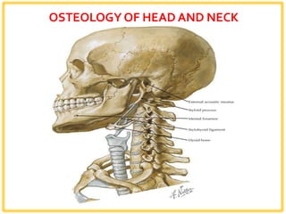
- 149. HYOID BONE
- 153. Attachments to hyoid bone
- 155. CERVICAL VERTEBRAE
- 158. Atlas
- 159. Axis