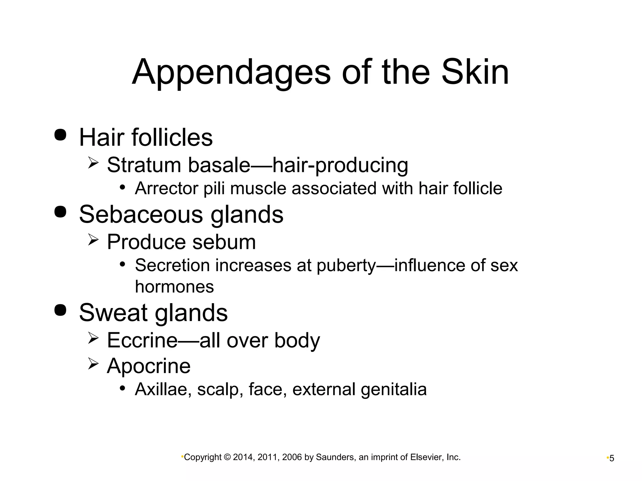
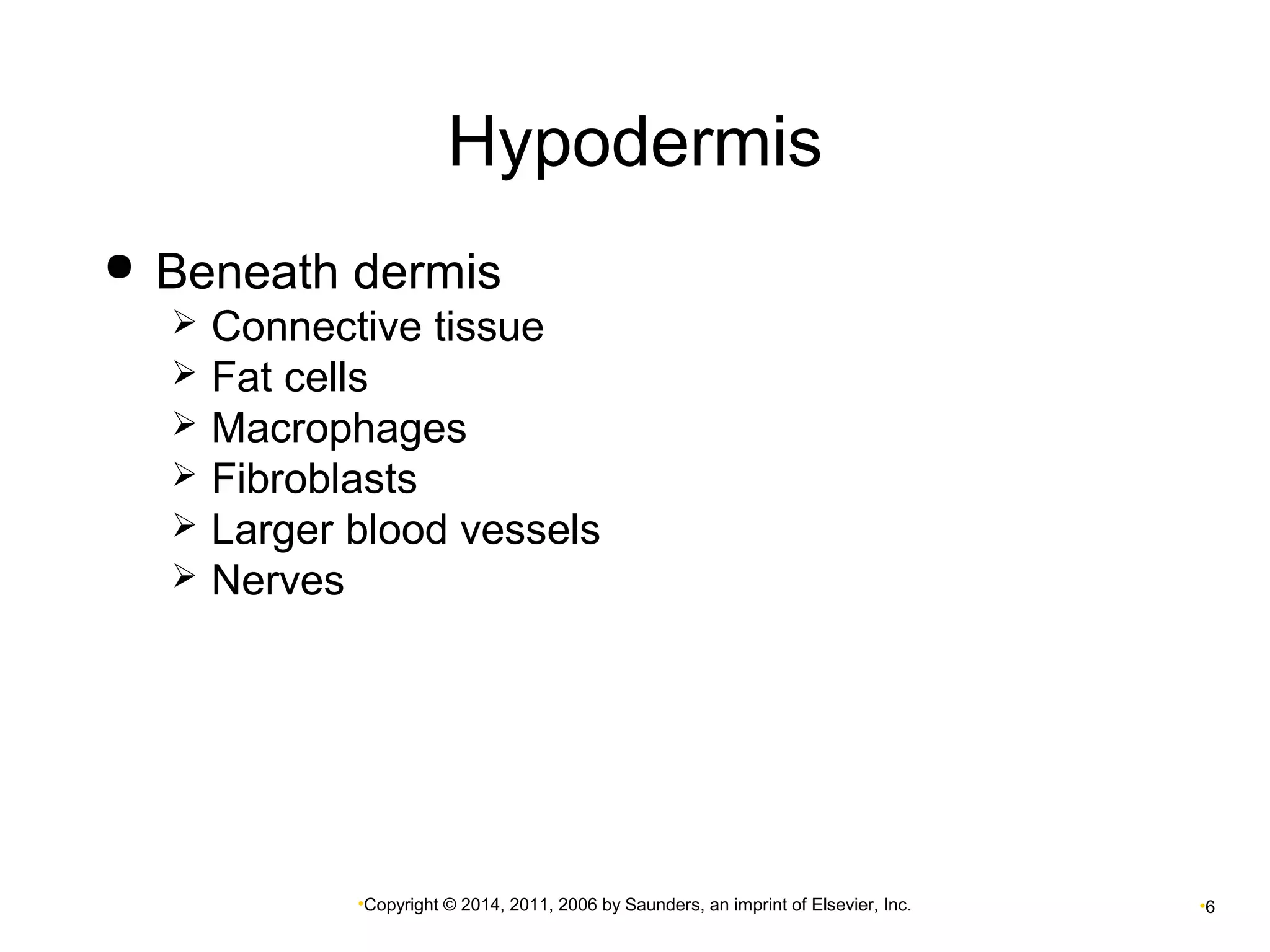
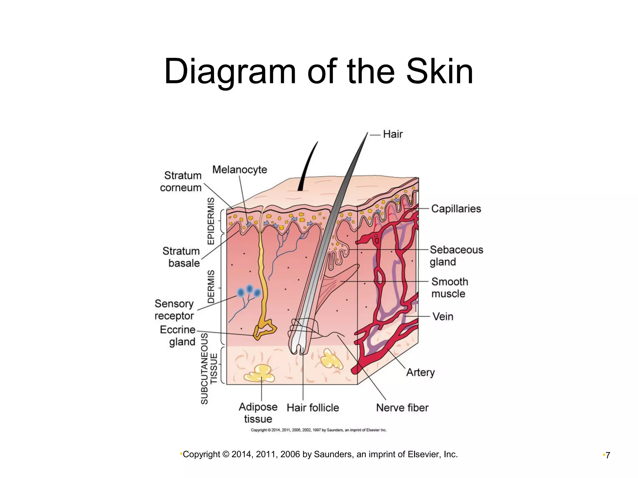
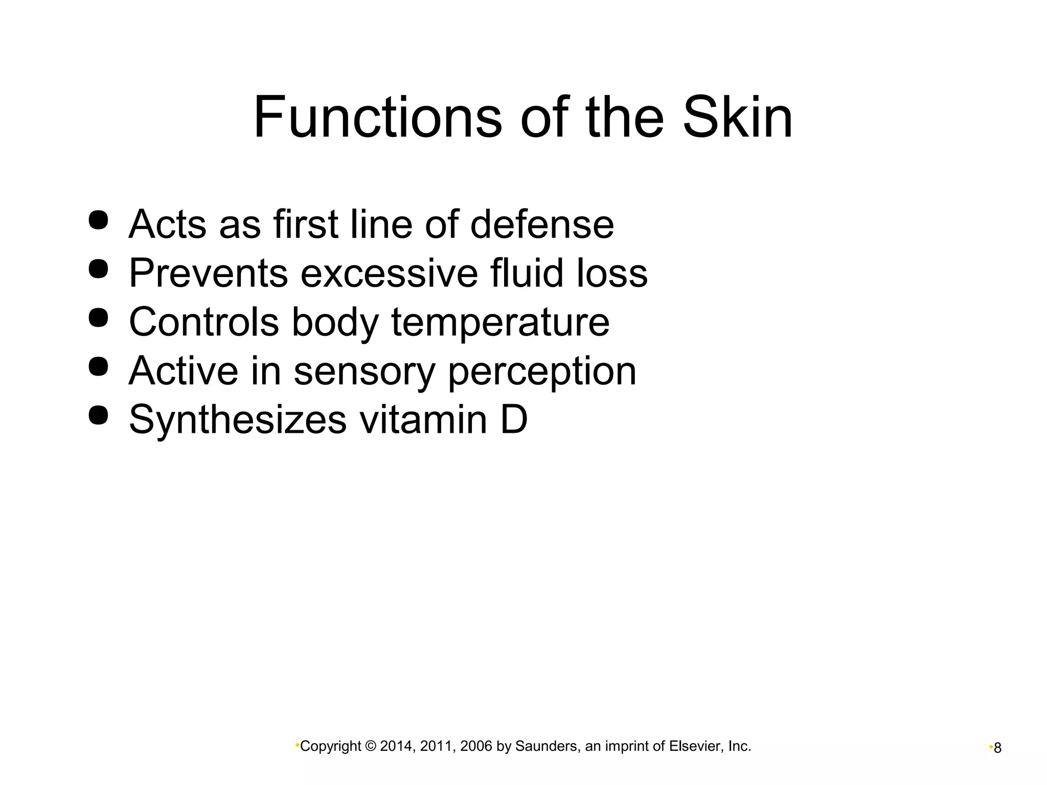
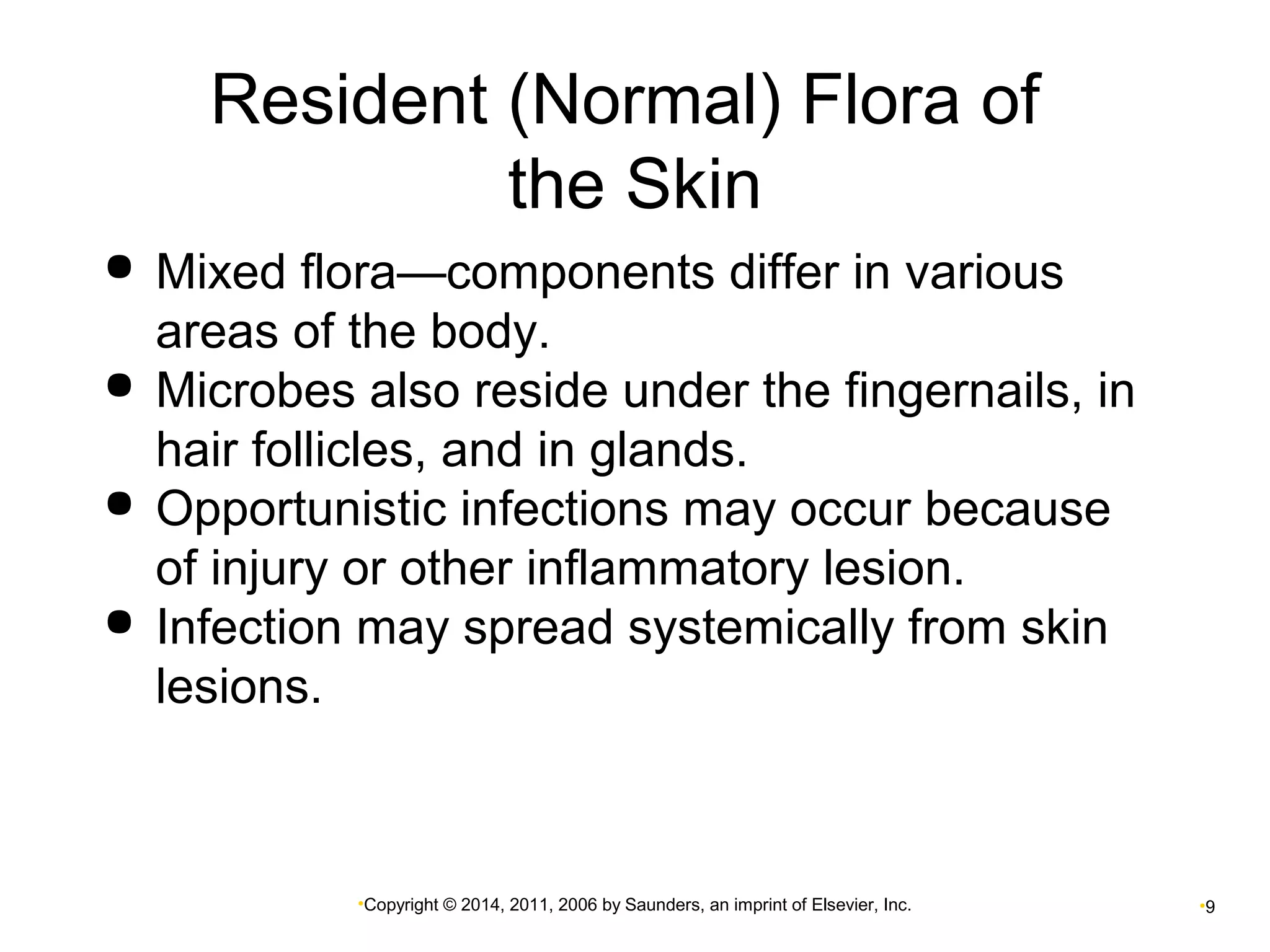
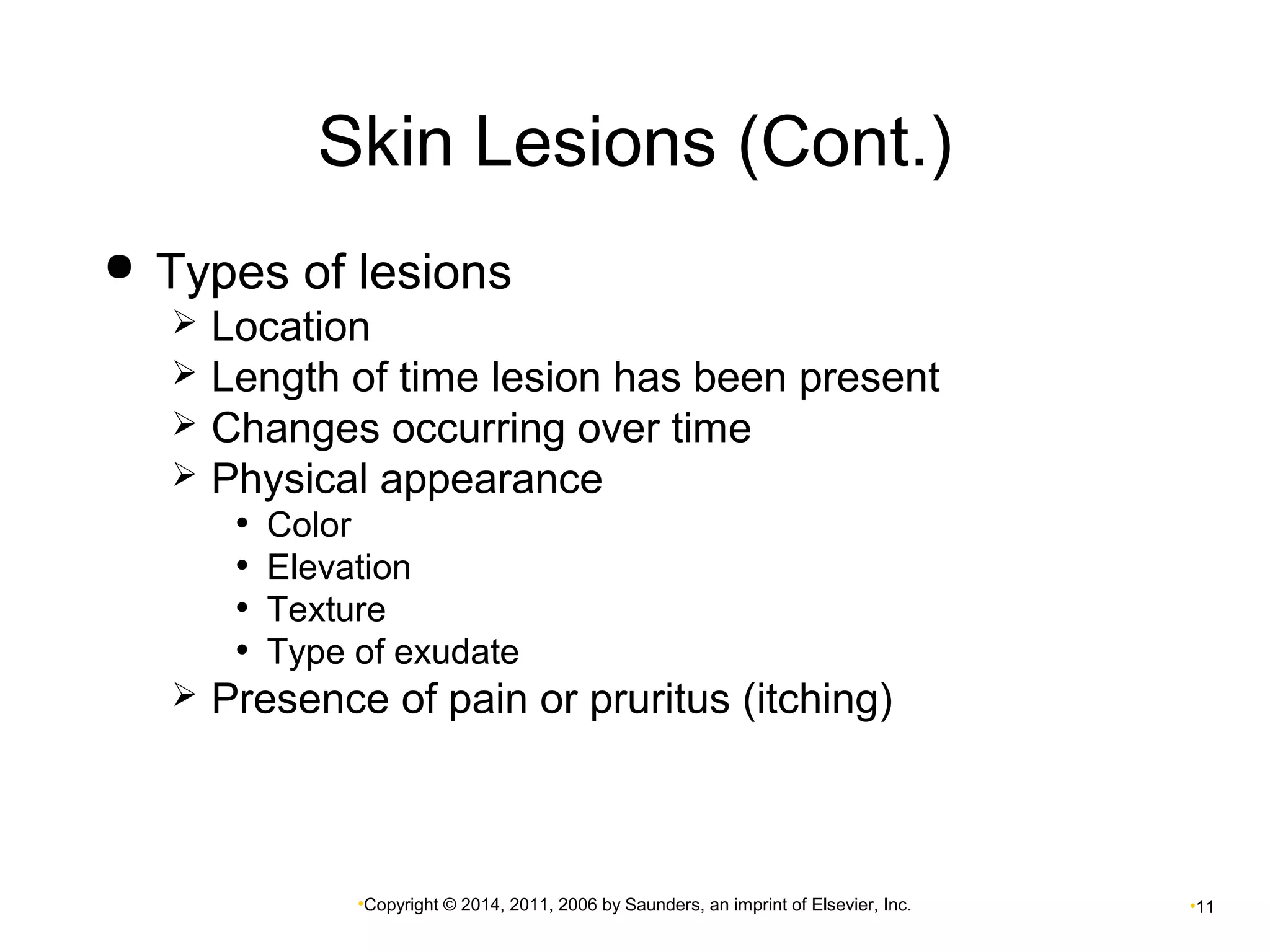
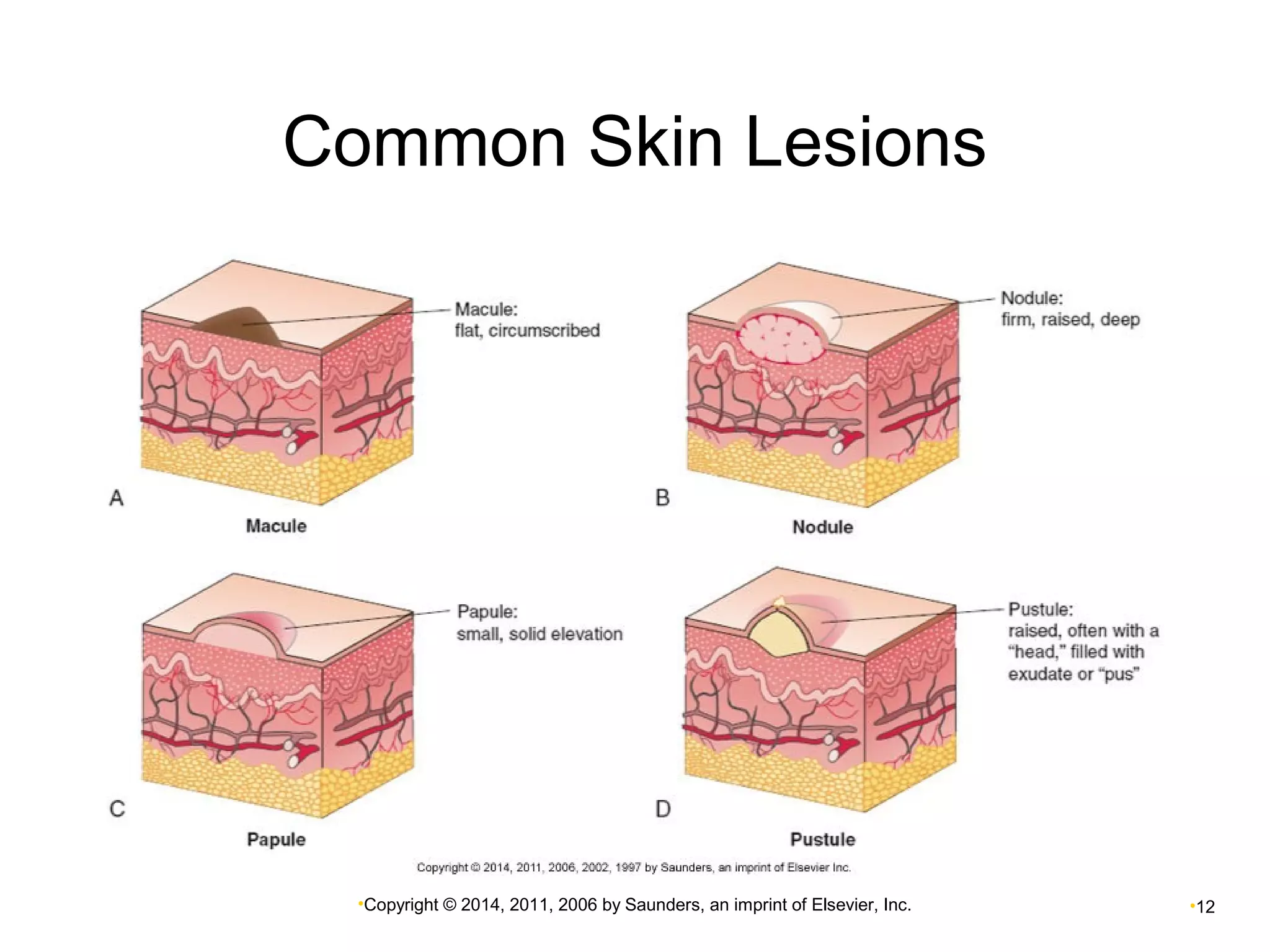
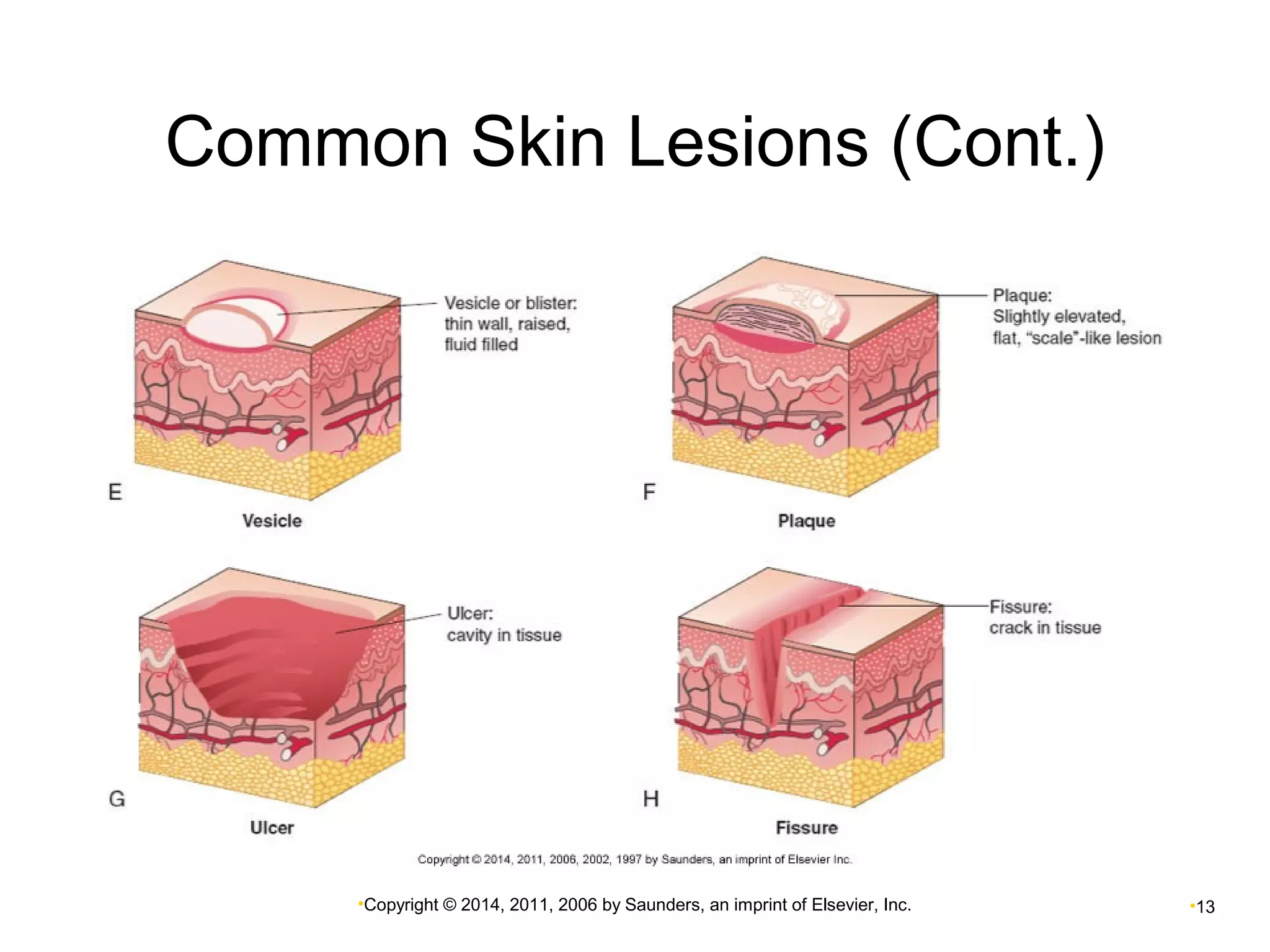
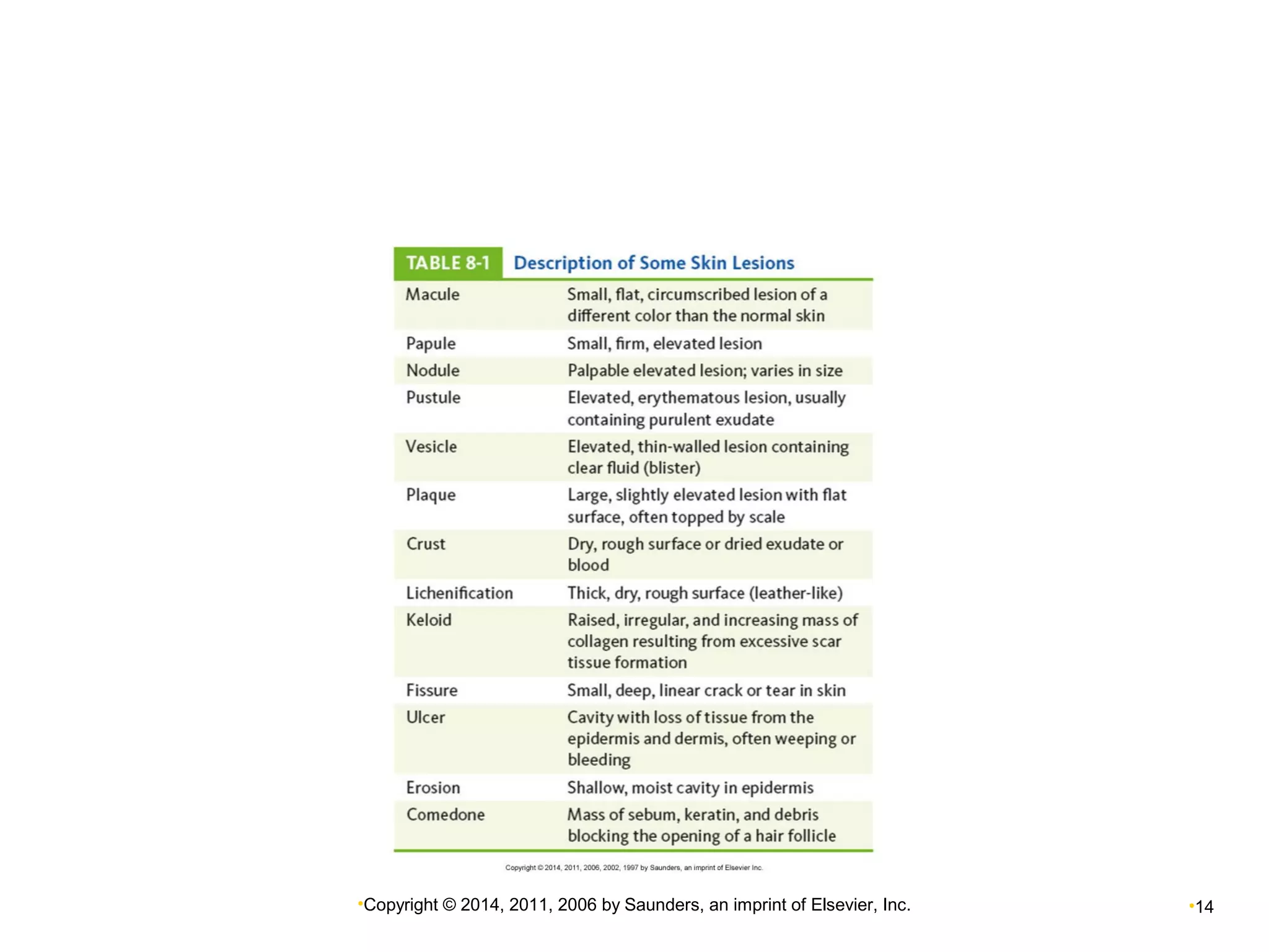
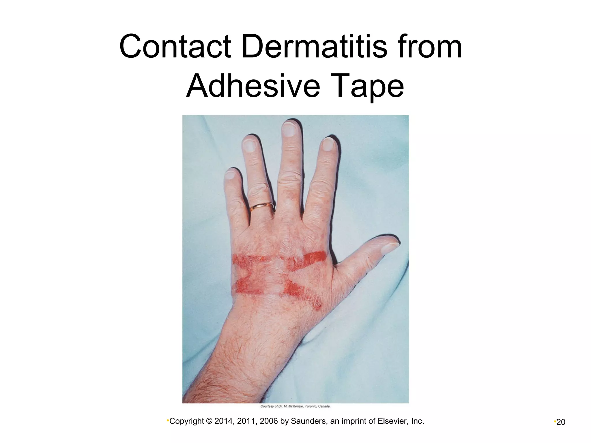
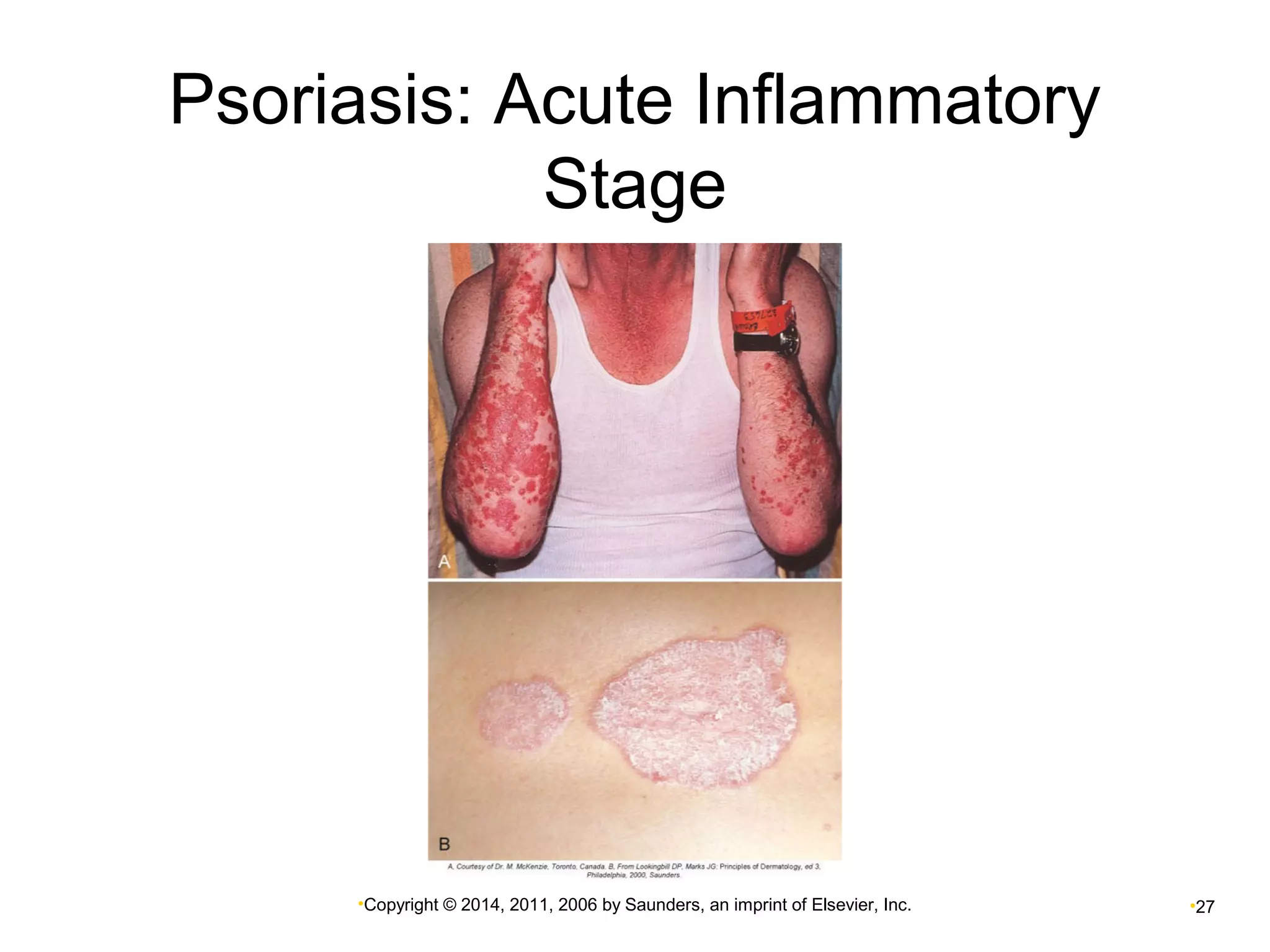
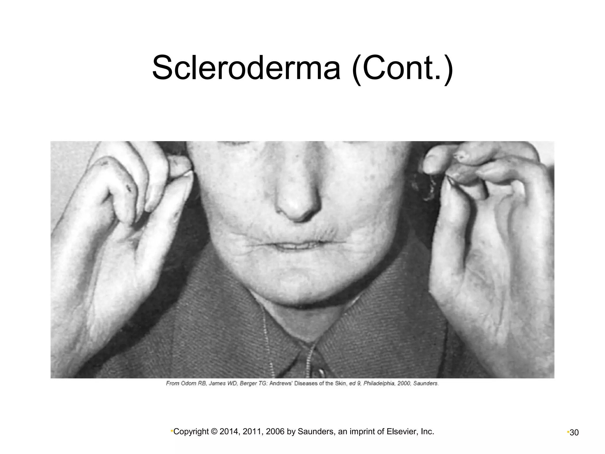
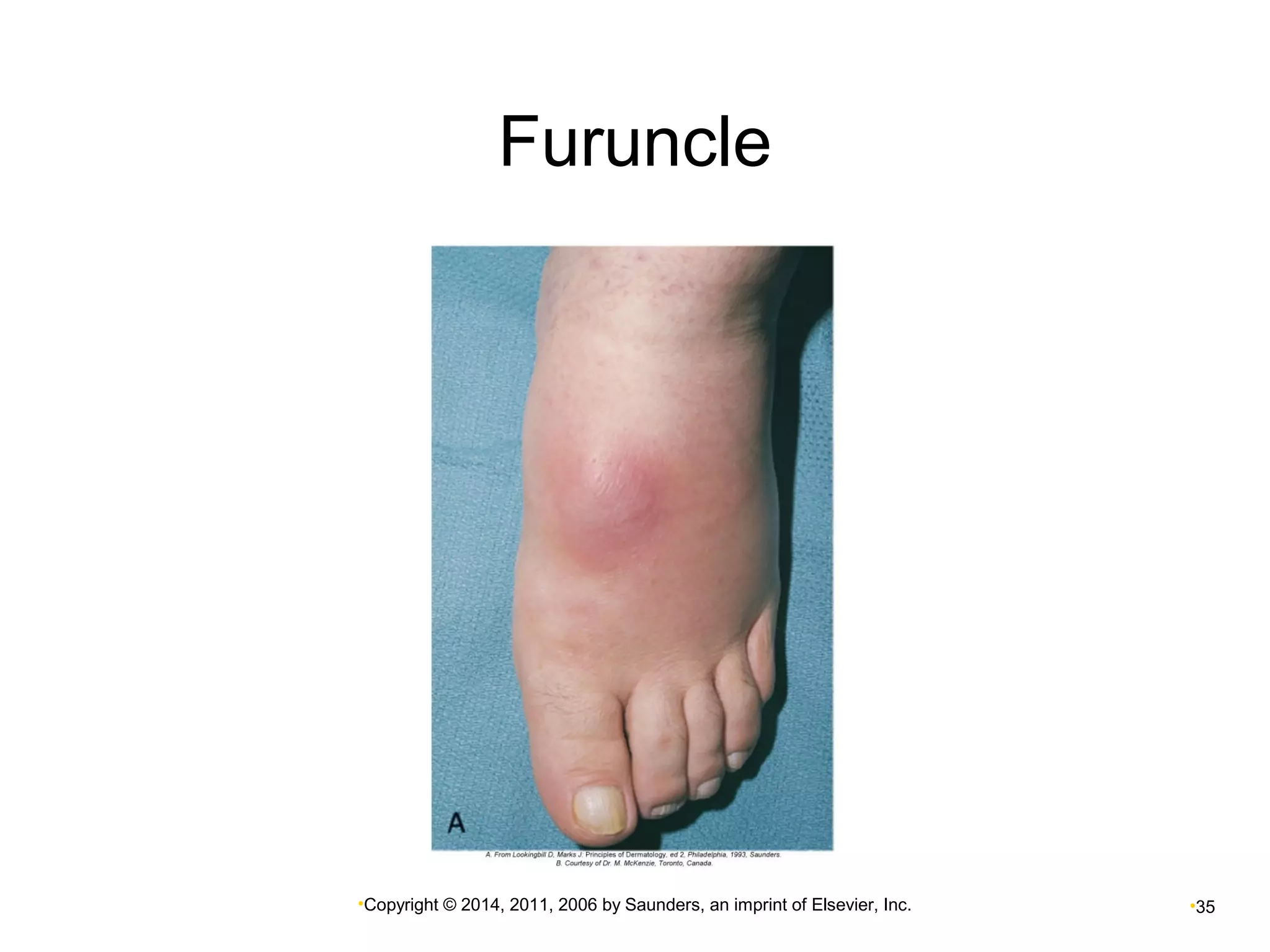
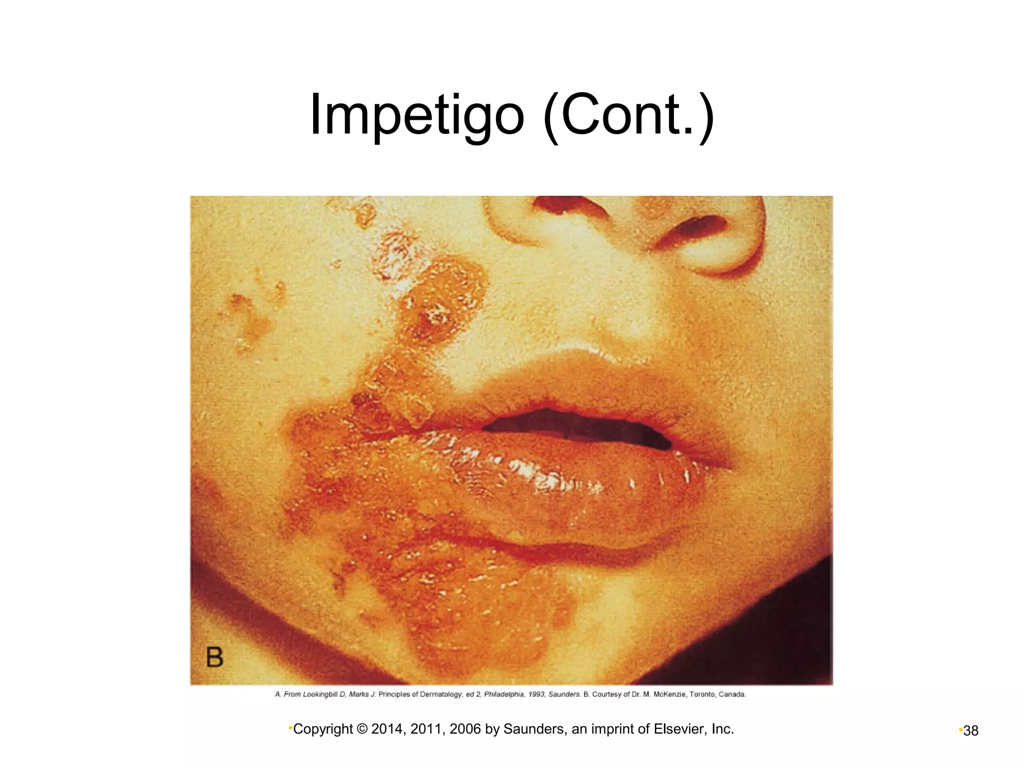
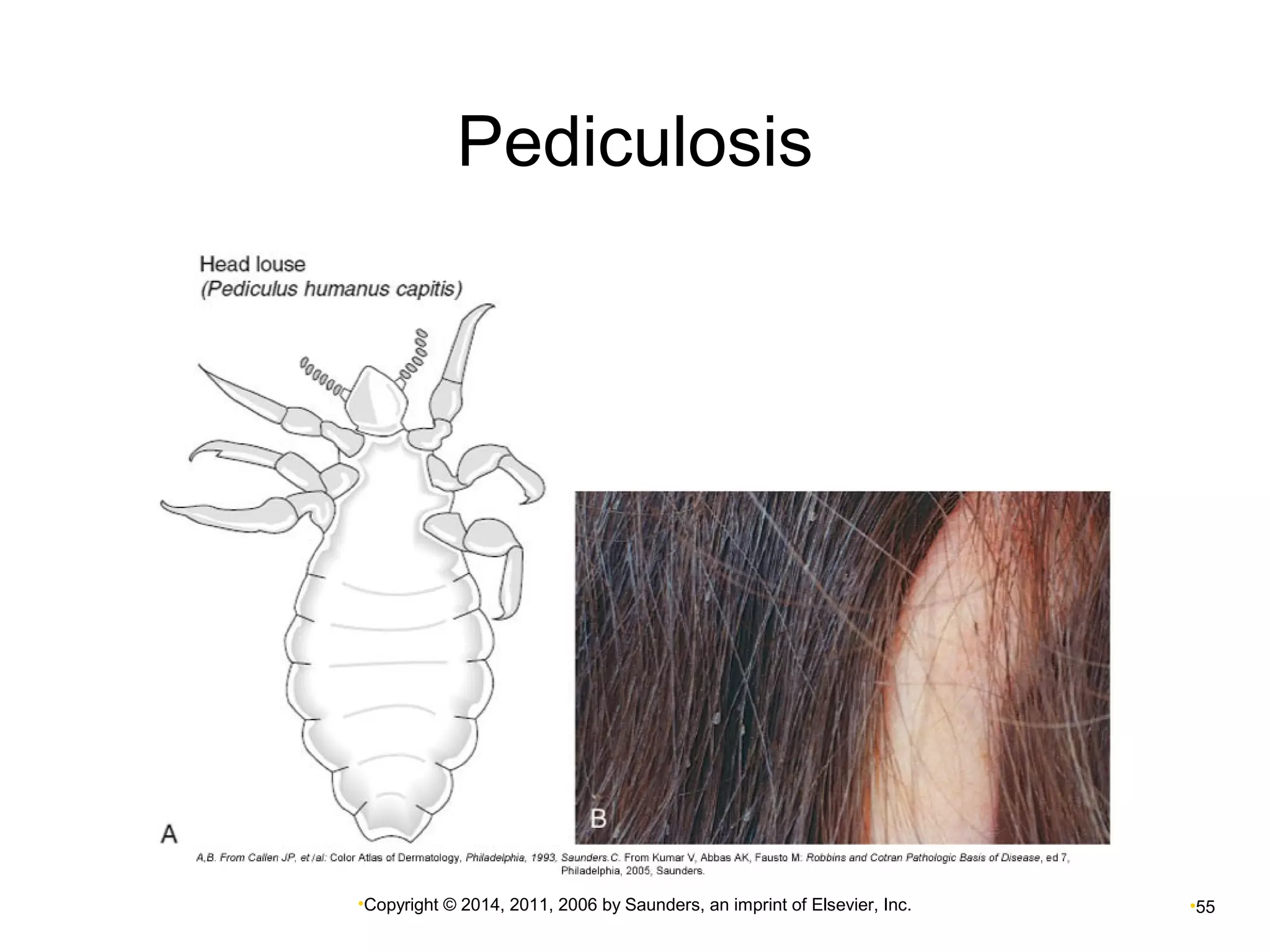
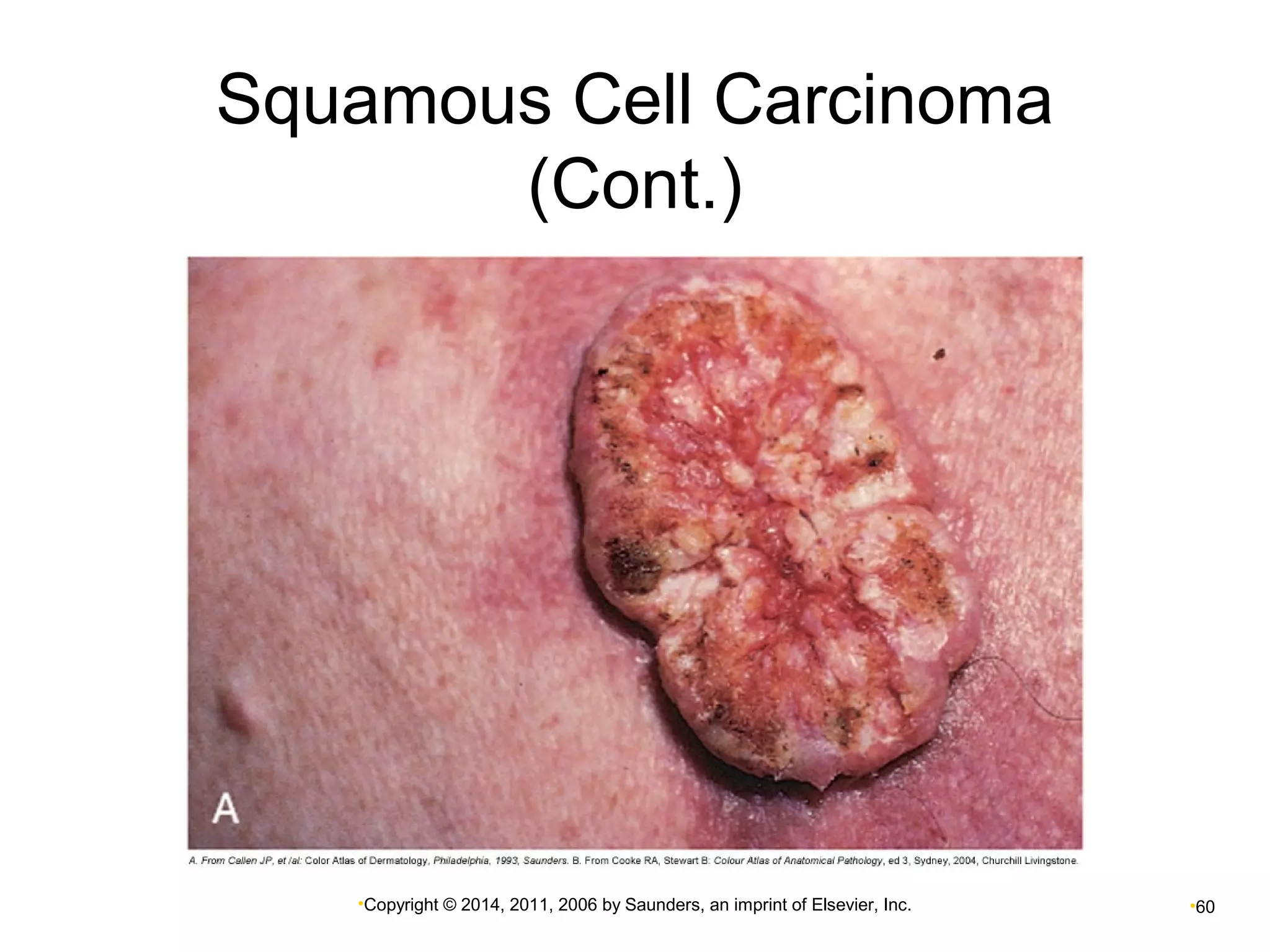
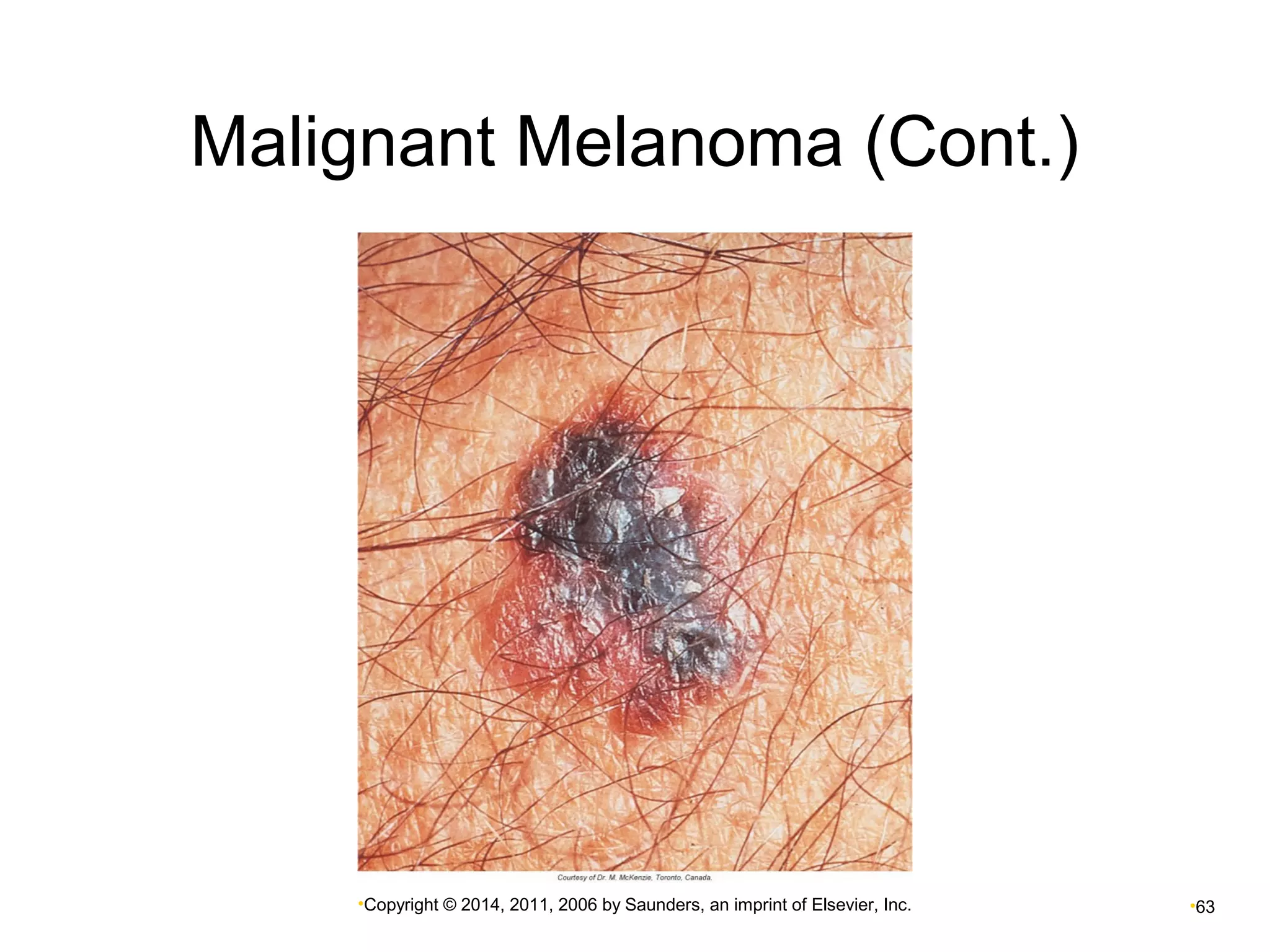
Chapter 8 
Skin Disorders
Review of Normal Skin 
 Layers of the skin 
 Epidermis—avascular 
 Dermis 
 Subcutaneous tissue (hypodermis) 
•Copyright © 2014, 2011, 2006 by Saunders, an imprint of Elsevier, Inc. •2
Epidermis 
 Five layers—vary in thickness 
 Keratin 
 Waterproofing of the skin 
•Copyright © 2014, 2011, 2006 by Saunders, an imprint of Elsevier, Inc. •3 
 Melanin 
 Skin pigment—determines skin color 
• Production depends on multiple genes and environment 
 Albinism 
 Lack of melatonin production 
 Vitiligo 
 Small areas of hypopigmentation 
 Melasma 
 Patches of darker skin
Dermis 
 Connective tissue 
 Contains elastic and collagen fibers 
 Flexibility and strength of the skin 
 Contains nerves and blood vessels 
 Includes sensory receptors for: 
• Pressure 
• Touch 
• Pain 
• Heat 
• Cold 
•Copyright © 2014, 2011, 2006 by Saunders, an imprint of Elsevier, Inc. •4
Appendages of the Skin 
 Hair follicles 
 Stratum basale—hair-producing 
• Arrector pili muscle associated with hair follicle 
 Sebaceous glands 
 Produce sebum 
• Secretion increases at puberty—influence of sex 
hormones 
 Sweat glands 
 Eccrine—all over body 
 Apocrine 
• Axillae, scalp, face, external genitalia 
•Copyright © 2014, 2011, 2006 by Saunders, an imprint of Elsevier, Inc. •5
Hypodermis 
 Beneath dermis 
 Connective tissue 
 Fat cells 
 Macrophages 
 Fibroblasts 
 Larger blood vessels 
 Nerves 
•Copyright © 2014, 2011, 2006 by Saunders, an imprint of Elsevier, Inc. •6
Diagram of the Skin 
•Copyright © 2014, 2011, 2006 by Saunders, an imprint of Elsevier, Inc. •7
Functions of the Skin 
 Acts as first line of defense 
 Prevents excessive fluid loss 
 Controls body temperature 
 Active in sensory perception 
 Synthesizes vitamin D 
•Copyright © 2014, 2011, 2006 by Saunders, an imprint of Elsevier, Inc. •8
Resident (Normal) Flora of 
the Skin 
 Mixed flora—components differ in various 
areas of the body. 
 Microbes also reside under the fingernails, in 
hair follicles, and in glands. 
 Opportunistic infections may occur because 
of injury or other inflammatory lesion. 
 Infection may spread systemically from skin 
lesions. 
•Copyright © 2014, 2011, 2006 by Saunders, an imprint of Elsevier, Inc. •9
Skin Lesions 
 The physical appearance of the lesion is 
necessary to make a diagnosis. 
 Skin lesions may be caused by: 
 Systemic disorders 
• Liver disease 
 Systemic infections 
• Chickenpox 
 Allergies to ingested food or drugs 
 Localized factors 
• Include exposure to toxins 
•Copyright © 2014, 2011, 2006 by Saunders, an imprint of Elsevier, Inc. •10
Skin Lesions (Cont.) 
 Types of lesions 
 Location 
 Length of time lesion has been present 
 Changes occurring over time 
 Physical appearance 
• Color 
• Elevation 
• Texture 
• Type of exudate 
 Presence of pain or pruritus (itching) 
•Copyright © 2014, 2011, 2006 by Saunders, an imprint of Elsevier, Inc. •11
Common Skin Lesions 
•Copyright © 2014, 2011, 2006 by Saunders, an imprint of Elsevier, Inc. •12
Common Skin Lesions (Cont.) 
•Copyright © 2014, 2011, 2006 by Saunders, an imprint of Elsevier, Inc. •13
•Copyright © 2014, 2011, 2006 by Saunders, an imprint of Elsevier, Inc. •14
Pruritus 
 Associated with 
 Allergic responses 
 Chemical irritation caused by insect bites 
 Infestations by parasites (e.g., scabies) 
 Mechanism not totally understood 
 Release of histamine in a hypersensitivity 
response causes marked pruritus 
 Infection may result from breaking the skin barrier. 
 Caused by scratching 
•Copyright © 2014, 2011, 2006 by Saunders, an imprint of Elsevier, Inc. •15
Diagnostic Tests for Skin Lesions 
 Culture and staining of specimens 
 Bacterial infections: microscopic and direct 
observations 
 Specific procedures for fungal or parasitic infections 
•Copyright © 2014, 2011, 2006 by Saunders, an imprint of Elsevier, Inc. •16 
 Biopsy 
 Detection of malignant changes 
• Safeguard prior to or following removal of skin lesions 
 Blood tests 
 Helpful in diagnosis of conditions caused by allergy 
or abnormal immune reaction 
 Skin testing using patch or scratch method
General Treatment Measures 
•Copyright © 2014, 2011, 2006 by Saunders, an imprint of Elsevier, Inc. •17 
 Pruritus 
 Topical agents to reduce sensation 
 May be treated by antihistamines or 
glucocorticoids 
 Avoidance of allergens 
 Reduce risk of recurrence 
 Infections 
 May require antibiotic treatment 
 Precancerous lesions 
 Surgery, laser therapy, electrodessication, 
cryosurgery
Inflammatory Disorders 
•Copyright © 2014, 2011, 2006 by Saunders, an imprint of Elsevier, Inc. •18
Contact Dermatitis 
 Exposure to an allergen 
 Metals, cosmetics, soaps, chemicals, plants 
 Sensitization occurs on first exposure. 
 Pruritic rash develops at site a few hours after 
exposure. 
 Direct chemical or mechanical irritation 
 Does not involve immune response 
 Is inflammatory because of direct exposure 
• Removal of irritant 
• Reduction of inflammation with topical glucocorticoids 
•Copyright © 2014, 2011, 2006 by Saunders, an imprint of Elsevier, Inc. •19
Contact Dermatitis from 
Adhesive Tape 
•Copyright © 2014, 2011, 2006 by Saunders, an imprint of Elsevier, Inc. •20
Urticaria (Hives) 
 Result of type I hypersensitivity 
 Ingestion of substances 
• Examples: shellfish, drugs, certain fruits 
 Lesions are highly pruritic. 
 Hives are often part of anaphylaxis! 
 Check for swelling around mouth and check 
airway. 
 Administer EpiPen or other first aid as required. 
•Copyright © 2014, 2011, 2006 by Saunders, an imprint of Elsevier, Inc. •21
•Copyright © 2014, 2011, 2006 by Saunders, an imprint of Elsevier, Inc. •22
Atopic Dermatitis (Eczema) 
 Atopic—inherited tendency 
 Common problem in infancy 
 Rash is erythematous, with serous exudate. 
 Commonly occurs on face, chest, and shoulders 
 In adults, rash is dry, scaly, and pruritic, often 
on flexor surfaces. 
•Copyright © 2014, 2011, 2006 by Saunders, an imprint of Elsevier, Inc. •23
Atopic Dermatitis (Eczema) 
(Cont.) 
 Chronic inflammation results from response 
to allergens. 
 Eosinophilia and increased serum IgE levels 
 Potential complication—secondary infections 
 Treatment 
 Topical glucocorticoids, antihistamines 
•Copyright © 2014, 2011, 2006 by Saunders, an imprint of Elsevier, Inc. •24
Infant with Extensive Atopic 
Dermatitis 
•Copyright © 2014, 2011, 2006 by Saunders, an imprint of Elsevier, Inc. •25
Psoriasis 
 Chronic inflammatory skin disorder 
 Onset usually in the teenage years 
 Psoriasis results from abnormal T cell 
activation. 
 Excessive proliferation of keratinocytes 
 Cellular proliferation is greatly increased. 
 Lesions found on face, scalp, elbows, knees 
 Itching or burning sensations 
 Treatment 
 Glucocorticoids, tar preparations, antimetabolites 
•Copyright © 2014, 2011, 2006 by Saunders, an imprint of Elsevier, Inc. •26
Psoriasis: Acute Inflammatory 
Stage 
•Copyright © 2014, 2011, 2006 by Saunders, an imprint of Elsevier, Inc. •27
Pemphigus 
 Autoimmune disorder 
 Autoantibodies disrupt cohesion between 
epidermal cells. 
 Causes blisters (bullae) to form 
 Skin sheds, leaving area painful and open to 
secondary infection. 
 May be life-threatening if extensive (e.g., Stevens- 
Johnson syndrome) 
 Systemic glucocorticoids and 
immunosuppressants 
•Copyright © 2014, 2011, 2006 by Saunders, an imprint of Elsevier, Inc. •28
Scleroderma 
 May occur as skin disorder 
 May be systemic and affect viscera 
 Primary cause unknown 
 Increased collagen deposition is observed in all 
cases. 
 Inflammation and fibrosis with decreased capillary 
networks 
• Hard, shiny, tight, immovable areas of skin 
• Impaired movement of mouth and eyes 
 May cause renal failure, intestinal obstruction, 
respiratory failure caused by distortion of 
tissues 
•Copyright © 2014, 2011, 2006 by Saunders, an imprint of Elsevier, Inc. •29
Scleroderma (Cont.) 
•Copyright © 2014, 2011, 2006 by Saunders, an imprint of Elsevier, Inc. •30
Skin Infections 
•Copyright © 2014, 2011, 2006 by Saunders, an imprint of Elsevier, Inc. •31
Skin Infections 
 May be caused by bacteria, viruses, fungi, 
other types of microbes, parasites 
 Caused by opportunistic microbes 
 Minor abrasions or cuts 
 Serious infections may develop. 
 Causative organism needs to be identified for 
appropriate treatment 
•Copyright © 2014, 2011, 2006 by Saunders, an imprint of Elsevier, Inc. •32
Bacterial Infections 
 Cellulitis (erysipelas) 
 Infection of the dermis and subcutaneous tissue 
 Usually secondary to an injury 
 May be iatrogenic 
 Causative organism 
• Usually Staphylococcus aureus 
• Sometimes Streptococcus 
 Frequently in lower trunks and legs 
• Especially in individuals with restricted circulation in the 
extremities; also in immunocompromised individuals 
• Area becomes red, swollen, and painful 
• Red streaks may develop, running along lymph vessels 
proximal to infected area 
•Copyright © 2014, 2011, 2006 by Saunders, an imprint of Elsevier, Inc. •33
Bacterial Infections (Cont.) 
 Furuncles (boils) 
 Usually caused by S. aureus 
• Begins at hair follicles 
• Face, neck, back 
• Frequently drains large amounts of purulent exudate 
 Autoinoculation 
• Squeezing boils can result in spread of infection to other 
areas of the skin. 
 Carbuncles 
• Collection of furuncles that coalesce to form a large 
infected mass 
•Copyright © 2014, 2011, 2006 by Saunders, an imprint of Elsevier, Inc. •34
Furuncle 
•Copyright © 2014, 2011, 2006 by Saunders, an imprint of Elsevier, Inc. •35
Impetigo 
 Common infection in infants and children 
 May also occur in adults 
 S. aureus—highly contagious in neonates 
 Lesions commonly on face 
 Transmission may occur through close 
physical contact or through fomites 
 Pruritus common 
 Leads to scratching and further spread of infection 
•Copyright © 2014, 2011, 2006 by Saunders, an imprint of Elsevier, Inc. •36
Impetigo: Treatment 
 Topical antibiotics in early stages 
 Systemic administration if lesions are 
extensive 
 Antibiotic-resistant strains of S. aureus are 
increasing in numbers. 
• Local outbreaks of infection may result. 
•Copyright © 2014, 2011, 2006 by Saunders, an imprint of Elsevier, Inc. •37
Impetigo (Cont.) 
•Copyright © 2014, 2011, 2006 by Saunders, an imprint of Elsevier, Inc. •38
Acute Necrotizing Fasciitis 
 Mixture of aerobic and anaerobic bacteria 
usually at site of infection 
 Severe inflammation and tissue necrosis 
 Usually caused by virulent strain of gram-positive, 
group A beta-hemolytic Streptococcus 
 Bacteria secrete toxins that break down fascia and 
connective tissue, causing massive tissue 
destruction. 
 Often a history of minor trauma or infection in 
the skin and subcutaneous tissue of an 
extremity 
•Copyright © 2014, 2011, 2006 by Saunders, an imprint of Elsevier, Inc. •39
Acute Necrotizing Fasciitis 
(Cont.) 
 Delay in treatment—greater tissue loss, 
potential amputation, higher probability of 
mortality 
 Systemic toxicity develops with fever, 
tachycardia, hypotension, mental confusion, 
disorientation, possible organ failure 
 Treatment 
 Aggressive antimicrobial therapy, fluid 
replacement 
 Excision of all infected tissue; amputation 
•Copyright © 2014, 2011, 2006 by Saunders, an imprint of Elsevier, Inc. •40
Leprosy (Hansen’s Disease) 
 Caused by Mycobacterium leprae 
 Chronic disease classified into three major 
types 
 Clinical signs and symptoms vary. 
 Generally affects skin, mucous membranes, and 
peripheral nerves 
 Damage can lead to loss of limbs. 
 Mechanism of pathogenicity largely unknown 
 Diagnosis through microscopic examination 
of skin biopsy 
 Treatment primarily with antibiotics 
•Copyright © 2014, 2011, 2006 by Saunders, an imprint of Elsevier, Inc. •41
Viral Infections 
•Copyright © 2014, 2011, 2006 by Saunders, an imprint of Elsevier, Inc. •42
Herpes Simplex 
 Herpes simplex type 1 (HSV-1) 
 Most common cause of cold sores or fever blisters 
 Herpes simplex type 2 (HSV-2)—genital 
herpes 
 Both types of HSV cause similar effects. 
 Primary infection may be asymptomatic 
 Virus remains latent in sensory nerve ganglia. 
 Recurrence may be triggered by: 
 Common cold, sun exposure, stress 
•Copyright © 2014, 2011, 2006 by Saunders, an imprint of Elsevier, Inc. •43
Herpes Simplex (Cont.) 
 Spread by direct contact with fluid from lesion 
 Spread of infection to others possible prior to 
appearance of lesions 
 Potential complication 
 Spread of virus to eye 
• Keratitis 
 Herpetic whitlow 
• Painful infection of the fingers 
•Copyright © 2014, 2011, 2006 by Saunders, an imprint of Elsevier, Inc. •44
Herpes Simplex (Cont.) 
•Copyright © 2014, 2011, 2006 by Saunders, an imprint of Elsevier, Inc. •45
Verrucae (Warts) 
 Human papillomavirus (HPV) types 1 to 4 
 Frequently develop in children and young adults 
 Plantar warts are common. 
 Spreads by viral shedding of the skin surface 
 May resolve spontaneously with time 
 Genital warts (HPV types 6 and 11) 
•Copyright © 2014, 2011, 2006 by Saunders, an imprint of Elsevier, Inc. •46
Plantar Warts 
•Copyright © 2014, 2011, 2006 by Saunders, an imprint of Elsevier, Inc. •47
Fungal Infections (Mycoses) 
 Most are superficial 
 Candida infection is associated with diabetes. 
 May spread systemically in immunocompromised 
individuals 
 Diagnosed from skin scrapings 
 Become fluorescent in ultraviolet light 
 Microscopic examination 
 Culturing of samples 
•Copyright © 2014, 2011, 2006 by Saunders, an imprint of Elsevier, Inc. •48
Tinea Pedis 
•Copyright © 2014, 2011, 2006 by Saunders, an imprint of Elsevier, Inc. •49
Tinea 
 Tinea capitis 
 Infection of the scalp 
 Common in school-age children 
 Erythema may be apparent. 
 Oral antifungal medication 
 Tinea corporis 
 Infection of the body, particularly of nonhairy parts 
 Round lesion with clear center (ringworm) 
 Pruritus may be present. 
 Topical antifungal medication 
•Copyright © 2014, 2011, 2006 by Saunders, an imprint of Elsevier, Inc. •50
Tinea (Cont.) 
 Tinea pedis 
 Athlete’s foot—involves the feet, particularly the toes 
 Associated with swimming pools and gymnasiums 
 May be part of normal flora that becomes 
opportunistic 
 Secondary bacterial infection may occur 
 Topical antifungal medication 
 Tinea unguium 
 Infection of the nails, particularly the toenails 
• Nails turn white, then brown. 
• Nail thickens and cracks. 
• Infection tends to spread to other nails. 
•Copyright © 2014, 2011, 2006 by Saunders, an imprint of Elsevier, Inc. •51
Other Infections 
•Copyright © 2014, 2011, 2006 by Saunders, an imprint of Elsevier, Inc. •52 
 Scabies 
 Invasion by mite Sarcoptes scabiei 
 Female burrows into epidermis 
• Lays eggs over a period of several weeks 
 Male dies after fertilizing the female 
 Female dies after laying the eggs. 
 Larvae migrate to skin surface. 
• Burrow into skin in search of nutrients 
• Intensively pruritic! 
 Larvae mature and cycle is repeated 
 Burrows appear on skin as tiny, light brown lines.
Scabies 
•Copyright © 2014, 2011, 2006 by Saunders, an imprint of Elsevier, Inc. •53
Other Infections (Cont.) 
 Pediculosis (lice) 
 Pediculus humanus corporis—body louse 
 Pediculus humanus capitis—head louse 
 Pediculus humanus pubis—pubic louse 
 Female lice lay eggs on hair shafts. 
 After hatching, louse bites human host, sucking 
blood for production of ova 
 Excoriations result from scratching. 
•Copyright © 2014, 2011, 2006 by Saunders, an imprint of Elsevier, Inc. •54
Pediculosis 
•Copyright © 2014, 2011, 2006 by Saunders, an imprint of Elsevier, Inc. •55
Skin Tumors 
•Copyright © 2014, 2011, 2006 by Saunders, an imprint of Elsevier, Inc. •56
Keratoses 
 Benign lesions usually associated with aging 
or skin damage. 
 Seborrheic keratoses 
 Proliferation of basal cells 
• Lead to oval elevation 
• May be smooth or rough 
 Actinic keratoses 
 On skin exposed to ultraviolet radiation 
 Commonly in fair-skinned persons 
 Lesion appears as pigmented, scaly patch 
•Copyright © 2014, 2011, 2006 by Saunders, an imprint of Elsevier, Inc. •57
Guidelines to Reduce Risk 
of Skin Cancers 
 Reducing sun exposure at midday and early 
afternoon 
 Covering up with clothing 
 Remaining in shade 
 Wearing broad-brimmed hats to protect face and 
neck 
 Applying sunscreen or sunblock 
 Protecting infants and children from exposure 
and sun damage to skin 
•Copyright © 2014, 2011, 2006 by Saunders, an imprint of Elsevier, Inc. •58
Squamous Cell Carcinoma 
 Painless, malignant tumor of the epidermis 
 Lesions most commonly found on exposed 
areas of the skin but also in oral cavity 
 Face and neck 
 Base of tongue 
 Excellent prognosis when lesion is removed 
within reasonable time 
 Invasive type arises from premalignant 
condition. 
•Copyright © 2014, 2011, 2006 by Saunders, an imprint of Elsevier, Inc. •59
Squamous Cell Carcinoma 
(Cont.) 
•Copyright © 2014, 2011, 2006 by Saunders, an imprint of Elsevier, Inc. •60
Malignant Melanoma 
 Highly metastatic form of skin cancer 
 Develops in melanocytes 
 From a nevus (mole) 
 Often appear as multicolored lesion with 
irregular border 
 Grow quickly 
 Change in shape, color, size, texture 
 May bleed 
 Treatment: surgical removal and radiation 
plus chemotherapy 
•Copyright © 2014, 2011, 2006 by Saunders, an imprint of Elsevier, Inc. •61
The ABCD of Melanoma 
 Melanoma is suspected in any nevus that 
shows: 
 Change in appearance 
 Change in border 
 Change in color 
 Increase in diameter 
•Copyright © 2014, 2011, 2006 by Saunders, an imprint of Elsevier, Inc. •62
Malignant Melanoma (Cont.) 
•Copyright © 2014, 2011, 2006 by Saunders, an imprint of Elsevier, Inc. •63
Kaposi’s Sarcoma 
 Occurs in those with AIDS and other 
immunodeficiencies 
 May affect viscera as well as skin 
 Malignant cells arise from endothelium in 
small blood vessels 
 Purplish macules 
 Nonpruritic, nonpainful 
 In immunocompromised patients, lesions 
develop rapidly over upper body. 
 Combination of radiation, chemotherapy, 
surgery, biological therapy 
•Copyright © 2014, 2011, 2006 by Saunders, an imprint of Elsevier, Inc. •64

Chapter 008

  • 1.
    Chapter 8 SkinDisorders
  • 2.
    Review of NormalSkin  Layers of the skin  Epidermis—avascular  Dermis  Subcutaneous tissue (hypodermis) •Copyright © 2014, 2011, 2006 by Saunders, an imprint of Elsevier, Inc. •2
  • 3.
    Epidermis  Fivelayers—vary in thickness  Keratin  Waterproofing of the skin •Copyright © 2014, 2011, 2006 by Saunders, an imprint of Elsevier, Inc. •3  Melanin  Skin pigment—determines skin color • Production depends on multiple genes and environment  Albinism  Lack of melatonin production  Vitiligo  Small areas of hypopigmentation  Melasma  Patches of darker skin
  • 4.
    Dermis  Connectivetissue  Contains elastic and collagen fibers  Flexibility and strength of the skin  Contains nerves and blood vessels  Includes sensory receptors for: • Pressure • Touch • Pain • Heat • Cold •Copyright © 2014, 2011, 2006 by Saunders, an imprint of Elsevier, Inc. •4
  • 5.
    Appendages of theSkin  Hair follicles  Stratum basale—hair-producing • Arrector pili muscle associated with hair follicle  Sebaceous glands  Produce sebum • Secretion increases at puberty—influence of sex hormones  Sweat glands  Eccrine—all over body  Apocrine • Axillae, scalp, face, external genitalia •Copyright © 2014, 2011, 2006 by Saunders, an imprint of Elsevier, Inc. •5
  • 6.
    Hypodermis  Beneathdermis  Connective tissue  Fat cells  Macrophages  Fibroblasts  Larger blood vessels  Nerves •Copyright © 2014, 2011, 2006 by Saunders, an imprint of Elsevier, Inc. •6
  • 7.
    Diagram of theSkin •Copyright © 2014, 2011, 2006 by Saunders, an imprint of Elsevier, Inc. •7
  • 8.
    Functions of theSkin  Acts as first line of defense  Prevents excessive fluid loss  Controls body temperature  Active in sensory perception  Synthesizes vitamin D •Copyright © 2014, 2011, 2006 by Saunders, an imprint of Elsevier, Inc. •8
  • 9.
    Resident (Normal) Floraof the Skin  Mixed flora—components differ in various areas of the body.  Microbes also reside under the fingernails, in hair follicles, and in glands.  Opportunistic infections may occur because of injury or other inflammatory lesion.  Infection may spread systemically from skin lesions. •Copyright © 2014, 2011, 2006 by Saunders, an imprint of Elsevier, Inc. •9
  • 10.
    Skin Lesions The physical appearance of the lesion is necessary to make a diagnosis.  Skin lesions may be caused by:  Systemic disorders • Liver disease  Systemic infections • Chickenpox  Allergies to ingested food or drugs  Localized factors • Include exposure to toxins •Copyright © 2014, 2011, 2006 by Saunders, an imprint of Elsevier, Inc. •10
  • 11.
    Skin Lesions (Cont.)  Types of lesions  Location  Length of time lesion has been present  Changes occurring over time  Physical appearance • Color • Elevation • Texture • Type of exudate  Presence of pain or pruritus (itching) •Copyright © 2014, 2011, 2006 by Saunders, an imprint of Elsevier, Inc. •11
  • 12.
    Common Skin Lesions •Copyright © 2014, 2011, 2006 by Saunders, an imprint of Elsevier, Inc. •12
  • 13.
    Common Skin Lesions(Cont.) •Copyright © 2014, 2011, 2006 by Saunders, an imprint of Elsevier, Inc. •13
  • 14.
    •Copyright © 2014,2011, 2006 by Saunders, an imprint of Elsevier, Inc. •14
  • 15.
    Pruritus  Associatedwith  Allergic responses  Chemical irritation caused by insect bites  Infestations by parasites (e.g., scabies)  Mechanism not totally understood  Release of histamine in a hypersensitivity response causes marked pruritus  Infection may result from breaking the skin barrier.  Caused by scratching •Copyright © 2014, 2011, 2006 by Saunders, an imprint of Elsevier, Inc. •15
  • 16.
    Diagnostic Tests forSkin Lesions  Culture and staining of specimens  Bacterial infections: microscopic and direct observations  Specific procedures for fungal or parasitic infections •Copyright © 2014, 2011, 2006 by Saunders, an imprint of Elsevier, Inc. •16  Biopsy  Detection of malignant changes • Safeguard prior to or following removal of skin lesions  Blood tests  Helpful in diagnosis of conditions caused by allergy or abnormal immune reaction  Skin testing using patch or scratch method
  • 17.
    General Treatment Measures •Copyright © 2014, 2011, 2006 by Saunders, an imprint of Elsevier, Inc. •17  Pruritus  Topical agents to reduce sensation  May be treated by antihistamines or glucocorticoids  Avoidance of allergens  Reduce risk of recurrence  Infections  May require antibiotic treatment  Precancerous lesions  Surgery, laser therapy, electrodessication, cryosurgery
  • 18.
    Inflammatory Disorders •Copyright© 2014, 2011, 2006 by Saunders, an imprint of Elsevier, Inc. •18
  • 19.
    Contact Dermatitis Exposure to an allergen  Metals, cosmetics, soaps, chemicals, plants  Sensitization occurs on first exposure.  Pruritic rash develops at site a few hours after exposure.  Direct chemical or mechanical irritation  Does not involve immune response  Is inflammatory because of direct exposure • Removal of irritant • Reduction of inflammation with topical glucocorticoids •Copyright © 2014, 2011, 2006 by Saunders, an imprint of Elsevier, Inc. •19
  • 20.
    Contact Dermatitis from Adhesive Tape •Copyright © 2014, 2011, 2006 by Saunders, an imprint of Elsevier, Inc. •20
  • 21.
    Urticaria (Hives) Result of type I hypersensitivity  Ingestion of substances • Examples: shellfish, drugs, certain fruits  Lesions are highly pruritic.  Hives are often part of anaphylaxis!  Check for swelling around mouth and check airway.  Administer EpiPen or other first aid as required. •Copyright © 2014, 2011, 2006 by Saunders, an imprint of Elsevier, Inc. •21
  • 22.
    •Copyright © 2014,2011, 2006 by Saunders, an imprint of Elsevier, Inc. •22
  • 23.
    Atopic Dermatitis (Eczema)  Atopic—inherited tendency  Common problem in infancy  Rash is erythematous, with serous exudate.  Commonly occurs on face, chest, and shoulders  In adults, rash is dry, scaly, and pruritic, often on flexor surfaces. •Copyright © 2014, 2011, 2006 by Saunders, an imprint of Elsevier, Inc. •23
  • 24.
    Atopic Dermatitis (Eczema) (Cont.)  Chronic inflammation results from response to allergens.  Eosinophilia and increased serum IgE levels  Potential complication—secondary infections  Treatment  Topical glucocorticoids, antihistamines •Copyright © 2014, 2011, 2006 by Saunders, an imprint of Elsevier, Inc. •24
  • 25.
    Infant with ExtensiveAtopic Dermatitis •Copyright © 2014, 2011, 2006 by Saunders, an imprint of Elsevier, Inc. •25
  • 26.
    Psoriasis  Chronicinflammatory skin disorder  Onset usually in the teenage years  Psoriasis results from abnormal T cell activation.  Excessive proliferation of keratinocytes  Cellular proliferation is greatly increased.  Lesions found on face, scalp, elbows, knees  Itching or burning sensations  Treatment  Glucocorticoids, tar preparations, antimetabolites •Copyright © 2014, 2011, 2006 by Saunders, an imprint of Elsevier, Inc. •26
  • 27.
    Psoriasis: Acute Inflammatory Stage •Copyright © 2014, 2011, 2006 by Saunders, an imprint of Elsevier, Inc. •27
  • 28.
    Pemphigus  Autoimmunedisorder  Autoantibodies disrupt cohesion between epidermal cells.  Causes blisters (bullae) to form  Skin sheds, leaving area painful and open to secondary infection.  May be life-threatening if extensive (e.g., Stevens- Johnson syndrome)  Systemic glucocorticoids and immunosuppressants •Copyright © 2014, 2011, 2006 by Saunders, an imprint of Elsevier, Inc. •28
  • 29.
    Scleroderma  Mayoccur as skin disorder  May be systemic and affect viscera  Primary cause unknown  Increased collagen deposition is observed in all cases.  Inflammation and fibrosis with decreased capillary networks • Hard, shiny, tight, immovable areas of skin • Impaired movement of mouth and eyes  May cause renal failure, intestinal obstruction, respiratory failure caused by distortion of tissues •Copyright © 2014, 2011, 2006 by Saunders, an imprint of Elsevier, Inc. •29
  • 30.
    Scleroderma (Cont.) •Copyright© 2014, 2011, 2006 by Saunders, an imprint of Elsevier, Inc. •30
  • 31.
    Skin Infections •Copyright© 2014, 2011, 2006 by Saunders, an imprint of Elsevier, Inc. •31
  • 32.
    Skin Infections May be caused by bacteria, viruses, fungi, other types of microbes, parasites  Caused by opportunistic microbes  Minor abrasions or cuts  Serious infections may develop.  Causative organism needs to be identified for appropriate treatment •Copyright © 2014, 2011, 2006 by Saunders, an imprint of Elsevier, Inc. •32
  • 33.
    Bacterial Infections Cellulitis (erysipelas)  Infection of the dermis and subcutaneous tissue  Usually secondary to an injury  May be iatrogenic  Causative organism • Usually Staphylococcus aureus • Sometimes Streptococcus  Frequently in lower trunks and legs • Especially in individuals with restricted circulation in the extremities; also in immunocompromised individuals • Area becomes red, swollen, and painful • Red streaks may develop, running along lymph vessels proximal to infected area •Copyright © 2014, 2011, 2006 by Saunders, an imprint of Elsevier, Inc. •33
  • 34.
    Bacterial Infections (Cont.)  Furuncles (boils)  Usually caused by S. aureus • Begins at hair follicles • Face, neck, back • Frequently drains large amounts of purulent exudate  Autoinoculation • Squeezing boils can result in spread of infection to other areas of the skin.  Carbuncles • Collection of furuncles that coalesce to form a large infected mass •Copyright © 2014, 2011, 2006 by Saunders, an imprint of Elsevier, Inc. •34
  • 35.
    Furuncle •Copyright ©2014, 2011, 2006 by Saunders, an imprint of Elsevier, Inc. •35
  • 36.
    Impetigo  Commoninfection in infants and children  May also occur in adults  S. aureus—highly contagious in neonates  Lesions commonly on face  Transmission may occur through close physical contact or through fomites  Pruritus common  Leads to scratching and further spread of infection •Copyright © 2014, 2011, 2006 by Saunders, an imprint of Elsevier, Inc. •36
  • 37.
    Impetigo: Treatment Topical antibiotics in early stages  Systemic administration if lesions are extensive  Antibiotic-resistant strains of S. aureus are increasing in numbers. • Local outbreaks of infection may result. •Copyright © 2014, 2011, 2006 by Saunders, an imprint of Elsevier, Inc. •37
  • 38.
    Impetigo (Cont.) •Copyright© 2014, 2011, 2006 by Saunders, an imprint of Elsevier, Inc. •38
  • 39.
    Acute Necrotizing Fasciitis  Mixture of aerobic and anaerobic bacteria usually at site of infection  Severe inflammation and tissue necrosis  Usually caused by virulent strain of gram-positive, group A beta-hemolytic Streptococcus  Bacteria secrete toxins that break down fascia and connective tissue, causing massive tissue destruction.  Often a history of minor trauma or infection in the skin and subcutaneous tissue of an extremity •Copyright © 2014, 2011, 2006 by Saunders, an imprint of Elsevier, Inc. •39
  • 40.
    Acute Necrotizing Fasciitis (Cont.)  Delay in treatment—greater tissue loss, potential amputation, higher probability of mortality  Systemic toxicity develops with fever, tachycardia, hypotension, mental confusion, disorientation, possible organ failure  Treatment  Aggressive antimicrobial therapy, fluid replacement  Excision of all infected tissue; amputation •Copyright © 2014, 2011, 2006 by Saunders, an imprint of Elsevier, Inc. •40
  • 41.
    Leprosy (Hansen’s Disease)  Caused by Mycobacterium leprae  Chronic disease classified into three major types  Clinical signs and symptoms vary.  Generally affects skin, mucous membranes, and peripheral nerves  Damage can lead to loss of limbs.  Mechanism of pathogenicity largely unknown  Diagnosis through microscopic examination of skin biopsy  Treatment primarily with antibiotics •Copyright © 2014, 2011, 2006 by Saunders, an imprint of Elsevier, Inc. •41
  • 42.
    Viral Infections •Copyright© 2014, 2011, 2006 by Saunders, an imprint of Elsevier, Inc. •42
  • 43.
    Herpes Simplex Herpes simplex type 1 (HSV-1)  Most common cause of cold sores or fever blisters  Herpes simplex type 2 (HSV-2)—genital herpes  Both types of HSV cause similar effects.  Primary infection may be asymptomatic  Virus remains latent in sensory nerve ganglia.  Recurrence may be triggered by:  Common cold, sun exposure, stress •Copyright © 2014, 2011, 2006 by Saunders, an imprint of Elsevier, Inc. •43
  • 44.
    Herpes Simplex (Cont.)  Spread by direct contact with fluid from lesion  Spread of infection to others possible prior to appearance of lesions  Potential complication  Spread of virus to eye • Keratitis  Herpetic whitlow • Painful infection of the fingers •Copyright © 2014, 2011, 2006 by Saunders, an imprint of Elsevier, Inc. •44
  • 45.
    Herpes Simplex (Cont.) •Copyright © 2014, 2011, 2006 by Saunders, an imprint of Elsevier, Inc. •45
  • 46.
    Verrucae (Warts) Human papillomavirus (HPV) types 1 to 4  Frequently develop in children and young adults  Plantar warts are common.  Spreads by viral shedding of the skin surface  May resolve spontaneously with time  Genital warts (HPV types 6 and 11) •Copyright © 2014, 2011, 2006 by Saunders, an imprint of Elsevier, Inc. •46
  • 47.
    Plantar Warts •Copyright© 2014, 2011, 2006 by Saunders, an imprint of Elsevier, Inc. •47
  • 48.
    Fungal Infections (Mycoses)  Most are superficial  Candida infection is associated with diabetes.  May spread systemically in immunocompromised individuals  Diagnosed from skin scrapings  Become fluorescent in ultraviolet light  Microscopic examination  Culturing of samples •Copyright © 2014, 2011, 2006 by Saunders, an imprint of Elsevier, Inc. •48
  • 49.
    Tinea Pedis •Copyright© 2014, 2011, 2006 by Saunders, an imprint of Elsevier, Inc. •49
  • 50.
    Tinea  Tineacapitis  Infection of the scalp  Common in school-age children  Erythema may be apparent.  Oral antifungal medication  Tinea corporis  Infection of the body, particularly of nonhairy parts  Round lesion with clear center (ringworm)  Pruritus may be present.  Topical antifungal medication •Copyright © 2014, 2011, 2006 by Saunders, an imprint of Elsevier, Inc. •50
  • 51.
    Tinea (Cont.) Tinea pedis  Athlete’s foot—involves the feet, particularly the toes  Associated with swimming pools and gymnasiums  May be part of normal flora that becomes opportunistic  Secondary bacterial infection may occur  Topical antifungal medication  Tinea unguium  Infection of the nails, particularly the toenails • Nails turn white, then brown. • Nail thickens and cracks. • Infection tends to spread to other nails. •Copyright © 2014, 2011, 2006 by Saunders, an imprint of Elsevier, Inc. •51
  • 52.
    Other Infections •Copyright© 2014, 2011, 2006 by Saunders, an imprint of Elsevier, Inc. •52  Scabies  Invasion by mite Sarcoptes scabiei  Female burrows into epidermis • Lays eggs over a period of several weeks  Male dies after fertilizing the female  Female dies after laying the eggs.  Larvae migrate to skin surface. • Burrow into skin in search of nutrients • Intensively pruritic!  Larvae mature and cycle is repeated  Burrows appear on skin as tiny, light brown lines.
  • 53.
    Scabies •Copyright ©2014, 2011, 2006 by Saunders, an imprint of Elsevier, Inc. •53
  • 54.
    Other Infections (Cont.)  Pediculosis (lice)  Pediculus humanus corporis—body louse  Pediculus humanus capitis—head louse  Pediculus humanus pubis—pubic louse  Female lice lay eggs on hair shafts.  After hatching, louse bites human host, sucking blood for production of ova  Excoriations result from scratching. •Copyright © 2014, 2011, 2006 by Saunders, an imprint of Elsevier, Inc. •54
  • 55.
    Pediculosis •Copyright ©2014, 2011, 2006 by Saunders, an imprint of Elsevier, Inc. •55
  • 56.
    Skin Tumors •Copyright© 2014, 2011, 2006 by Saunders, an imprint of Elsevier, Inc. •56
  • 57.
    Keratoses  Benignlesions usually associated with aging or skin damage.  Seborrheic keratoses  Proliferation of basal cells • Lead to oval elevation • May be smooth or rough  Actinic keratoses  On skin exposed to ultraviolet radiation  Commonly in fair-skinned persons  Lesion appears as pigmented, scaly patch •Copyright © 2014, 2011, 2006 by Saunders, an imprint of Elsevier, Inc. •57
  • 58.
    Guidelines to ReduceRisk of Skin Cancers  Reducing sun exposure at midday and early afternoon  Covering up with clothing  Remaining in shade  Wearing broad-brimmed hats to protect face and neck  Applying sunscreen or sunblock  Protecting infants and children from exposure and sun damage to skin •Copyright © 2014, 2011, 2006 by Saunders, an imprint of Elsevier, Inc. •58
  • 59.
    Squamous Cell Carcinoma  Painless, malignant tumor of the epidermis  Lesions most commonly found on exposed areas of the skin but also in oral cavity  Face and neck  Base of tongue  Excellent prognosis when lesion is removed within reasonable time  Invasive type arises from premalignant condition. •Copyright © 2014, 2011, 2006 by Saunders, an imprint of Elsevier, Inc. •59
  • 60.
    Squamous Cell Carcinoma (Cont.) •Copyright © 2014, 2011, 2006 by Saunders, an imprint of Elsevier, Inc. •60
  • 61.
    Malignant Melanoma Highly metastatic form of skin cancer  Develops in melanocytes  From a nevus (mole)  Often appear as multicolored lesion with irregular border  Grow quickly  Change in shape, color, size, texture  May bleed  Treatment: surgical removal and radiation plus chemotherapy •Copyright © 2014, 2011, 2006 by Saunders, an imprint of Elsevier, Inc. •61
  • 62.
    The ABCD ofMelanoma  Melanoma is suspected in any nevus that shows:  Change in appearance  Change in border  Change in color  Increase in diameter •Copyright © 2014, 2011, 2006 by Saunders, an imprint of Elsevier, Inc. •62
  • 63.
    Malignant Melanoma (Cont.) •Copyright © 2014, 2011, 2006 by Saunders, an imprint of Elsevier, Inc. •63
  • 64.
    Kaposi’s Sarcoma Occurs in those with AIDS and other immunodeficiencies  May affect viscera as well as skin  Malignant cells arise from endothelium in small blood vessels  Purplish macules  Nonpruritic, nonpainful  In immunocompromised patients, lesions develop rapidly over upper body.  Combination of radiation, chemotherapy, surgery, biological therapy •Copyright © 2014, 2011, 2006 by Saunders, an imprint of Elsevier, Inc. •64

Editor's Notes

  • #27 You may wish to discuss the common condition of psoriatic arthritis and the challenges for treatment of both problems.
  • #53 Discuss breaking the cycle of infection in this parasitic disease. Note that this infection is highly communicable in any setting and does not respect social status!