The document provides a comprehensive overview of the skeletal system, detailing its two main subdivisions: the axial and appendicular skeleton, as well as the functions of bones including support, protection, movement, storage, and blood cell formation. It discusses the classification of bones based on shape and structure, emphasizing long, short, flat, and irregular bones, along with their anatomical features and growth processes. Additionally, it covers bone remodeling, types of bone cells, homeostatic imbalances, and the mechanism of bone fracture repair.


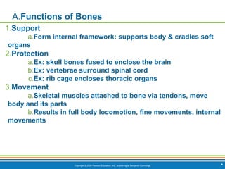


![Copyright © 2009 Pearson Education, Inc., publishing as Benjamin Cummings *
Summary of Joint Classes
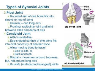
[Insert Table 5.3 here]
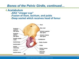
Table 5.3](https://image.slidesharecdn.com/ch-240714220035-4205758a/85/Ch-5-Lecture-Skeletal-System-marieb-ppt-pdf-135-320.jpg)


![CTEV [ clubfoot] DR ARUN LAL ,DR MOHAMED ASHRAF travancore medical college k...](https://cdn.slidesharecdn.com/ss_thumbnails/ctevclubfootdrarunlaldrmohamedashraftravancoremedicalcollegekollamkeralaindia-260208063247-18fc466c-thumbnail.jpg?width=640&height=640&fit=bounds)

![PERI-PROSTHETIC FRACTURE NAIL-PLATE CONSTRUCT [NPC].pptx](https://cdn.slidesharecdn.com/ss_thumbnails/drarunkumardrmohamedashrafperiprostheticfrasturenail-plateconstructnpc-260209164459-7e9d15a1-thumbnail.jpg?width=640&height=640&fit=bounds)


