Download to read offline


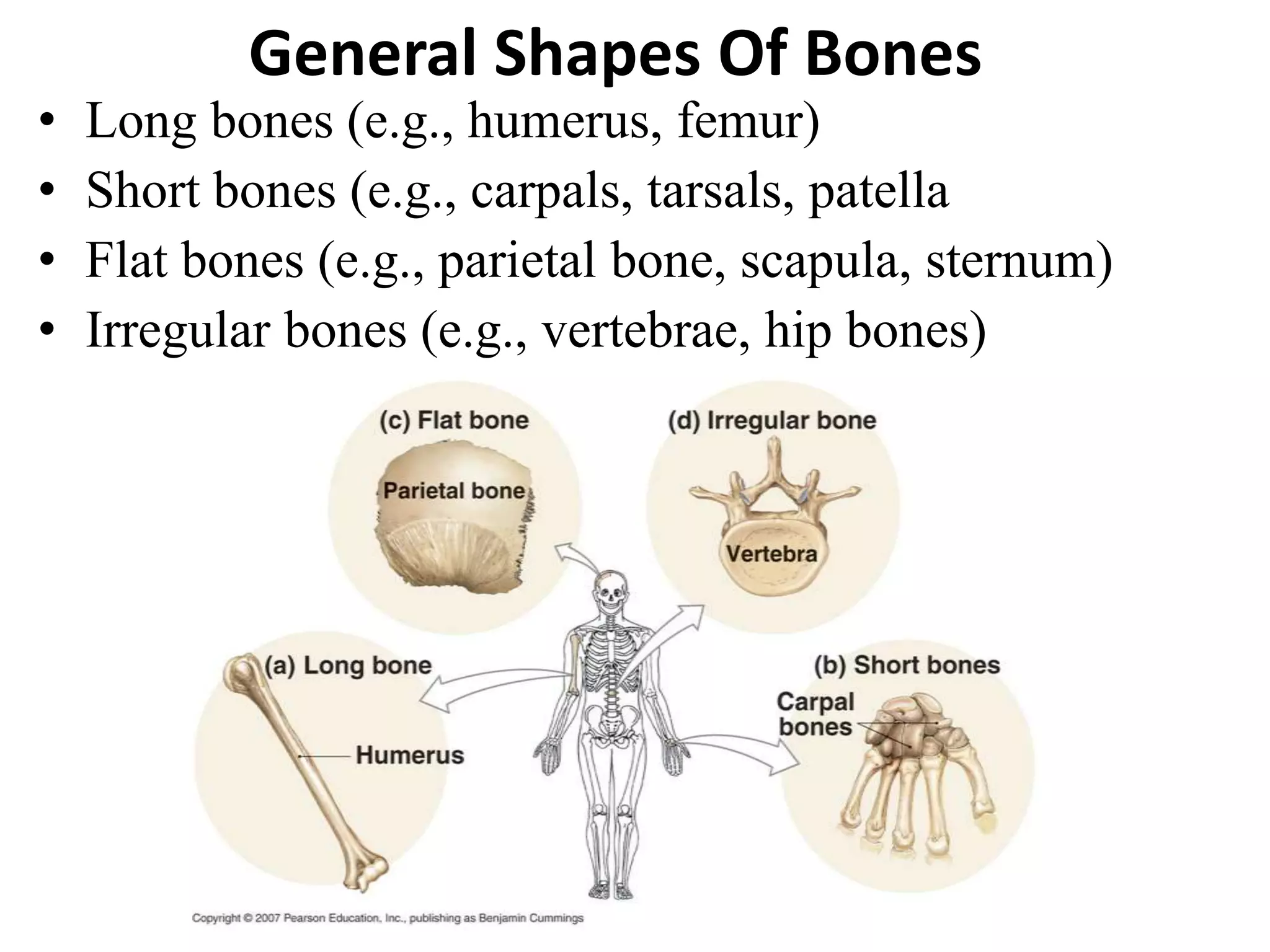


The skeletal system contains 206 bones that are categorized into four basic shapes: long bones, short bones, flat bones, and irregular bones. The skeletal system functions to provide support, protect organs, regulate blood calcium levels, and allow for muscle attachment. Bones form through two processes - intramembranous ossification which forms flat bones, and endochondral ossification which replaces cartilage. The muscular system contains three main types of muscle: skeletal, cardiac, and smooth muscle. Skeletal muscle is attached to bones by tendons and forms the bulk of muscle mass controlled voluntarily.


