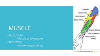
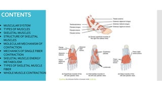
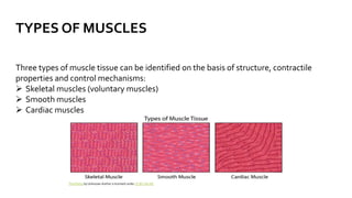
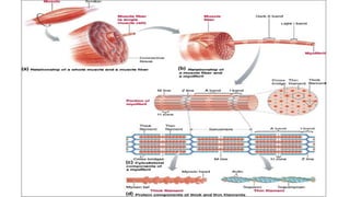
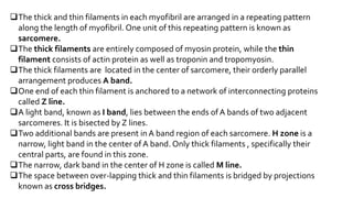
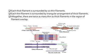
MUSCLE
PRESENTEDTO:
PROF. DR. SADIA REHMAN
PRESENTED BY:
HUMAIRA BIBI (GROUP 04)
This Photo by Unknown Author is licensed under CC BY-SA-NC
CONTENTS
 MUSCULAR SYSTEM
 TYPES OF MUSCLES
 SKELETAL MUSCLES
 STRUCTURE OF SKELETAL
MUSCLES
 MOLECULAR MECHANISM OF
CONTACTION
 MECHANICS OF SINGLE FIBER
CONTRACTION
 SKELETAL MUSCLE ENERGY
METABOLISM
 TYPES OF SKELETAL MUSCLE
FIBER
 WHOLE MUSCLE CONTRACTION
This Photo by Unknown Author is licensed under CC BY-SA
MUSCULAR SYSTEM
Muscle is a soft tissue found in both animals and humans.The cells of
muscles comprise protein filaments of actin and myosin that slide past
one another, which produces contraction and changes both the length
and shape of cells
 The term muscle is derived from Latin word “musculus” which refers to a
little mouse.
The human muscular system includes more than 600 muscles, which
make up about 40 to 50 percent of the body weight.
The energy required for the functioning of muscles is predominantly
powered by the oxidation of fats and carbohydrates particularly and from
the stored energy molecules ATP.
Muscles are linked to bones with the help of tendons.
TYPES OF MUSCLES
Three types of muscle tissue can be identified on the basis of structure, contractile
properties and control mechanisms:
 Skeletal muscles (voluntary muscles)
 Smooth muscles
 Cardiac muscles
This Photo by Unknown Author is licensed under CC BY-SA-NC
SKELETAL MUSCLES
 Skeletal muscle is a muscle tissue attached to the bones and its contraction is
responsible for supporting and moving the skeleton.
 The contraction of skeletal muscle is initiated by impulses in the neurons to the
muscle and is usually under voluntary control
 As they are under the control of central nervous system, they are also lnown as
voluntary muscles.
 They are long, cylindrical shaped and are multinucleated
 Consist of dark and light bands. Hence, they are striated muscles.
 A single skeletal muscle cell is known as muscle fiber.
 Adult skeletal muscle fibers have diameter between 10 and 100 micro-meter, and
lengths that may extend up to 20cm.
STRUCTURE OF SKELETAL MUSCLES
Each muscle fiber is formed during development by the fusion of a number of
undifferentiated, mononucleated cells, called myoblasts.
If skeletal muscle fibers are destroyed after birth, they cannot be replaced by the
division of other existing muscle fibers
New fibers can be formed, from undifferentiated cells known as satellite cells, which
are located adjacent to the muscle fibers and undergo differentiation.
When seen through light microscope, they have series of light and dark bands
perpendicular to the long axis of fiber. Because of this characteristic banding, they are
known as striated muscles.
This pattern results from the arrangement of thick and thin filaments in the cytoplasm
into approximately cylindrical bundles(1 to 2 micrometer in diameter) known as
myofibrils.
The thick and thin filaments in each myofibril are arranged in a repeating pattern
along the length of myofibril. One unit of this repeating pattern is known as
sarcomere.
The thick filaments are entirely composed of myosin protein, while the thin
filament consists of actin protein as well as troponin and tropomyosin.
The thick filaments are located in the center of sarcomere, their orderly parallel
arrangement produces A band.
One end of each thin filament is anchored to a network of interconnecting proteins
called Z line.
A light band, known as I band, lies between the ends ofA bands of two adjacent
sarcomeres. It is bisected by Z lines.
Two additional bands are present in A band region of each sarcomere. H zone is a
narrow, light band in the center of A band. Only thick filaments , specifically their
central parts, are found in this zone.
The narrow, dark band in the center of H zone is called M line.
The space between over-lapping thick and thin filaments is bridged by projections
known as cross bridges.
 Each thick filament is surrounded by six thin filaments.
 Each thin filament is surrounded by triangular arrangement of thick filaments.
 Altogether, there are twice as many thin as thick filaments in the region of
filament overlap.
Muscle Introduction and molecular structure.ppt

Muscle Introduction and molecular structure.ppt

  • 1.
    MUSCLE PRESENTEDTO: PROF. DR. SADIAREHMAN PRESENTED BY: HUMAIRA BIBI (GROUP 04) This Photo by Unknown Author is licensed under CC BY-SA-NC
  • 2.
    CONTENTS  MUSCULAR SYSTEM TYPES OF MUSCLES  SKELETAL MUSCLES  STRUCTURE OF SKELETAL MUSCLES  MOLECULAR MECHANISM OF CONTACTION  MECHANICS OF SINGLE FIBER CONTRACTION  SKELETAL MUSCLE ENERGY METABOLISM  TYPES OF SKELETAL MUSCLE FIBER  WHOLE MUSCLE CONTRACTION This Photo by Unknown Author is licensed under CC BY-SA
  • 3.
    MUSCULAR SYSTEM Muscle isa soft tissue found in both animals and humans.The cells of muscles comprise protein filaments of actin and myosin that slide past one another, which produces contraction and changes both the length and shape of cells  The term muscle is derived from Latin word “musculus” which refers to a little mouse. The human muscular system includes more than 600 muscles, which make up about 40 to 50 percent of the body weight. The energy required for the functioning of muscles is predominantly powered by the oxidation of fats and carbohydrates particularly and from the stored energy molecules ATP. Muscles are linked to bones with the help of tendons.
  • 4.
    TYPES OF MUSCLES Threetypes of muscle tissue can be identified on the basis of structure, contractile properties and control mechanisms:  Skeletal muscles (voluntary muscles)  Smooth muscles  Cardiac muscles This Photo by Unknown Author is licensed under CC BY-SA-NC
  • 5.
    SKELETAL MUSCLES  Skeletalmuscle is a muscle tissue attached to the bones and its contraction is responsible for supporting and moving the skeleton.  The contraction of skeletal muscle is initiated by impulses in the neurons to the muscle and is usually under voluntary control  As they are under the control of central nervous system, they are also lnown as voluntary muscles.  They are long, cylindrical shaped and are multinucleated  Consist of dark and light bands. Hence, they are striated muscles.  A single skeletal muscle cell is known as muscle fiber.  Adult skeletal muscle fibers have diameter between 10 and 100 micro-meter, and lengths that may extend up to 20cm.
  • 6.
    STRUCTURE OF SKELETALMUSCLES Each muscle fiber is formed during development by the fusion of a number of undifferentiated, mononucleated cells, called myoblasts. If skeletal muscle fibers are destroyed after birth, they cannot be replaced by the division of other existing muscle fibers New fibers can be formed, from undifferentiated cells known as satellite cells, which are located adjacent to the muscle fibers and undergo differentiation. When seen through light microscope, they have series of light and dark bands perpendicular to the long axis of fiber. Because of this characteristic banding, they are known as striated muscles. This pattern results from the arrangement of thick and thin filaments in the cytoplasm into approximately cylindrical bundles(1 to 2 micrometer in diameter) known as myofibrils.
  • 8.
    The thick andthin filaments in each myofibril are arranged in a repeating pattern along the length of myofibril. One unit of this repeating pattern is known as sarcomere. The thick filaments are entirely composed of myosin protein, while the thin filament consists of actin protein as well as troponin and tropomyosin. The thick filaments are located in the center of sarcomere, their orderly parallel arrangement produces A band. One end of each thin filament is anchored to a network of interconnecting proteins called Z line. A light band, known as I band, lies between the ends ofA bands of two adjacent sarcomeres. It is bisected by Z lines. Two additional bands are present in A band region of each sarcomere. H zone is a narrow, light band in the center of A band. Only thick filaments , specifically their central parts, are found in this zone. The narrow, dark band in the center of H zone is called M line. The space between over-lapping thick and thin filaments is bridged by projections known as cross bridges.
  • 9.
     Each thickfilament is surrounded by six thin filaments.  Each thin filament is surrounded by triangular arrangement of thick filaments.  Altogether, there are twice as many thin as thick filaments in the region of filament overlap.