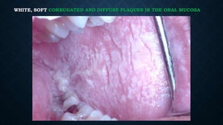
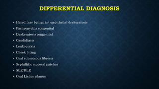
WHITE SPONGE NEVUS
Dr Randy Chance BMSC BDS
INTRO
• a rare, autosomal-dominant disorder that affects the uncornified
stratified squamous epithelia
• First described by Hyde in 1909, but Cannon coined the term in
1935
• It’s a type of genodermatoses with problems related to normal
epithelial maturation and desquamation- it is unscrapable
• No gender or racial predilection
• Prevalence rate below 1 in 200,000
• Present from childhood
OTHER NAMES
• Cannon’s disease
• Familial white folded mucosal dysplasia
• Hereditary mucosal leukokeratosis
• Nevus of Cannon
• White folded gingivostomatitis
• Exfoliative Leukoedma
CLINICAL FEATURES
• Usually occurs before 20 years of age and often in early childhood
• It exhibits no race or gender predilection (or slight female
predilection)
• Variable penetrance and expressivity
• Extra-oral sites such as the mucous membrane of the nasal cavity,
esophagus, larynx, rectum and vagina may be involved
WHITE, SOFT CORRUGATED AND DIFFUSE PLAQUES IN THE ORAL MUCOSA
SIGNIFICANT PREDILECTION FOR THE CHEEK MUCOSA
Other sites:
Ventral tongue
Labial mucosa
Alveolar ridge
Floor of the mouth
OTHER CLINICAL FEATURES
• Painless
• Symmetrical
• Bilateral
• Cannot be scraped off
• Differential diagnoses are numerous
• No systemic alterations
PATHOGENESIS
• genetic based pathogenesis, a benign course
• HPV 16??
Missense mutations in K4 and K13 genes (mucosal keratins)
Interference with intermediate filament assembly
Easily damaged intermediate filament
Cytokine flooding of underlying basal cells from mild trauma
Excessive basal cell proliferation leading to mucosal
hyperkeratosis
DIFFERENTIAL DIAGNOSIS
• Hereditary benign intraepithelial dyskeratosis
• Pachyonychia congenital
• Dyskeratosis congenital
• Candidiasis
• Leukoplakia
• Cheek biting
• Oral submucous fibrosis
• Syphillitic mucosal patches
• SLE/DLE
• Oral Lichen planus
LAB PATHOLOGY
• Prominent hyperparakeratosis
• Marked acanthosis
• Vacuolization in keratinocytes
• Perinuclear eosinophilic condensation of epithelial
cells
PROBLEMS AND TREATMENT
• Superimposed infection with Candida and/or Staphylococcus
• Tetracycline mouthwash – 0.25% aqueous tetracycline solution, 5ml
twice daily.
• Amoxycillin PO – initially 250mg three times daily for 4 weeks
followed by 250mg once each week to maintain the improvement.
• Erythromycin
• Antifungal, retinoids, metronidazole, CHX mouthwash
THE END
• Check out and join my facebook page for regular oral pathology/medicine posts
https://www.facebook.com/orpath
And my youtube channel for videos of oral pathology and medicine.
Youtube channel can be accessed via the facebook page.
Thank you. 

White sponge nevus

  • 1.
    WHITE SPONGE NEVUS DrRandy Chance BMSC BDS
  • 2.
    INTRO • a rare,autosomal-dominant disorder that affects the uncornified stratified squamous epithelia • First described by Hyde in 1909, but Cannon coined the term in 1935 • It’s a type of genodermatoses with problems related to normal epithelial maturation and desquamation- it is unscrapable • No gender or racial predilection • Prevalence rate below 1 in 200,000 • Present from childhood
  • 4.
    OTHER NAMES • Cannon’sdisease • Familial white folded mucosal dysplasia • Hereditary mucosal leukokeratosis • Nevus of Cannon • White folded gingivostomatitis • Exfoliative Leukoedma
  • 5.
    CLINICAL FEATURES • Usuallyoccurs before 20 years of age and often in early childhood • It exhibits no race or gender predilection (or slight female predilection) • Variable penetrance and expressivity • Extra-oral sites such as the mucous membrane of the nasal cavity, esophagus, larynx, rectum and vagina may be involved
  • 6.
    WHITE, SOFT CORRUGATEDAND DIFFUSE PLAQUES IN THE ORAL MUCOSA
  • 7.
    SIGNIFICANT PREDILECTION FORTHE CHEEK MUCOSA Other sites: Ventral tongue Labial mucosa Alveolar ridge Floor of the mouth
  • 8.
    OTHER CLINICAL FEATURES •Painless • Symmetrical • Bilateral • Cannot be scraped off • Differential diagnoses are numerous • No systemic alterations
  • 9.
    PATHOGENESIS • genetic basedpathogenesis, a benign course • HPV 16?? Missense mutations in K4 and K13 genes (mucosal keratins) Interference with intermediate filament assembly Easily damaged intermediate filament Cytokine flooding of underlying basal cells from mild trauma Excessive basal cell proliferation leading to mucosal hyperkeratosis
  • 10.
    DIFFERENTIAL DIAGNOSIS • Hereditarybenign intraepithelial dyskeratosis • Pachyonychia congenital • Dyskeratosis congenital • Candidiasis • Leukoplakia • Cheek biting • Oral submucous fibrosis • Syphillitic mucosal patches • SLE/DLE • Oral Lichen planus
  • 11.
  • 18.
    • Prominent hyperparakeratosis •Marked acanthosis • Vacuolization in keratinocytes • Perinuclear eosinophilic condensation of epithelial cells
  • 20.
    PROBLEMS AND TREATMENT •Superimposed infection with Candida and/or Staphylococcus • Tetracycline mouthwash – 0.25% aqueous tetracycline solution, 5ml twice daily. • Amoxycillin PO – initially 250mg three times daily for 4 weeks followed by 250mg once each week to maintain the improvement. • Erythromycin • Antifungal, retinoids, metronidazole, CHX mouthwash
  • 21.
    THE END • Checkout and join my facebook page for regular oral pathology/medicine posts https://www.facebook.com/orpath And my youtube channel for videos of oral pathology and medicine. Youtube channel can be accessed via the facebook page. Thank you. 