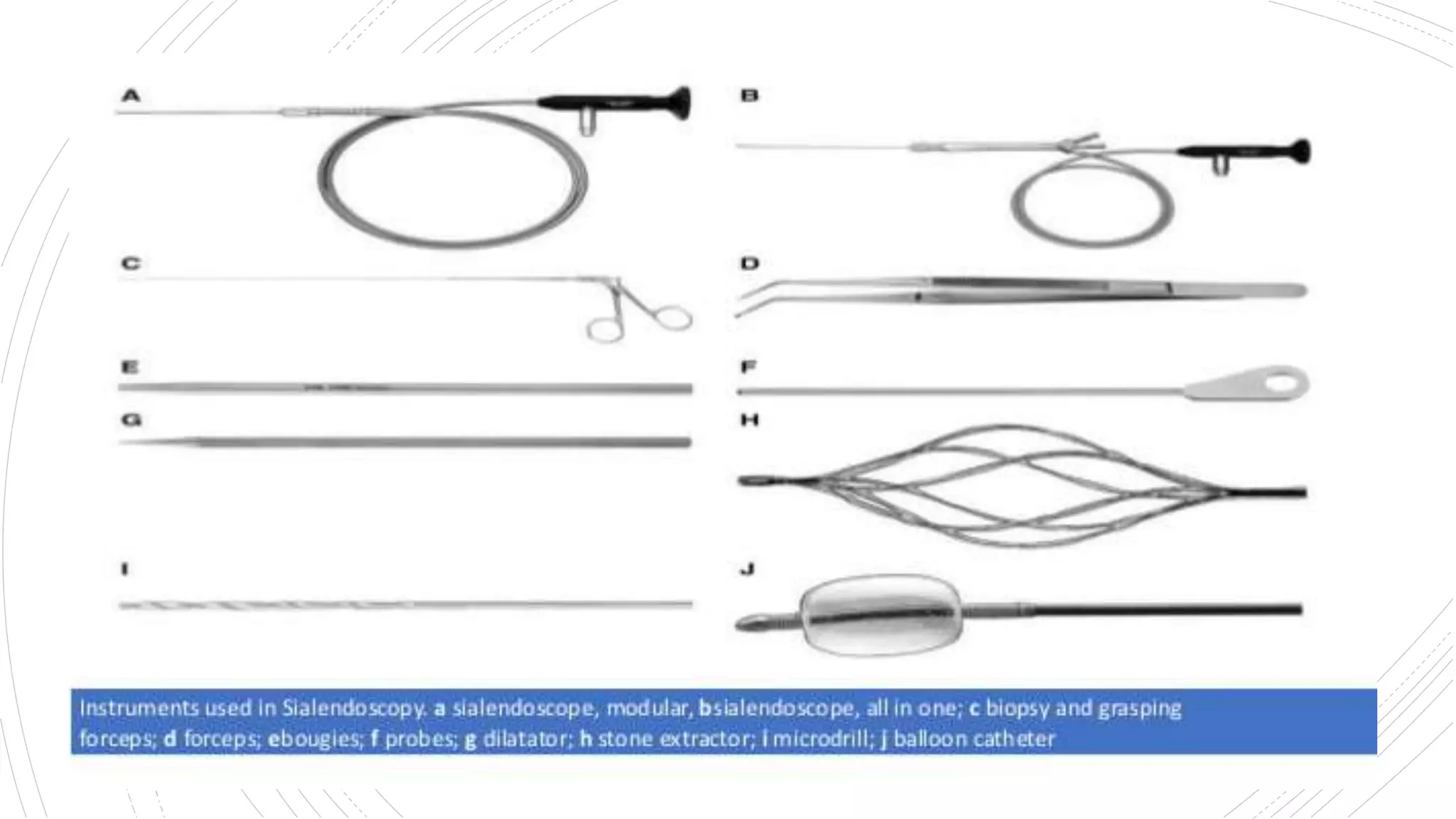
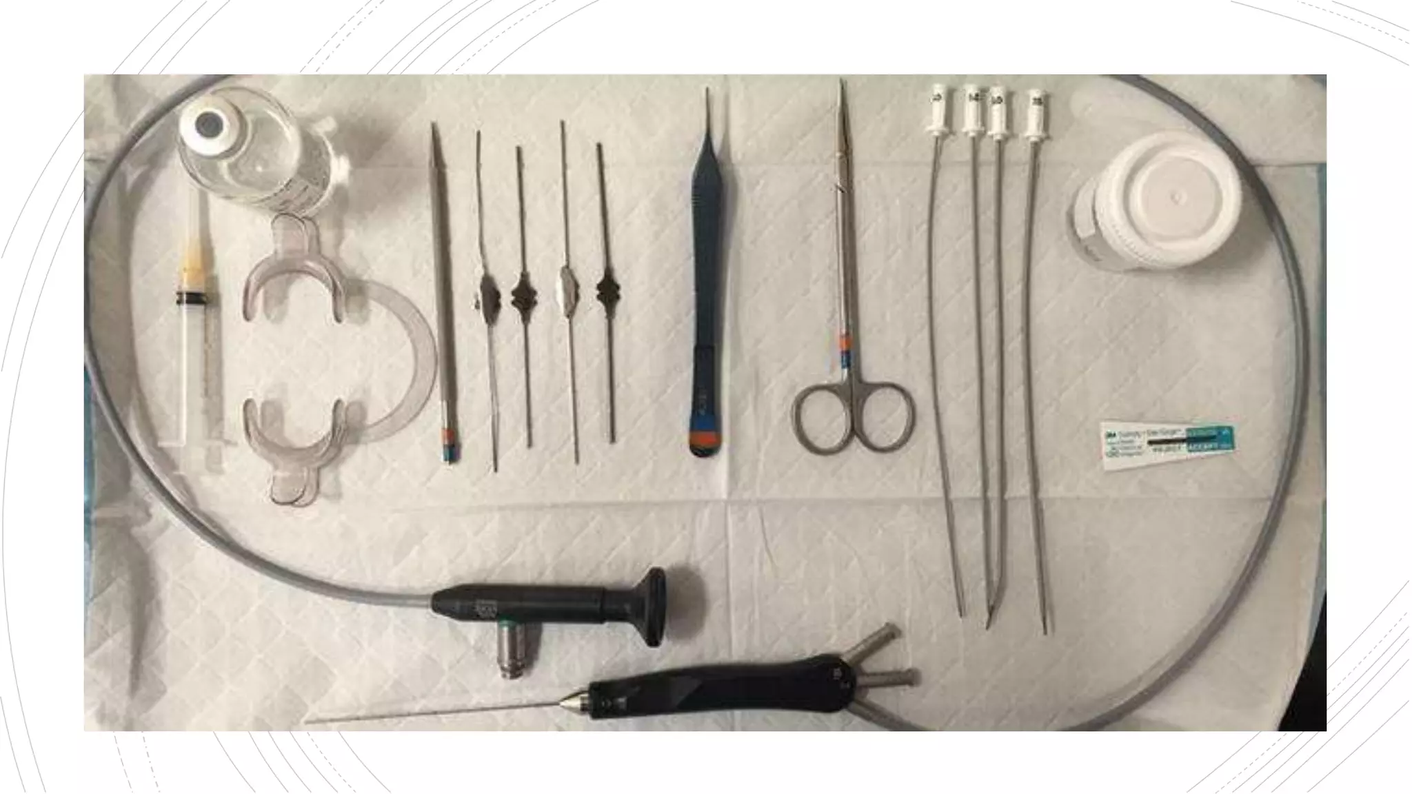
Sialendoscopy
Dr Safika Zaman
PGT
Dept of ENT & Head neck surgery
RKMSP
Milestones
 1990: Konigsberger and Gundlach separately
performed sialoendoscopy when they introduced an
endoscope into the major salivary glands.
 1991, Katz introduced a 0.8-millimeter flexible
endoscope to diagnose and treat salivary gland stones.
 1994, Nahlieli used a rigid mini-endoscope to diagnose
and treat major salivary gland obstructions
Introduction
 Paradigm shift towards organ and function
preservation.
 Open to endoscopic approach.
 Diagnostic & therapeutic tool.
Imaging
techniques
Source- Scott Brown’s text book – 8th edition
Indications
of
sialendoscopy
Therapeutic: Secondary to obstructive pathology includes
Sialoliths
Strictures
Ductal polyps
Mucous plugs
Diagnostic: to understand pathology of other salivery
gland disease.
Juvenile
recurrent
parotititis
 Steroid can be delivered directly at site.
Sialolithiasis
 Sialolithiasis is a common disorder characterized by
the formation of a calculus usually within the ductal
system of a gland.
 Sialoliths can arise in both the major and minor
salivary glands.
 Obstruction of a major duct will give rise to pain and
swelling.
 Submandibular glands- 83% , than the parotid (10%) or
sublingual (7%) glands.
Data Source- Scott Brown’s text book – 8th
edition
sialendescope
Tip diameter – 0.9- 1.6 mm.
Mini instruments – grasping forceps, micro drills, wire
basket, high pressure balloon dilators, guide wire, biopsy
forceps
https://www.joms.org/article/S0278-2391(03)00691-
8/pdf#
Submandibular
gland  Approximately 4–6 cm long with an average diameter of
1.5 mm.
 Orifice diameter ranges between 0.5 and 0.1mm.
 Opening just lateral to the lingual frenum.
 Genu- and the angle varies between 24 and 178.
 Stones distal to genu are difficult to manipulate.
Submandibular
anatomy
Parotid
anatomy
Stensen’s duct: Secondary and the tertiary ducts arising from
the medial and the lateral lobe of the parotid form the
Stensen’s duct.
Passes through the buccal fat pad, buccopharyngeal fascia and
buccinator muscle
Opening-laterally to the second maxillary molar
The duct is 4–7 cm long.
Average diameter of 1.4 cm.
The orifice is 0.5 mm and 1.2 mm at the transbuccinator
muscular sphincteric passage
Parotid
anatomy
Theductal
system
Technique
LA
•With 10% - 20%
lignocaine spray
probe
•Dialatation of papilla
with salivery probe.
dental
•Introduction of
dental tampons.
Cont.
endoscope
•Introduction of sialendoscope
guidewire
•Introduction of guidewire in
the working channel.
intervention
•Interventional sialendoscopy
depending on etiology.
Decisionmaking
Sialoendoscopy: Review and
Nuances of Technique
Srinivasa Rama Chandra
Basketstone
removal
Endoscopic
dilatation
Sialendoscopy
assistedsurgical
approach
 Indication- large ,palpable fixed duct and parenchymal
stone.
 Calculi not responding to minimally invasive approach.
Combind
techniques
Advantage
 Less morbidity.
 Shorter hospital stay.
 Both diagnostic and therapeutic.
Complications
 Local pain
 Temporary lingual nerve paraesthesia
 Ranula
 Infection around papilla
 Post op ductal stricture
 Breaking of endoscopic tool inside the duct.
Contraindication
 Acute inflammation.
 Complete distal duct stenosis.
 Symptomatic intra-parenchymal stone.
 Limited mouth opening.
Limitations
 Time consuming
 Costly
 Massively fibrosed gland and in multiple stones
Thank You

Sialoendoscopy