Downloaded 206 times


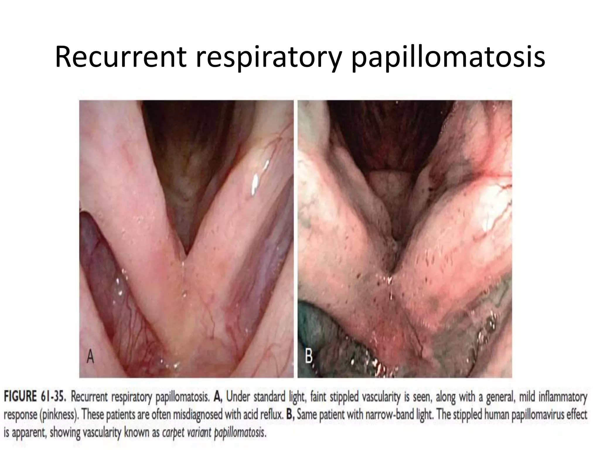

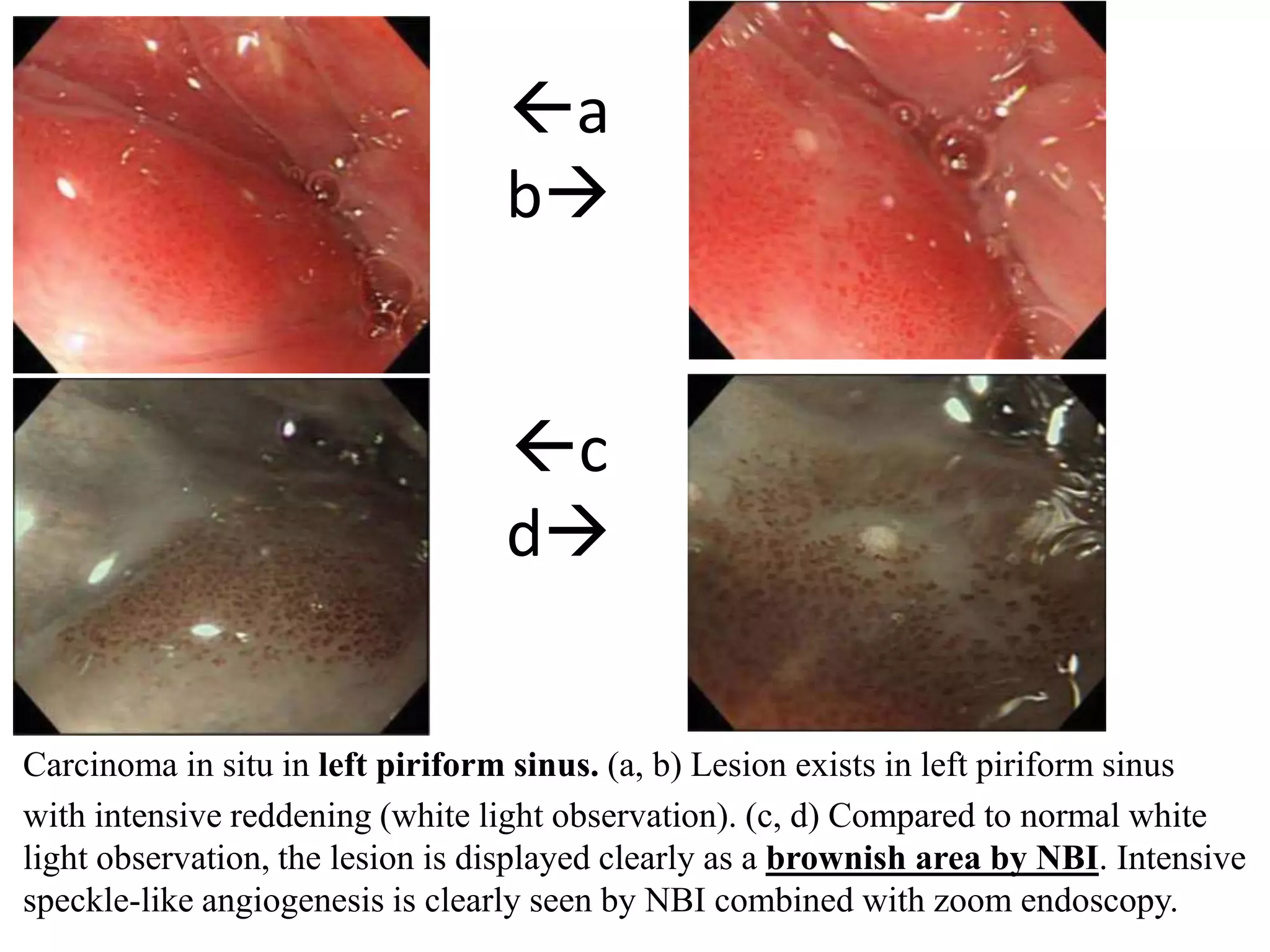

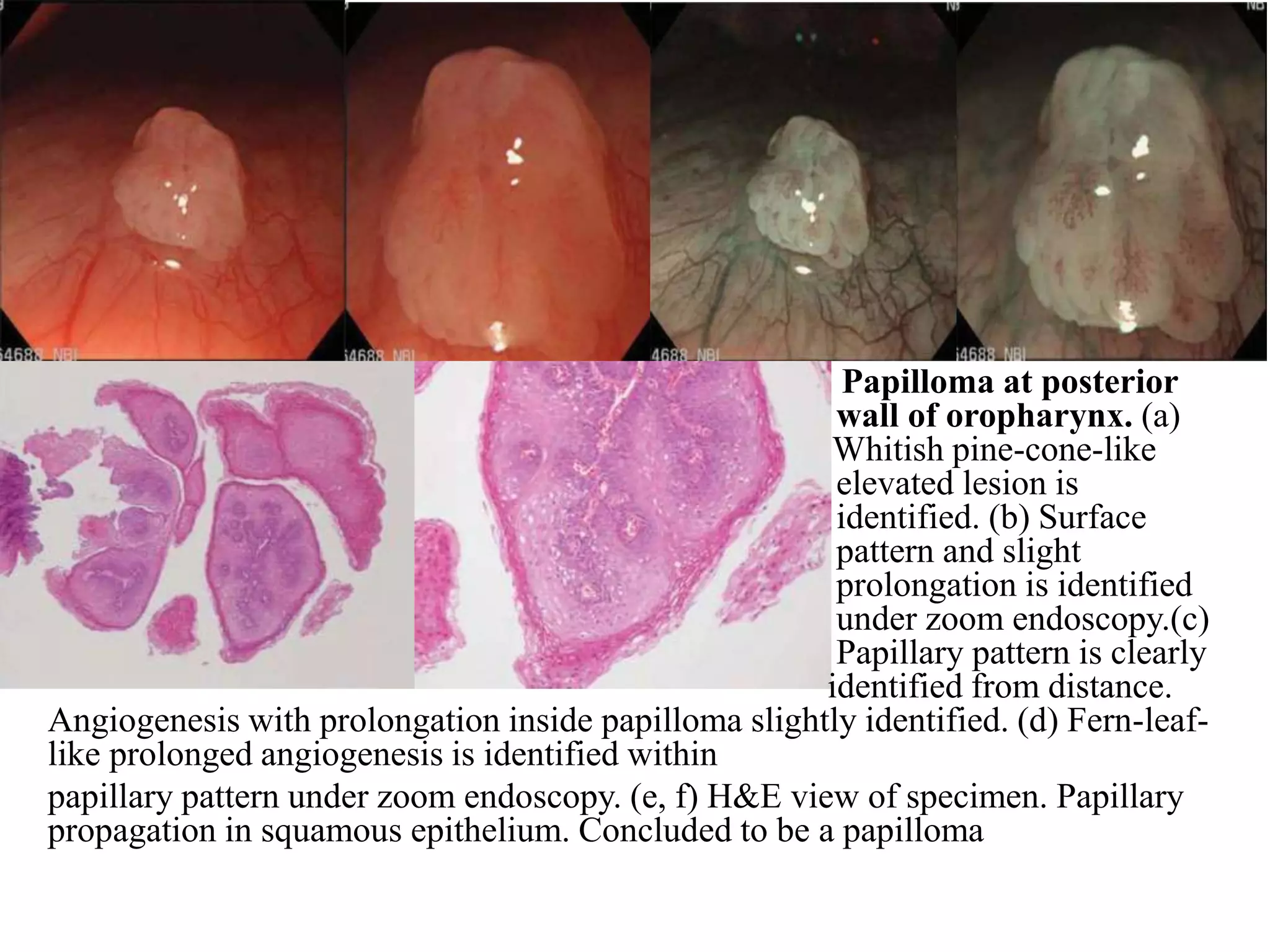


Narrow-band imaging (NBI) is an endoscopic imaging technique that uses specific blue and green wavelengths of light to enhance visualization of mucosal and vascular patterns. It helps identify subtle abnormalities by highlighting areas with high hemoglobin concentration. In the larynx, NBI has been used to identify recurrent respiratory papillomatosis and screen for malignancies. It provides sharper contrast than white light imaging, allowing for better detection of lesions and guidance of biopsy to suspicious areas. NBI is available for laryngoscopes and gastroscopes and is being explored for its utility in evaluating laryngeal and hypopharyngeal lesions.


![Apporach to lung biopsy [Auto-saved].pptx latest](https://cdn.slidesharecdn.com/ss_thumbnails/apporachtolungbiopsyauto-saved-251211225655-93258539-thumbnail.jpg?width=640&height=640&fit=bounds)


