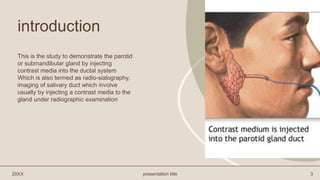
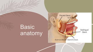
Sialography
Ananya girish babu
Lecture
Objectives
 INTRODCUTION
 BASIC ANATOMY
 INIDCATION AND
CONTRAINDICATION
 CONTRAST MEDIUM
 EQUIPMENTS
 PATIENT PREPERATION
 PROCEDURE
 AFTER CARE AND RISK
FACTORS
 MCQ RELATED
introduction
This is the study to demonstrate the parotid
or submandibular gland by injecting
contrast media into the ductal system
Which is also termed as radio-sialography,
imaging of salivary duct which involve
usually by injecting a contrast media to the
gland under radiographic examination
20XX presentation title 3
Basic
anatomy
• The major salivary glands are the largest and most important salivary glands. They produce most of the
saliva in your mouth.
• There are three pairs of major salivary glands:
1. the parotid glands
2. the submandibular glands
3. the sublingual glands.
Parotid Glands
The parotid glands are the largest salivary glands. They are located just in front of the ears. The saliva
produced in these glands is secreted into the mouth from a duct near your upper second molar.
Submandibular Glands
About the size of a walnut, the submandibular glands are located below the jaw. The saliva produced in
these glands is secreted into the mouth from under the tongue.
Sublingual Glands
The sublingual glands are the smallest of the major salivary glands. These almond-shaped structures are
located under the floor of the mouth and below either side of the tongue.
20XX presentation title 5
Indication
• Calculi
• Chronic inflammatory disease
• Mass lesion
• Obstructive lesion
• Penetrating trauma
• Strictures
• Fistula
• Tumor
• Prior evaluation of CT salivary gland
20XX presentation title 6
CONTRAST MEDIA
1. Allergic to ionic contrast media
2. Acute sialadenitis
20XX presentation title 7
CONTRAINDICATION
Water soluble ionic contrast media
[triovideo 280, conray 280]
Or
Non ionic contrast media such as omnipaque
350
20XX presentation title 8
Equipment's
• Lacrimal canula 22G
• Lacrimal dilator – liebreichs double ended lacrimal probe
• 2 cc syringe
• Lemon or vitamin C tablet
Equipments
LACRIMAL CANULA
22G
LACRIMAL DILATOR
– LIEBREICHS
DOUBLE ENDED
LACRIMAL PROBER
2 CC SYRINGE
20XX presentation title 9
20XX presentation title 10
Procedure
1.Preliminary film –plain radiography should be taken before the procedure
Frontal view with face rotation 5-10°
Lateral view with face rotation 15-20°
2. Locating the duct opening
(a) Parotid duct opens opposite 2nd upper molar tooth on the buccal surface of the cheek.
(b) Submandibular duct opens at the base of the frenulum of the tongue.
3. Dilate the punctum with lacrimal dilator.
20XX presentation title 11
4. Technique
Two techniques for cannulating the ducts are by using:
(a) Intracath technique.
(b) Lacrimal cannula technique - In the lacrimal cannula method, contrast is injected into the cannula
which is introduced through the duct opening. Contrast is injected till the patient complains of pain by a
prearranged signal. About 0.5-1.0 cc of contrast is required.
5. Film exposure
Positioning for parotids:
• Frontal view is taken with face rotated 5-10 degrees towards the side of study.
• Lateral view is taken with 15-20 degrees cranial tube tilt.
Positioning for submandibular gland
• Lateral view is taken with 15-20 degrees cranial tube tilt.
Films are taken during injection. The catheter is left in place till the adequacy of films is ensured.
20XX presentation title 12
6. Aftercare: none
If sialadenitis occurs after the procedure, it should be treated with antibiotics and anti-inflammatory
drugs.
7. Complications
• Sialadenitis and abscess
• Stricture of the ducts.
8. Disadvantages of sialogram
• Masses less than 1cm may not be detected.
• Contrast does not always penetrate the deep lobe of parotid gland.
20XX presentation title 13
Lets answer the questions
1. Which among is not a indication of sialography
a. Mass lesion b. chronic inflammatory disease c. acute sialadenitis d. calculi
Ans: b
2. Study to demonstrate the parotoid gland and submandibular gland
a. Sialography b. dacrocystography c. barium studies
Ans: a
3. Where did the parotid duct present ----------------
Ans :opposite 2nd upper molar tooth on the buccal
4. Name of dilator used in sialography---------
Ans : liebreich’s double ended lacrimal dilator
5. Contrast media used in sialography -----------
Ans : water soluble ionic and non ionic
20XX presentation title 14
thank you
Ananya girish babu

Sialography.pptx

  • 1.
  • 2.
    Objectives  INTRODCUTION  BASICANATOMY  INIDCATION AND CONTRAINDICATION  CONTRAST MEDIUM  EQUIPMENTS  PATIENT PREPERATION  PROCEDURE  AFTER CARE AND RISK FACTORS  MCQ RELATED
  • 3.
    introduction This is thestudy to demonstrate the parotid or submandibular gland by injecting contrast media into the ductal system Which is also termed as radio-sialography, imaging of salivary duct which involve usually by injecting a contrast media to the gland under radiographic examination 20XX presentation title 3
  • 4.
  • 5.
    • The majorsalivary glands are the largest and most important salivary glands. They produce most of the saliva in your mouth. • There are three pairs of major salivary glands: 1. the parotid glands 2. the submandibular glands 3. the sublingual glands. Parotid Glands The parotid glands are the largest salivary glands. They are located just in front of the ears. The saliva produced in these glands is secreted into the mouth from a duct near your upper second molar. Submandibular Glands About the size of a walnut, the submandibular glands are located below the jaw. The saliva produced in these glands is secreted into the mouth from under the tongue. Sublingual Glands The sublingual glands are the smallest of the major salivary glands. These almond-shaped structures are located under the floor of the mouth and below either side of the tongue. 20XX presentation title 5
  • 6.
    Indication • Calculi • Chronicinflammatory disease • Mass lesion • Obstructive lesion • Penetrating trauma • Strictures • Fistula • Tumor • Prior evaluation of CT salivary gland 20XX presentation title 6
  • 7.
    CONTRAST MEDIA 1. Allergicto ionic contrast media 2. Acute sialadenitis 20XX presentation title 7 CONTRAINDICATION Water soluble ionic contrast media [triovideo 280, conray 280] Or Non ionic contrast media such as omnipaque 350
  • 8.
    20XX presentation title8 Equipment's • Lacrimal canula 22G • Lacrimal dilator – liebreichs double ended lacrimal probe • 2 cc syringe • Lemon or vitamin C tablet
  • 9.
    Equipments LACRIMAL CANULA 22G LACRIMAL DILATOR –LIEBREICHS DOUBLE ENDED LACRIMAL PROBER 2 CC SYRINGE 20XX presentation title 9
  • 10.
    20XX presentation title10 Procedure 1.Preliminary film –plain radiography should be taken before the procedure Frontal view with face rotation 5-10° Lateral view with face rotation 15-20° 2. Locating the duct opening (a) Parotid duct opens opposite 2nd upper molar tooth on the buccal surface of the cheek. (b) Submandibular duct opens at the base of the frenulum of the tongue. 3. Dilate the punctum with lacrimal dilator.
  • 11.
    20XX presentation title11 4. Technique Two techniques for cannulating the ducts are by using: (a) Intracath technique. (b) Lacrimal cannula technique - In the lacrimal cannula method, contrast is injected into the cannula which is introduced through the duct opening. Contrast is injected till the patient complains of pain by a prearranged signal. About 0.5-1.0 cc of contrast is required. 5. Film exposure Positioning for parotids: • Frontal view is taken with face rotated 5-10 degrees towards the side of study. • Lateral view is taken with 15-20 degrees cranial tube tilt. Positioning for submandibular gland • Lateral view is taken with 15-20 degrees cranial tube tilt. Films are taken during injection. The catheter is left in place till the adequacy of films is ensured.
  • 12.
    20XX presentation title12 6. Aftercare: none If sialadenitis occurs after the procedure, it should be treated with antibiotics and anti-inflammatory drugs. 7. Complications • Sialadenitis and abscess • Stricture of the ducts. 8. Disadvantages of sialogram • Masses less than 1cm may not be detected. • Contrast does not always penetrate the deep lobe of parotid gland.
  • 13.
  • 14.
    Lets answer thequestions 1. Which among is not a indication of sialography a. Mass lesion b. chronic inflammatory disease c. acute sialadenitis d. calculi Ans: b 2. Study to demonstrate the parotoid gland and submandibular gland a. Sialography b. dacrocystography c. barium studies Ans: a 3. Where did the parotid duct present ---------------- Ans :opposite 2nd upper molar tooth on the buccal 4. Name of dilator used in sialography--------- Ans : liebreich’s double ended lacrimal dilator 5. Contrast media used in sialography ----------- Ans : water soluble ionic and non ionic 20XX presentation title 14
  • 15.