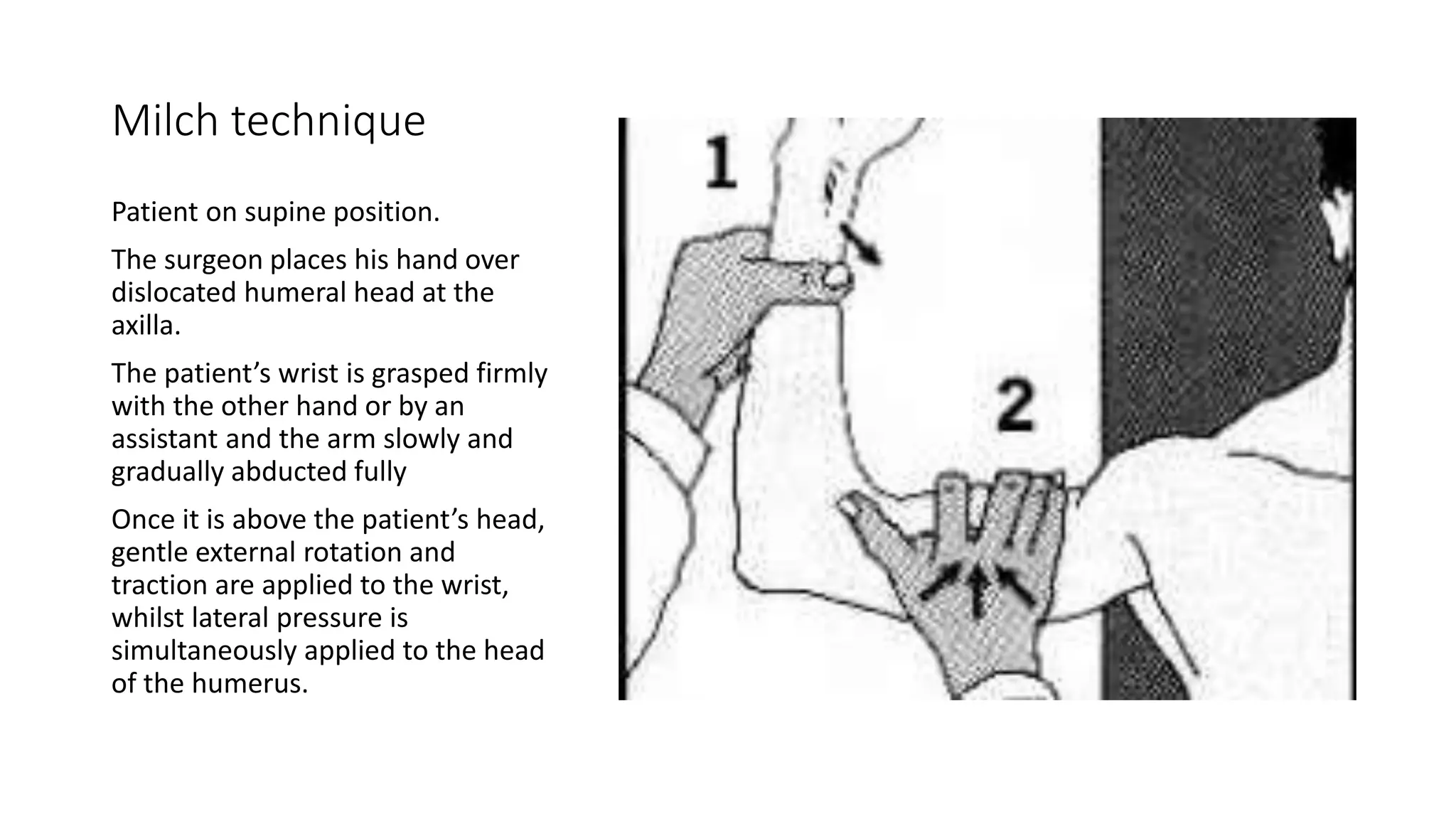
The document discusses anatomy, mechanism of injury, classification, diagnosis, and management of anterior shoulder dislocations. Key points include:
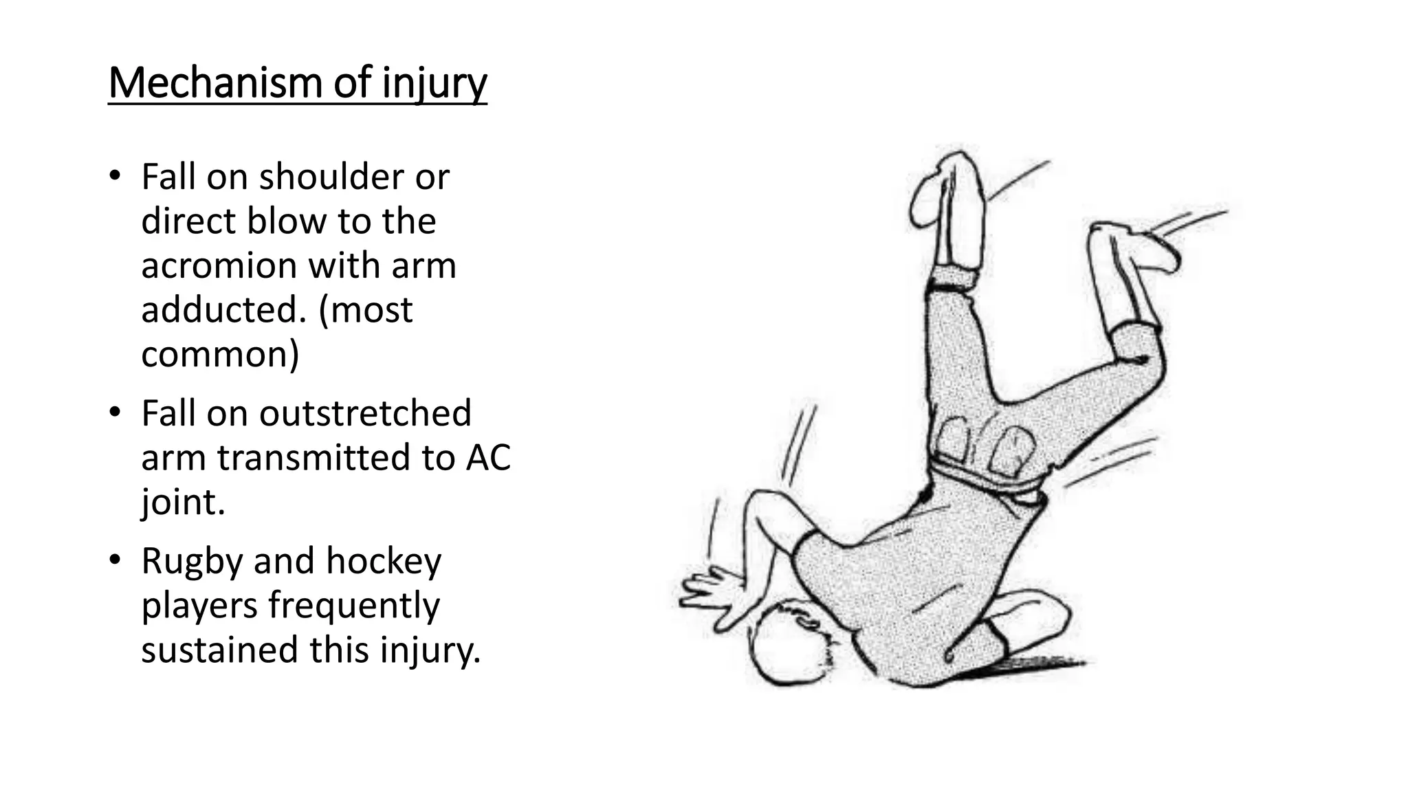
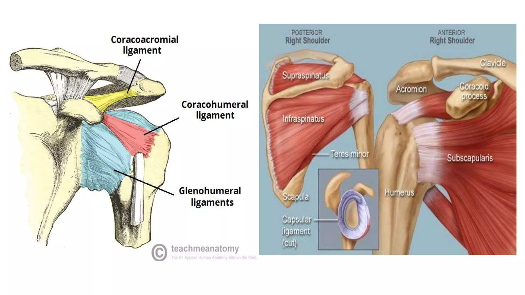
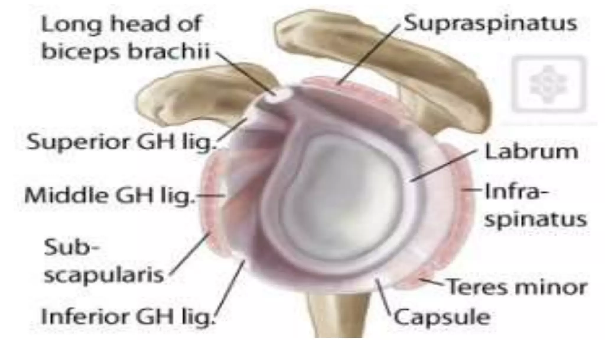
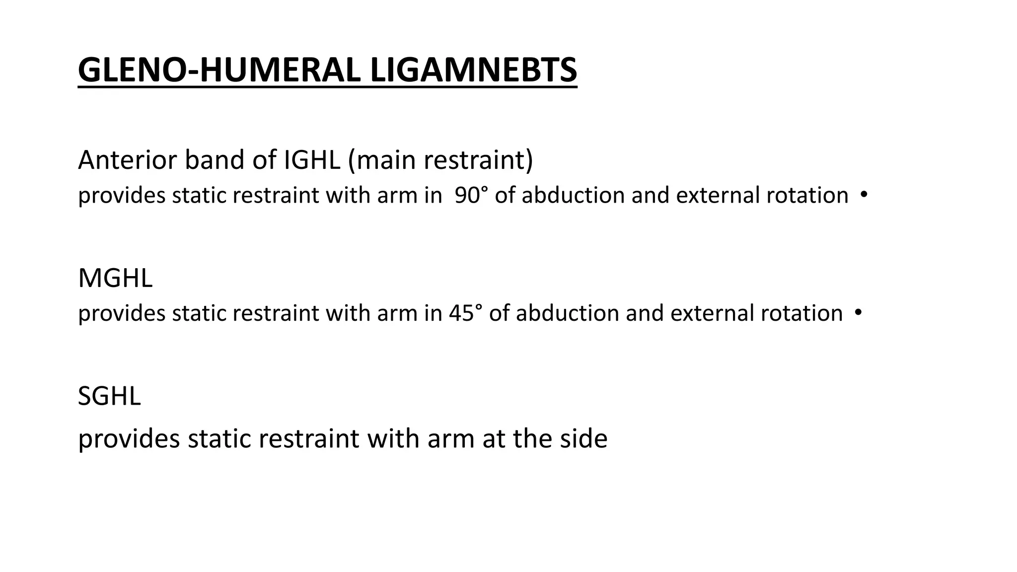
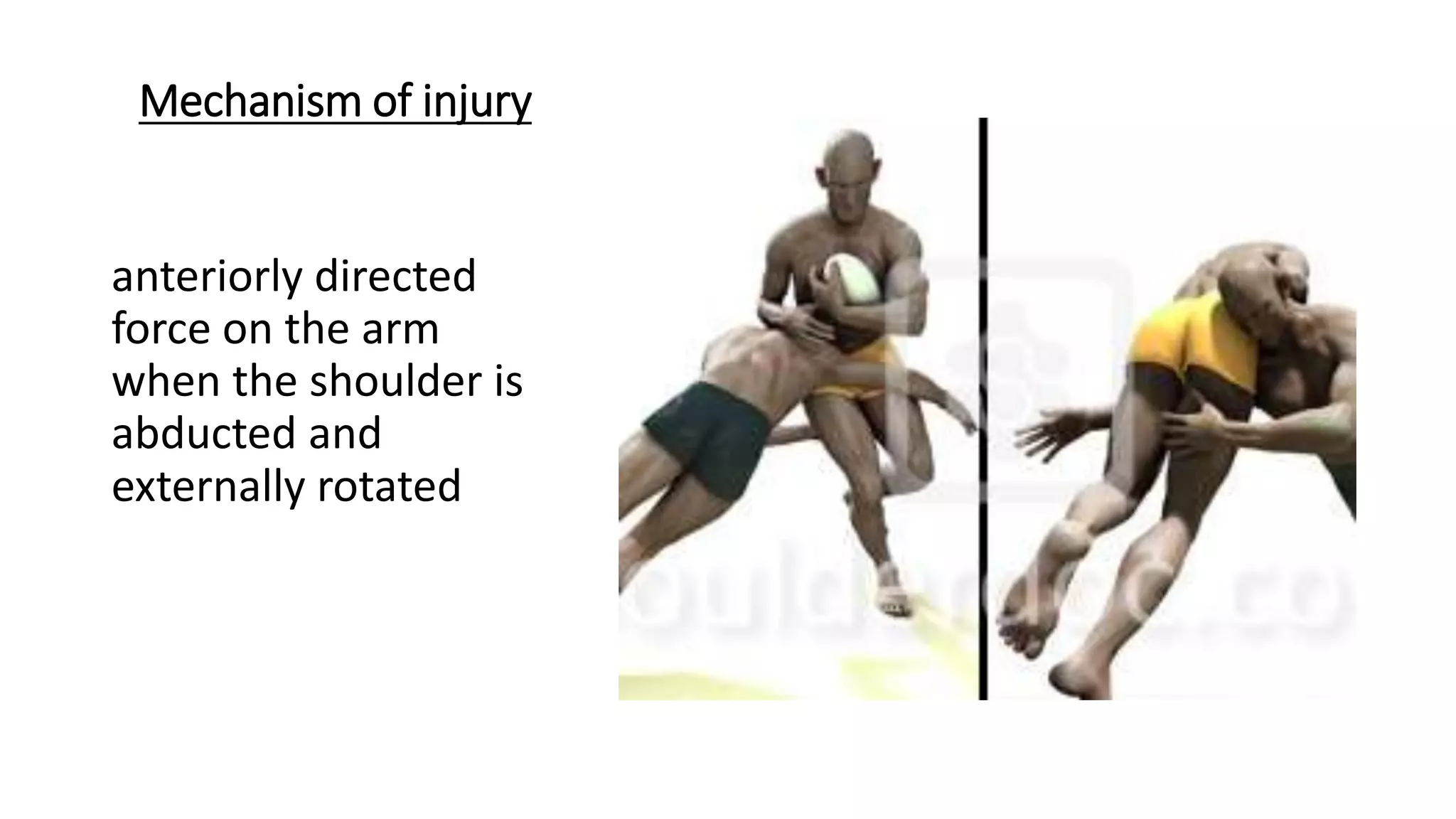
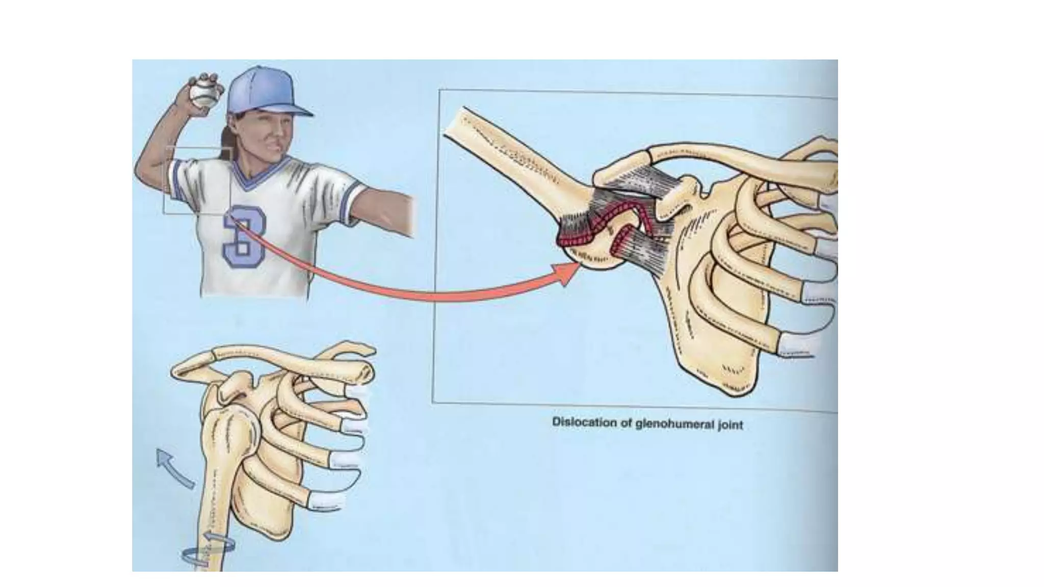
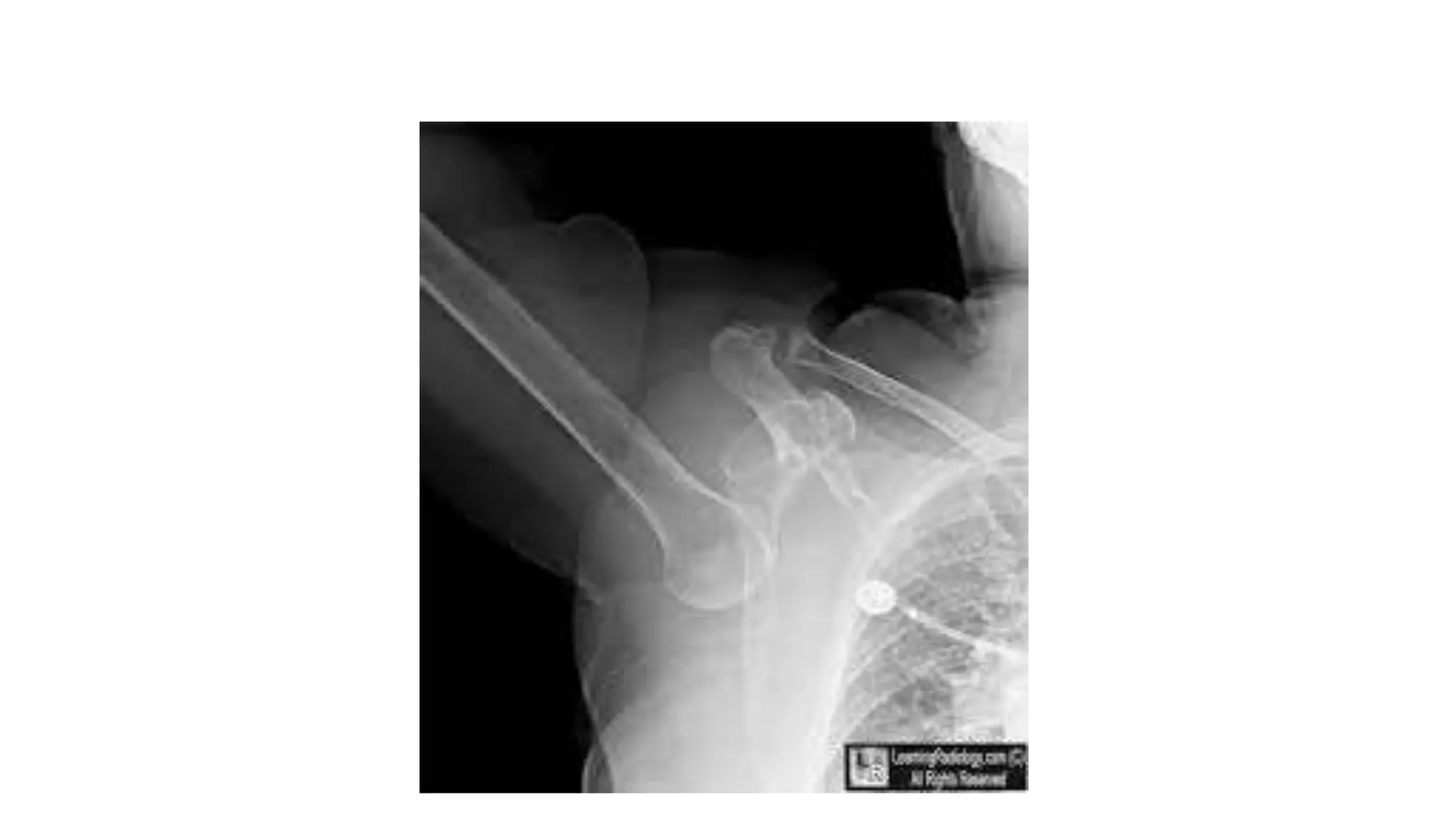
- Anterior shoulder dislocations are caused by an anteriorly directed force on the arm when abducted and externally rotated, which can tear the anterior labrum and ligaments.
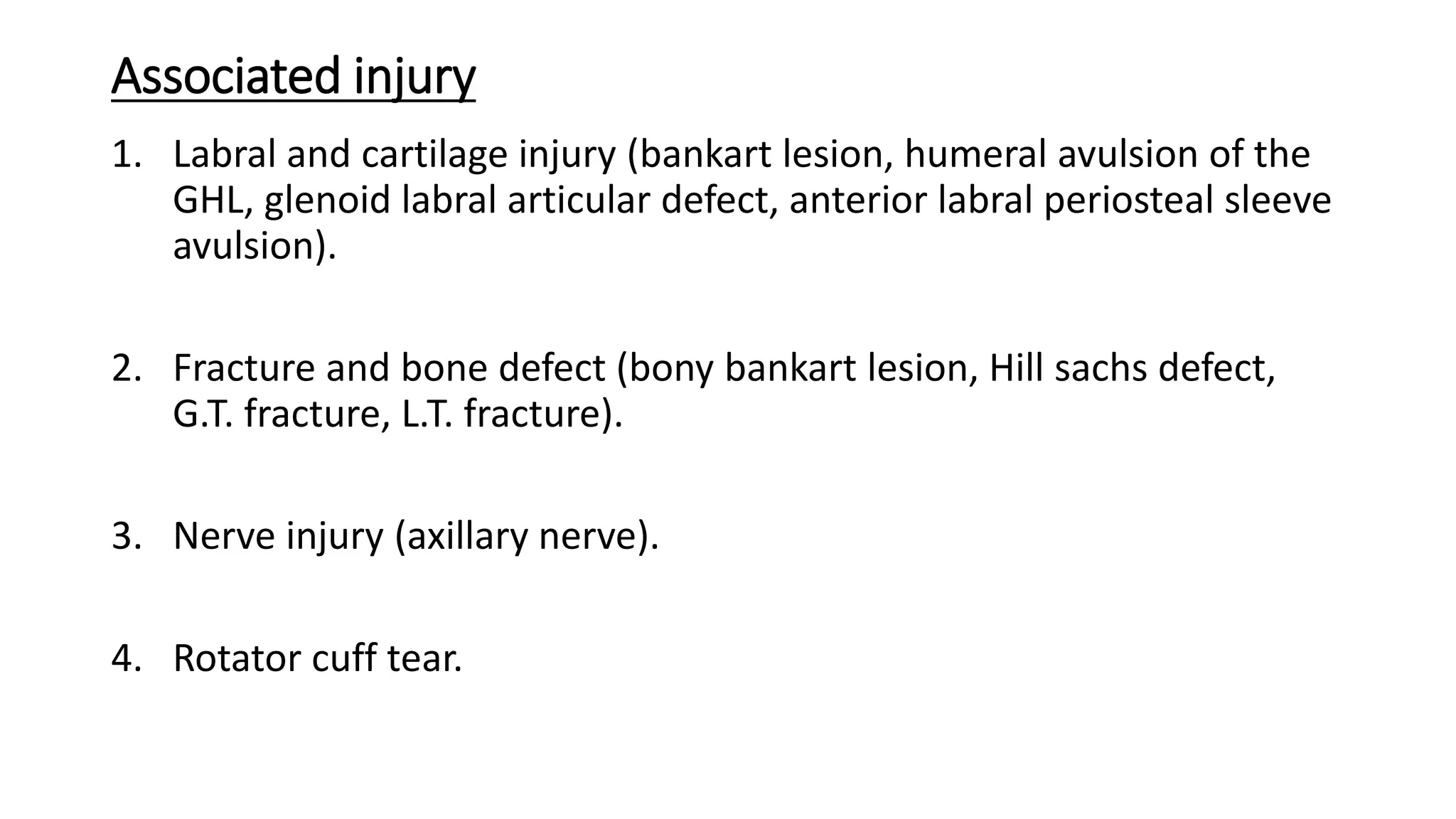
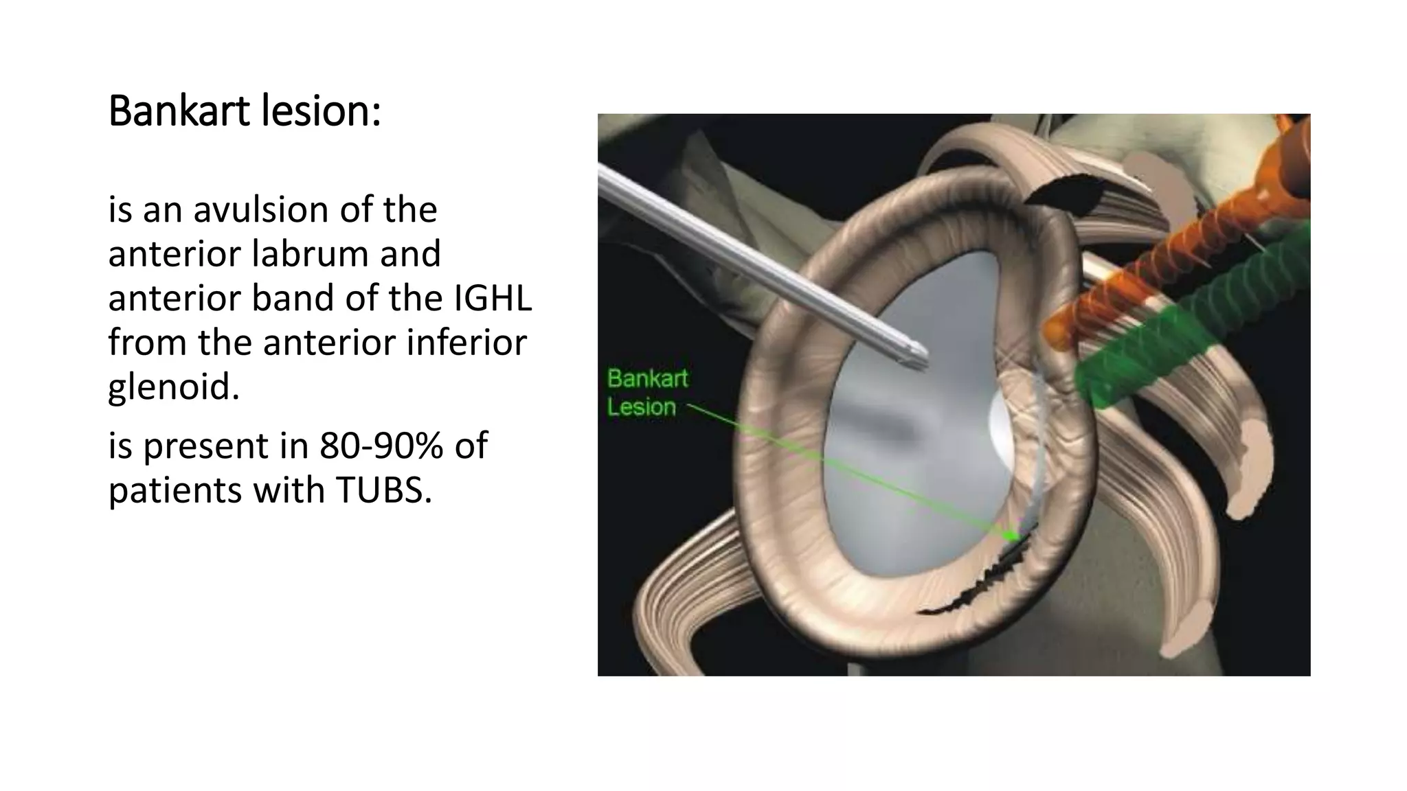
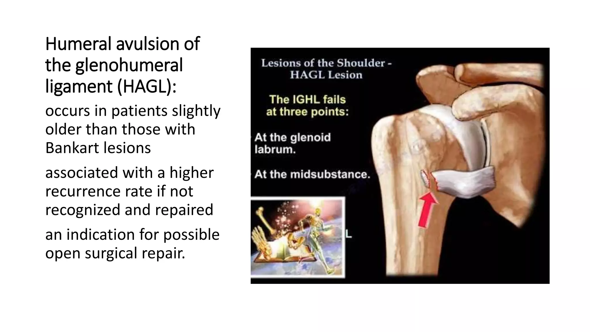
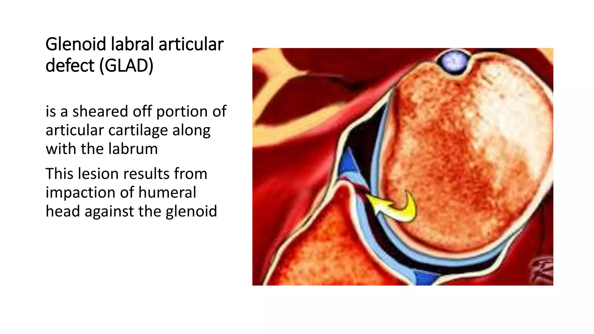
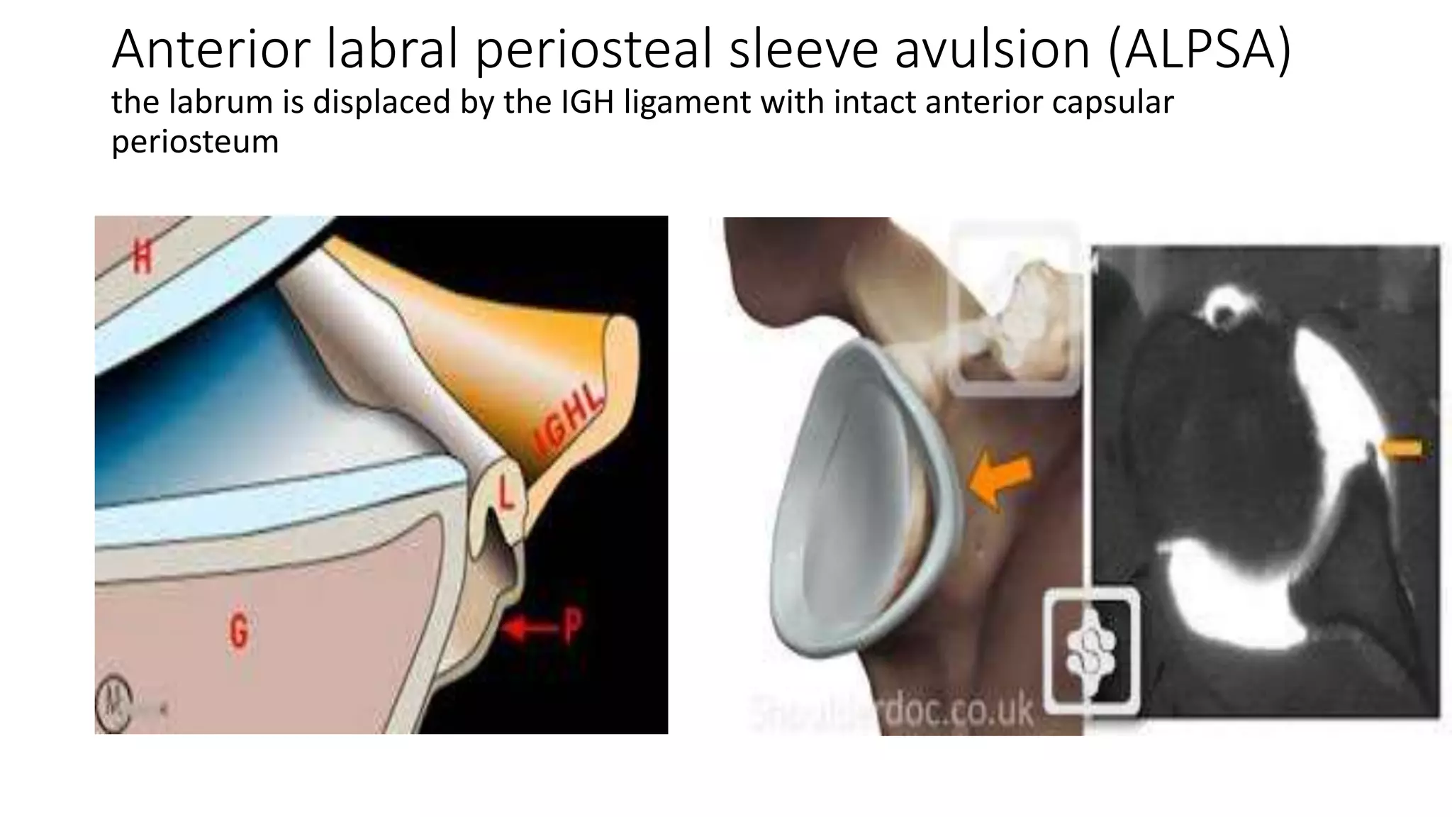
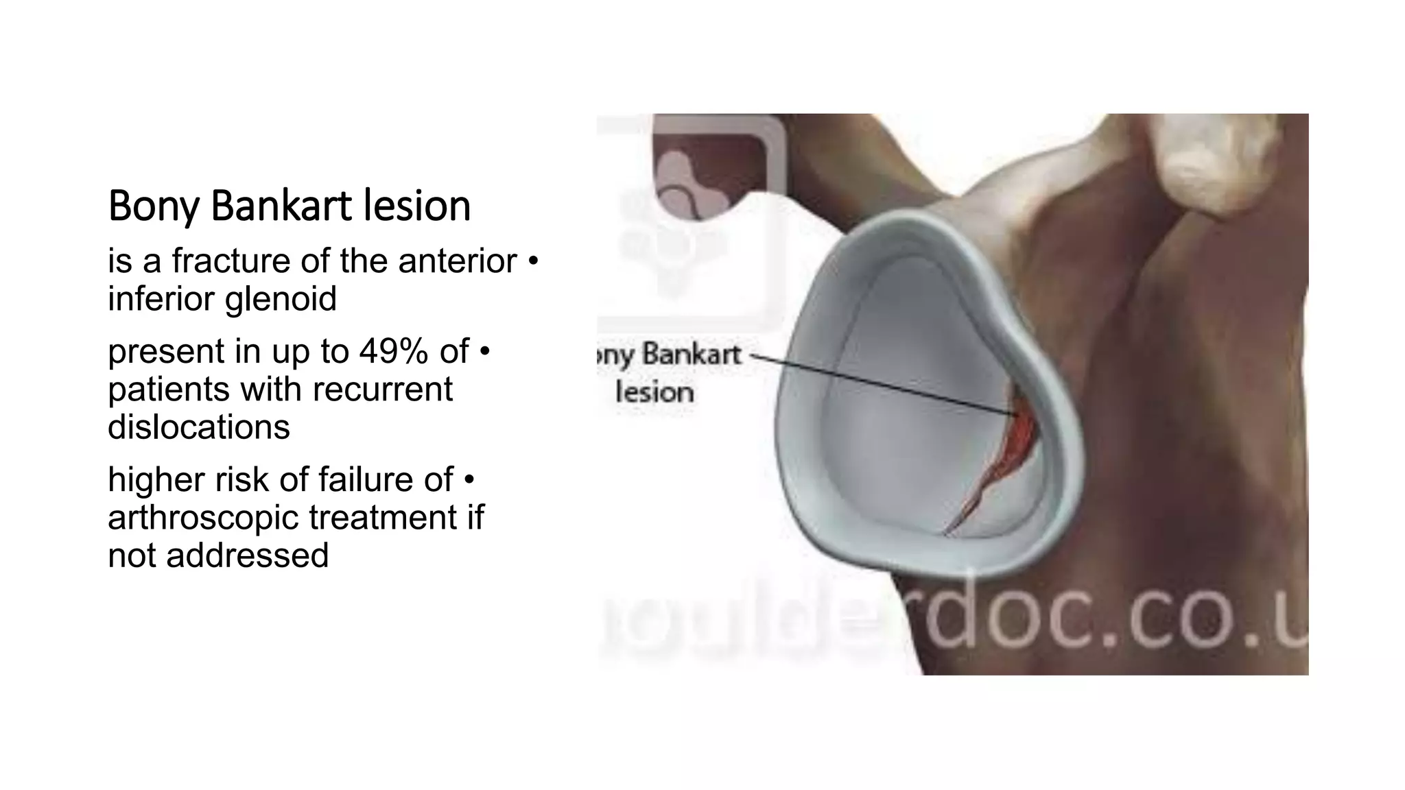
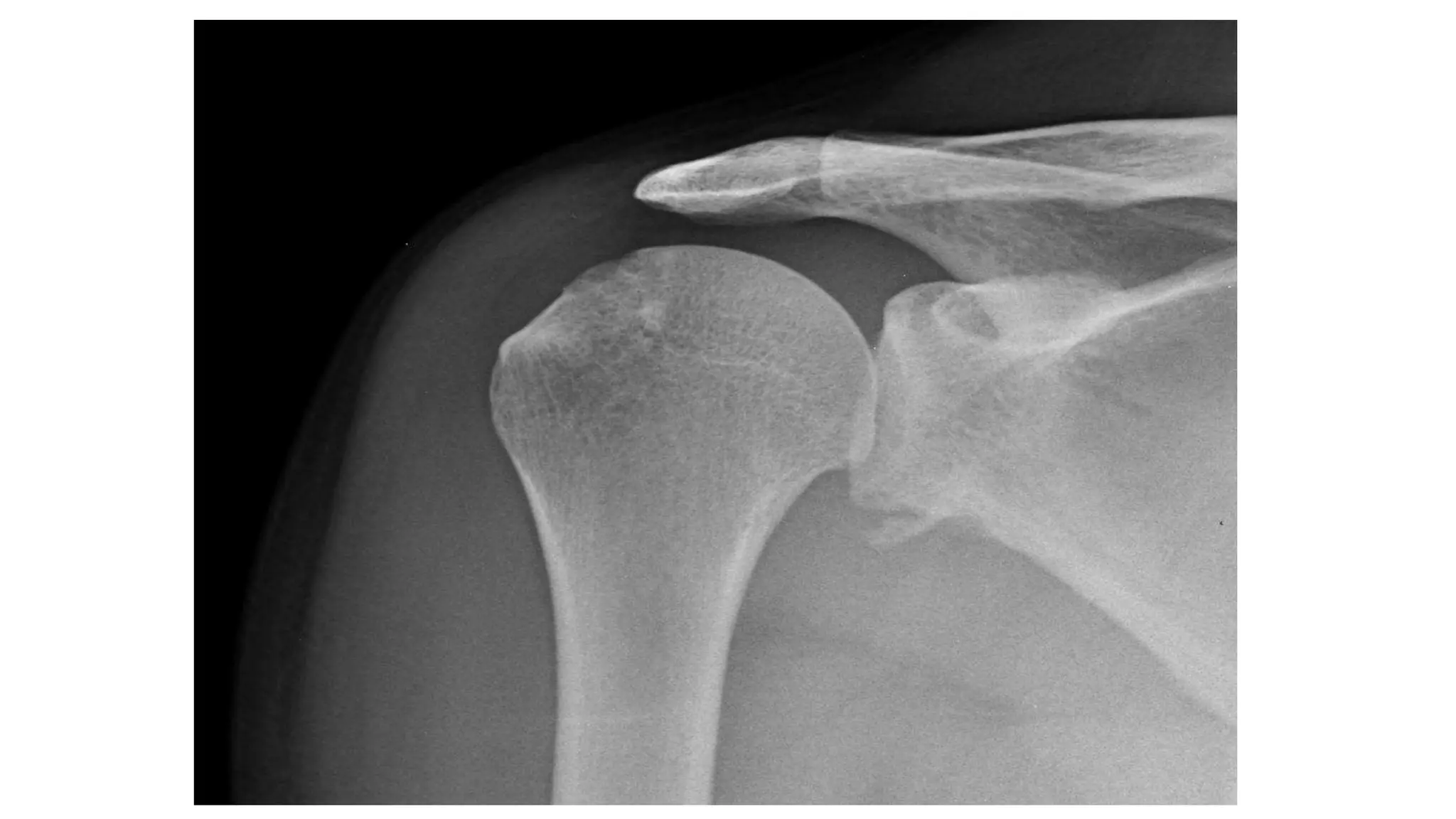
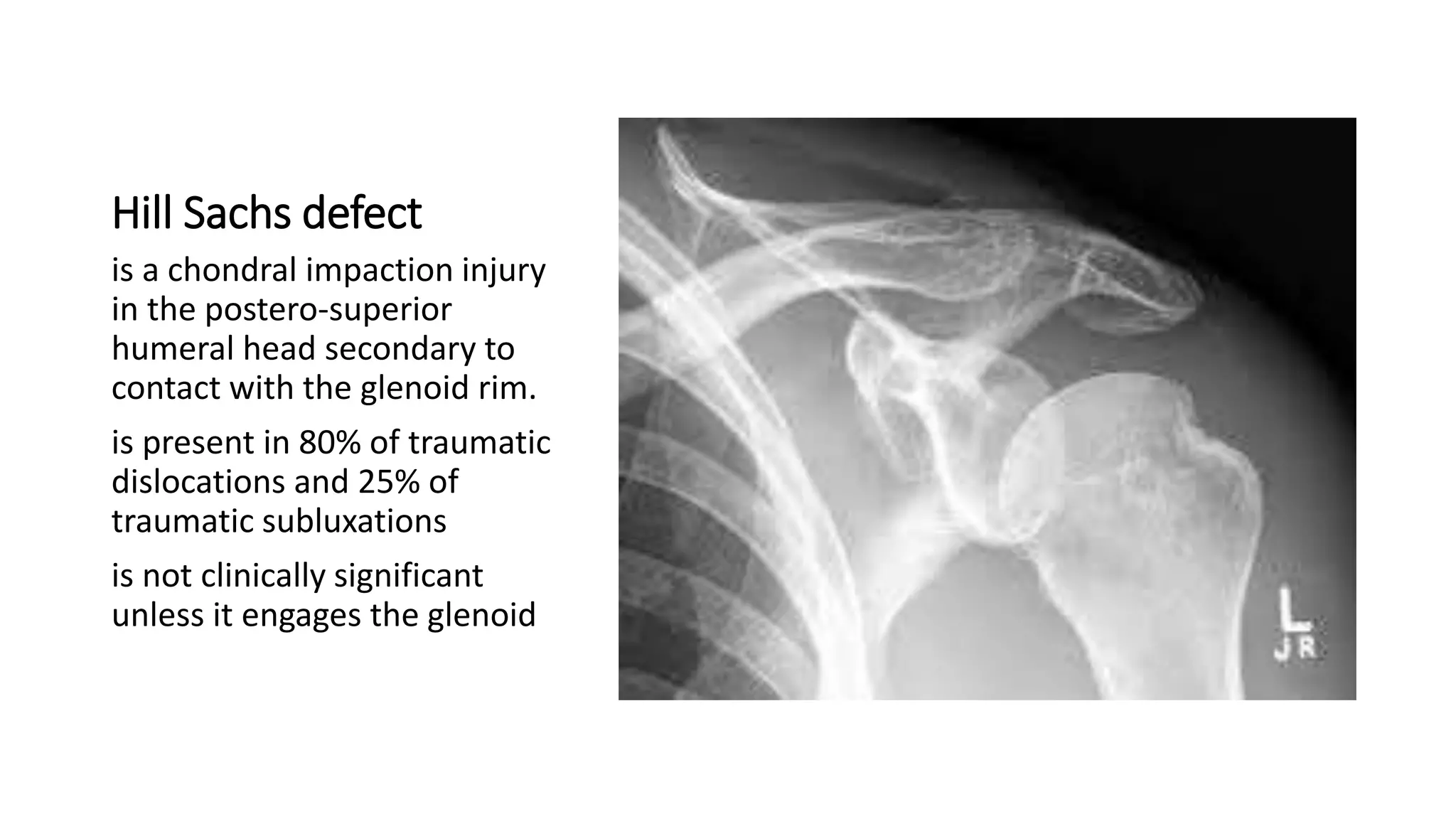
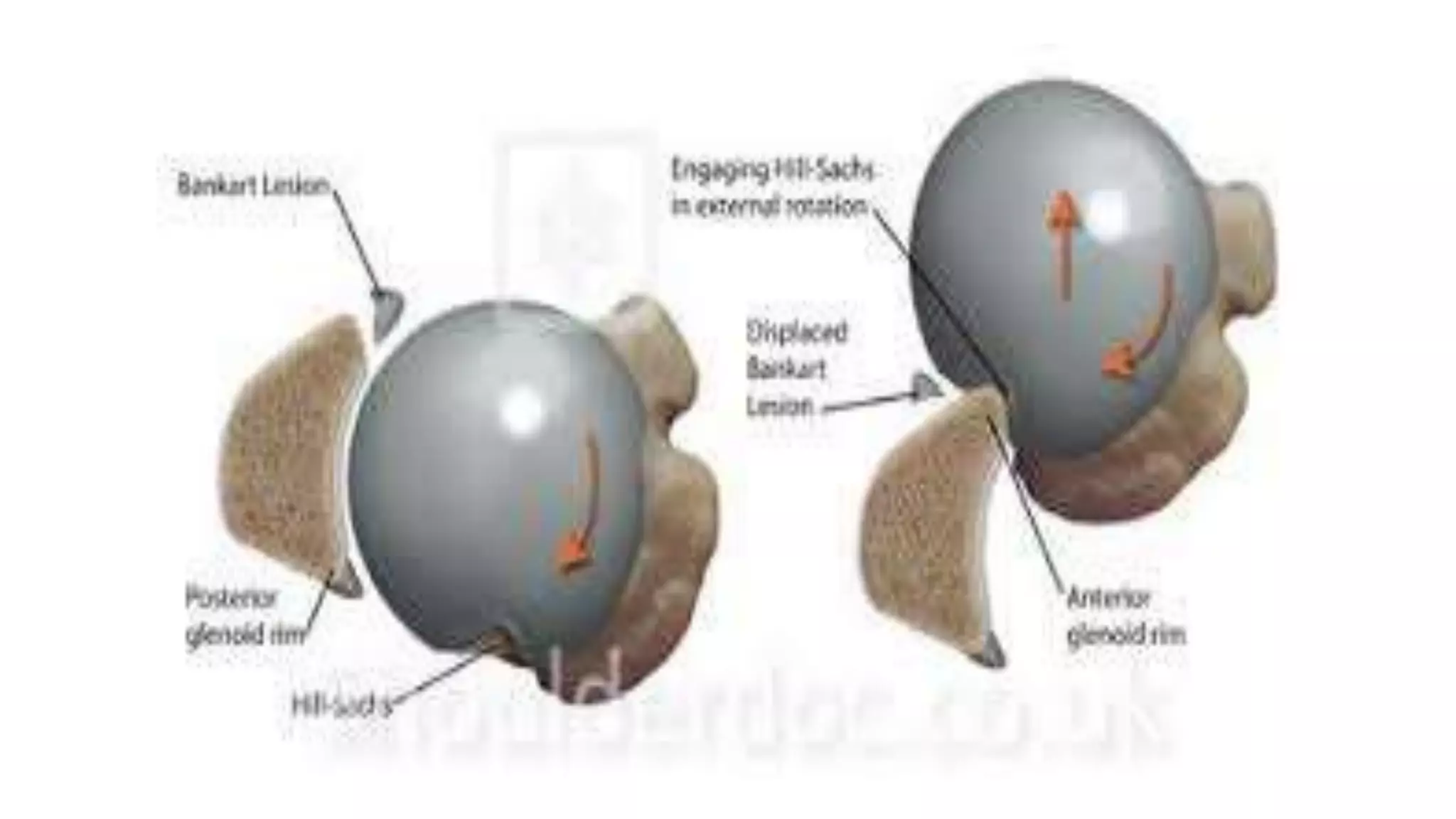
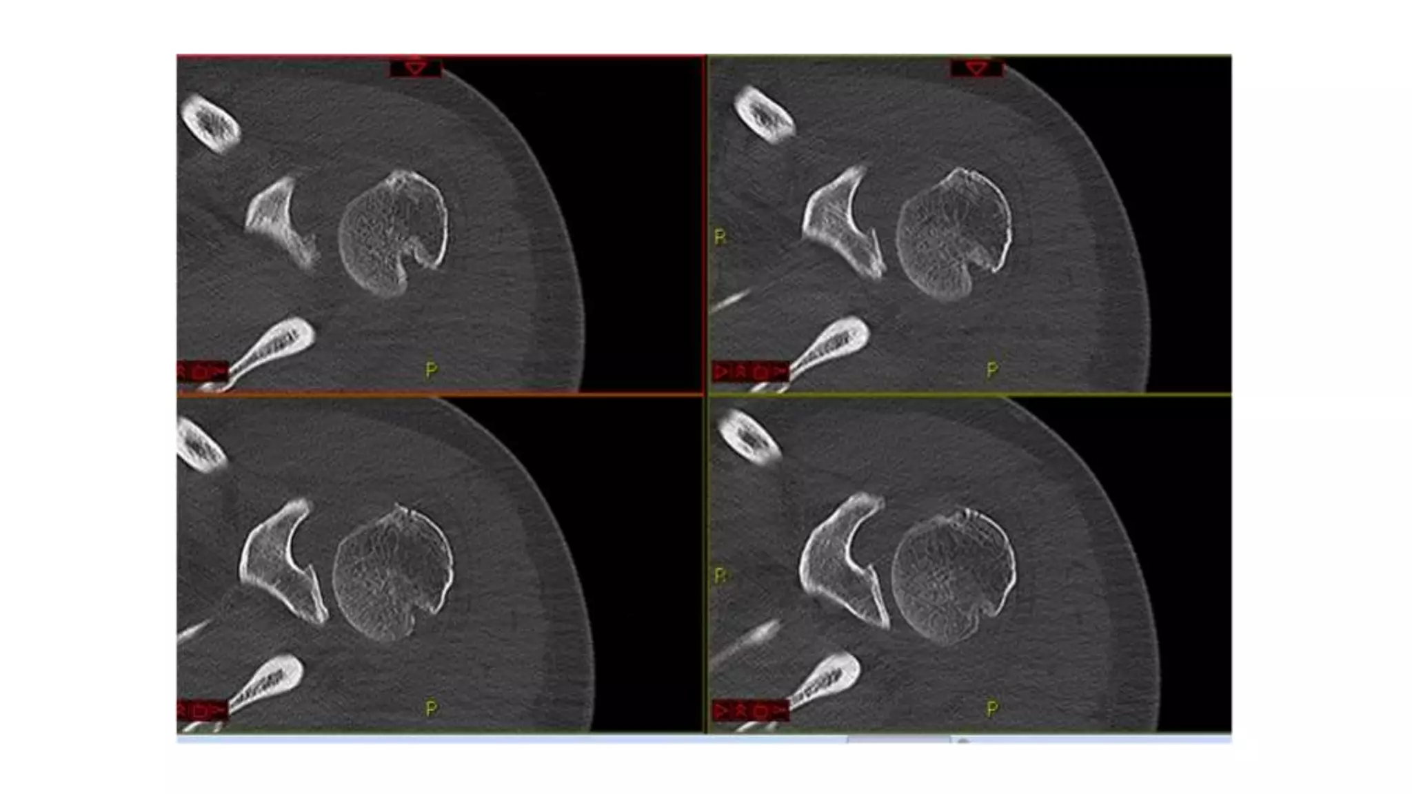
- Associated injuries may include bone fractures like bony Bankart lesions or Hill-Sachs defects. Labral injuries include Bankart lesions in 80-90% of cases.
- Treatment depends on any associated injuries and classification. Most cases are treated non-operatively with sling immobilization followed by physical therapy. Surgery is required for repair of labral tears or bone defects.
Presentation of common upper limb fractures and dislocations. Covering all the injuries from many sides (Definition - Classification - Mechanisms of injury - Clinical features - Radiological studies - Management - Complications)
paediatric injuries around the elbow
supracondylar elbow injuries
pulled elbow in paediatric age r
radiological signs around elbow in supracondylar fracture humerus
The upper limb consists of various joints that enable movement and provide flexibility. These joints can be classified into different types based on their structure and function.
Understanding the anatomy and function of these joints is crucial for assessing and managing conditions related to the upper limb, as well as for rehabilitation and therapeutic interventions. Joint injuries, arthritis, and other disorders may affect the functionality of these joints, and appropriate medical care may be necessary for optimal outcomes.
Seminar of U.V. Spectroscopy by SAMIR PANDASAMIR PANDA
Spectroscopy is a branch of science dealing the study of interaction of electromagnetic radiation with matter.
Ultraviolet-visible spectroscopy refers to absorption spectroscopy or reflect spectroscopy in the UV-VIS spectral region.
Ultraviolet-visible spectroscopy is an analytical method that can measure the amount of light received by the analyte.
This pdf is about the Schizophrenia.
For more details visit on YouTube; @SELF-EXPLANATORY;
https://www.youtube.com/channel/UCAiarMZDNhe1A3Rnpr_WkzA/videos
Thanks...!
(May 29th, 2024) Advancements in Intravital Microscopy- Insights for Preclini...Scintica Instrumentation
Intravital microscopy (IVM) is a powerful tool utilized to study cellular behavior over time and space in vivo. Much of our understanding of cell biology has been accomplished using various in vitro and ex vivo methods; however, these studies do not necessarily reflect the natural dynamics of biological processes. Unlike traditional cell culture or fixed tissue imaging, IVM allows for the ultra-fast high-resolution imaging of cellular processes over time and space and were studied in its natural environment. Real-time visualization of biological processes in the context of an intact organism helps maintain physiological relevance and provide insights into the progression of disease, response to treatments or developmental processes.
In this webinar we give an overview of advanced applications of the IVM system in preclinical research. IVIM technology is a provider of all-in-one intravital microscopy systems and solutions optimized for in vivo imaging of live animal models at sub-micron resolution. The system’s unique features and user-friendly software enables researchers to probe fast dynamic biological processes such as immune cell tracking, cell-cell interaction as well as vascularization and tumor metastasis with exceptional detail. This webinar will also give an overview of IVM being utilized in drug development, offering a view into the intricate interaction between drugs/nanoparticles and tissues in vivo and allows for the evaluation of therapeutic intervention in a variety of tissues and organs. This interdisciplinary collaboration continues to drive the advancements of novel therapeutic strategies.
Multi-source connectivity as the driver of solar wind variability in the heli...Sérgio Sacani
The ambient solar wind that flls the heliosphere originates from multiple
sources in the solar corona and is highly structured. It is often described
as high-speed, relatively homogeneous, plasma streams from coronal
holes and slow-speed, highly variable, streams whose source regions are
under debate. A key goal of ESA/NASA’s Solar Orbiter mission is to identify
solar wind sources and understand what drives the complexity seen in the
heliosphere. By combining magnetic feld modelling and spectroscopic
techniques with high-resolution observations and measurements, we show
that the solar wind variability detected in situ by Solar Orbiter in March
2022 is driven by spatio-temporal changes in the magnetic connectivity to
multiple sources in the solar atmosphere. The magnetic feld footpoints
connected to the spacecraft moved from the boundaries of a coronal hole
to one active region (12961) and then across to another region (12957). This
is refected in the in situ measurements, which show the transition from fast
to highly Alfvénic then to slow solar wind that is disrupted by the arrival of
a coronal mass ejection. Our results describe solar wind variability at 0.5 au
but are applicable to near-Earth observatories.
Slide 1: Title Slide
Extrachromosomal Inheritance
Slide 2: Introduction to Extrachromosomal Inheritance
Definition: Extrachromosomal inheritance refers to the transmission of genetic material that is not found within the nucleus.
Key Components: Involves genes located in mitochondria, chloroplasts, and plasmids.
Slide 3: Mitochondrial Inheritance
Mitochondria: Organelles responsible for energy production.
Mitochondrial DNA (mtDNA): Circular DNA molecule found in mitochondria.
Inheritance Pattern: Maternally inherited, meaning it is passed from mothers to all their offspring.
Diseases: Examples include Leber’s hereditary optic neuropathy (LHON) and mitochondrial myopathy.
Slide 4: Chloroplast Inheritance
Chloroplasts: Organelles responsible for photosynthesis in plants.
Chloroplast DNA (cpDNA): Circular DNA molecule found in chloroplasts.
Inheritance Pattern: Often maternally inherited in most plants, but can vary in some species.
Examples: Variegation in plants, where leaf color patterns are determined by chloroplast DNA.
Slide 5: Plasmid Inheritance
Plasmids: Small, circular DNA molecules found in bacteria and some eukaryotes.
Features: Can carry antibiotic resistance genes and can be transferred between cells through processes like conjugation.
Significance: Important in biotechnology for gene cloning and genetic engineering.
Slide 6: Mechanisms of Extrachromosomal Inheritance
Non-Mendelian Patterns: Do not follow Mendel’s laws of inheritance.
Cytoplasmic Segregation: During cell division, organelles like mitochondria and chloroplasts are randomly distributed to daughter cells.
Heteroplasmy: Presence of more than one type of organellar genome within a cell, leading to variation in expression.
Slide 7: Examples of Extrachromosomal Inheritance
Four O’clock Plant (Mirabilis jalapa): Shows variegated leaves due to different cpDNA in leaf cells.
Petite Mutants in Yeast: Result from mutations in mitochondrial DNA affecting respiration.
Slide 8: Importance of Extrachromosomal Inheritance
Evolution: Provides insight into the evolution of eukaryotic cells.
Medicine: Understanding mitochondrial inheritance helps in diagnosing and treating mitochondrial diseases.
Agriculture: Chloroplast inheritance can be used in plant breeding and genetic modification.
Slide 9: Recent Research and Advances
Gene Editing: Techniques like CRISPR-Cas9 are being used to edit mitochondrial and chloroplast DNA.
Therapies: Development of mitochondrial replacement therapy (MRT) for preventing mitochondrial diseases.
Slide 10: Conclusion
Summary: Extrachromosomal inheritance involves the transmission of genetic material outside the nucleus and plays a crucial role in genetics, medicine, and biotechnology.
Future Directions: Continued research and technological advancements hold promise for new treatments and applications.
Slide 11: Questions and Discussion
Invite Audience: Open the floor for any questions or further discussion on the topic.
Nutraceutical market, scope and growth: Herbal drug technologyLokesh Patil
As consumer awareness of health and wellness rises, the nutraceutical market—which includes goods like functional meals, drinks, and dietary supplements that provide health advantages beyond basic nutrition—is growing significantly. As healthcare expenses rise, the population ages, and people want natural and preventative health solutions more and more, this industry is increasing quickly. Further driving market expansion are product formulation innovations and the use of cutting-edge technology for customized nutrition. With its worldwide reach, the nutraceutical industry is expected to keep growing and provide significant chances for research and investment in a number of categories, including vitamins, minerals, probiotics, and herbal supplements.
The increased availability of biomedical data, particularly in the public domain, offers the opportunity to better understand human health and to develop effective therapeutics for a wide range of unmet medical needs. However, data scientists remain stymied by the fact that data remain hard to find and to productively reuse because data and their metadata i) are wholly inaccessible, ii) are in non-standard or incompatible representations, iii) do not conform to community standards, and iv) have unclear or highly restricted terms and conditions that preclude legitimate reuse. These limitations require a rethink on data can be made machine and AI-ready - the key motivation behind the FAIR Guiding Principles. Concurrently, while recent efforts have explored the use of deep learning to fuse disparate data into predictive models for a wide range of biomedical applications, these models often fail even when the correct answer is already known, and fail to explain individual predictions in terms that data scientists can appreciate. These limitations suggest that new methods to produce practical artificial intelligence are still needed.
In this talk, I will discuss our work in (1) building an integrative knowledge infrastructure to prepare FAIR and "AI-ready" data and services along with (2) neurosymbolic AI methods to improve the quality of predictions and to generate plausible explanations. Attention is given to standards, platforms, and methods to wrangle knowledge into simple, but effective semantic and latent representations, and to make these available into standards-compliant and discoverable interfaces that can be used in model building, validation, and explanation. Our work, and those of others in the field, creates a baseline for building trustworthy and easy to deploy AI models in biomedicine.
Bio
Dr. Michel Dumontier is the Distinguished Professor of Data Science at Maastricht University, founder and executive director of the Institute of Data Science, and co-founder of the FAIR (Findable, Accessible, Interoperable and Reusable) data principles. His research explores socio-technological approaches for responsible discovery science, which includes collaborative multi-modal knowledge graphs, privacy-preserving distributed data mining, and AI methods for drug discovery and personalized medicine. His work is supported through the Dutch National Research Agenda, the Netherlands Organisation for Scientific Research, Horizon Europe, the European Open Science Cloud, the US National Institutes of Health, and a Marie-Curie Innovative Training Network. He is the editor-in-chief for the journal Data Science and is internationally recognized for his contributions in bioinformatics, biomedical informatics, and semantic technologies including ontologies and linked data.
THE IMPORTANCE OF MARTIAN ATMOSPHERE SAMPLE RETURN.Sérgio Sacani
The return of a sample of near-surface atmosphere from Mars would facilitate answers to several first-order science questions surrounding the formation and evolution of the planet. One of the important aspects of terrestrial planet formation in general is the role that primary atmospheres played in influencing the chemistry and structure of the planets and their antecedents. Studies of the martian atmosphere can be used to investigate the role of a primary atmosphere in its history. Atmosphere samples would also inform our understanding of the near-surface chemistry of the planet, and ultimately the prospects for life. High-precision isotopic analyses of constituent gases are needed to address these questions, requiring that the analyses are made on returned samples rather than in situ.
3. Anatomy :-
The clavicle is an irregular S-shape that holds the scapula and shoulder joint in a
functional position away from the chest wall.
Middle 1/3 is narrowest (most common location of fracture).
Subcutanous bone.
First bone to ossify and last to fuse.
It articulates with the manubrium of the sternum medially through SC joint, and to
the acromion of the scapula laterally through AC joint.
It is also stabilizied by the coracoclavicular CC ligaments (conoid and trapezoid)
4.
5. Mechanism of injury
Fall onto the shoulder
(87%)
direct blow to lateral aspect
of shoulder 6%
fall on an outstretched arm
or direct trauma
7. Group I (Middle third) 80%
Non-Displaced :- Less than 100%
displacement
Non-operative
displaced :-Greater than 100%
displacement
Nonunion rate of 4.5%
operative
8. Group II - Neer Classification of Lateral third
(10-15%)
9. Group II - Neer
Classification of Lateral
third (10-15%)
Type 1 minimal
displacement
(interligamentus)
Stable because conoid and
trapezoid ligaments remain
intact
Nonoperative
10. Group II - Neer
Classification of Lateral
third (10-15%)
Type II displaced 2ndry to a fracture line
medial to the CC ligament
Type II A :-
conoid and trapezoid attached (fracture
medial to CC ligaments)
Medial clavicle unstable
Up to 56% nonunion rate with nonoperative
management
Operative
11. Group II - Neer
Classification of Lateral
third (10-15%)
Type II B :-
conoid torn, trapezoid attached (fracture
between the CC ligaments)
Medial clavicle unstable
Up to 30-45% nonunion rate with
nonoperative management
Operative
12. Group II - Neer
Classification of Lateral
third (10-15%)
Type III
fracture of the articular surface
Conoid and trapezoid intact therefore stable
injury
Non-operative
13. Group II - Neer
Classification of Lateral
third (10-15%)
Type IV
periosteal sleeve fracture (children)
Conoid and trapezoid ligaments remain
attached to periosteum and overall the
fracture pattern is stable
Non-operative
14. Group II - Neer
Classification of Lateral
third (10-15%)
Type V
Comminuted fracture
Conoid and trapezoid ligaments remain
attached to comminuted fragment
Medial clavicle unstable
Operative
15. Group III - Medial
third (5-8%)
Anterior displacement :-
Most often non-operative
Rarely symptomatic
Posterior displacement :-
Rare injury (2-3%)
Often physeal fracture-dislocation (age <
25)
Stability dependent on costoclavicular
ligaments
Must assess airway and great vessel
compromise
Serendipity radiographs and CT scan to
evaluate
Surgical management with thoracic
surgeon on standby
22. Management
Non-operative :-
• no attempt at reduction should be made.
• sling immobilization with gentle ROM exercises at 2-4 weeks and
strengthening at 6-10 weeks.
Indication :-
1) nondisplaced Group I (middle third).
2) stable Group II fractures (Type I, III, IV).
3) nondisplaced Group III (medial third).
4) pediatric distal clavicle fractures (skeletally immature).
23. Operative :-
• ORIF
• CC ligaments repair vs reconstruction indicated in type IIb fracture.
Indication :-
Absolute :-
1) unstable Group II fractures (Type IIA, Type IIB, Type V).
2) open fxs.
3) displaced fracture with skin tenting.
4) subclavian artery or vein injury.
5) floating shoulder (clavicle and scapula neck fx).
6) symptomatic nonunion.
7) posteriorly displaced Group III fxs.
8) displaced Group I (middle third) with >2cm shortening.
24. relative and controversial indications :-
1) brachial plexus injury (questionable b/c 66% have spontaneous
return).
2) closed head injury.
3) seizure disorder.
4) polytrauma patient.
28. Anatomy :-
• AC joint is a synovial joint with a fibrocartilaginous disk.
• It has thin capsule that is stabilized by sup. Inf. Ant. and post.
Ligaments.
• The most robust ligaments is superior AC ligament which is
responsible for horizontal AC joint stability.
• Vertical stability is provided by the CC ligaments.
• Normal AC joint are ,5 to ,6 mm in width.
• Normal CC distance is 1,1 cm to 1,3 cm.
29.
30. Mechanism of injury
• Fall on shoulder or
direct blow to the
acromion with arm
adducted. (most
common)
• Fall on outstretched
arm transmitted to AC
joint.
• Rugby and hockey
players frequently
sustained this injury.
34. Imaging
• AP view
• 15 cephalic tilt (zanca view) to evaluate joint displacement and intra-
articular fracture
• Axillary view is mandatory to determine AP displacement
37. Management
Based on type :-
Type 1 and 2 :- non-operative (sling, analgesia and early range of
motion). Return to sports when pain free
Type 3 :- controversy.
Type 4, 5, 6 :- surgical repair with reconstruction of the CC ligaments.
39. Anatomy :-
• Shoulder girdle consist of clavicle, scapula, and proximal humerus,
and their associated ligaments and muscles
• Scapula has consisting of :-
1. Body: triangular, flattened region that articulate with the thorax.
The anterior surface is covered by the subscapularis muscle (one of
the rotator cuff muscle)
2. Spine: a projection that runs horizontally from across the posterior
aspect of the body, forming 2 fossae, which are the origins of the
supra and infra-spinatus and teres minor muscles. It terminates in
the acromion
3. Acromion: lateral end of spine that extend to the heighest and most
lateral point of shoulder.
40. 4. Coracoid: an anterior protrusion that is the origin of the short head
of biceps and brachialis muscles, and the insertion of the CC ligaments.
5. Glenoid: a shallow, saucer-shaped projection from the lateral aspect
of the scapula, deepened by a fibrocartilaginous labrum, which
articulates with the humeral head
• The glenoid is oriented anteriorly by around 30 degree.
41.
42. Shoulder stability
Static stability
1. Bone shape: the glenoid
provides a shallow concave
surface to the humeral head.
2. Labrum: this fibrocartilaginous
rim markedly increases the
glenoid depth.
3. Capsular ligaments: consist of
superior, middle, inferior (ant.
&post. Band) glenohumeral
ligaments
4. Joint capsule.
Dynamic stability
1. Rotator cuff muscles: form a
cuff of muscle that encloses
the glenohumeral joint.
Contraction of these m. result
in concavity compression
which maintains shoulder
congruence and stability
during movement.
2. Long head of biceps: its
position above and anterior to
the humeral head acts to resist
anterior dislocation.
43.
44.
45. GLENO-HUMERAL LIGAMNEBTS
Anterior band of IGHL (main restraint)
•provides static restraint with arm in 90° of abduction and external rotation
MGHL
•provides static restraint with arm in 45° of abduction and external rotation
SGHL
provides static restraint with arm at the side
49. Bankart lesion:
is an avulsion of the
anterior labrum and
anterior band of the IGHL
from the anterior inferior
glenoid.
is present in 80-90% of
patients with TUBS.
50. Humeral avulsion of
the glenohumeral
ligament (HAGL):
occurs in patients slightly
older than those with
Bankart lesions
associated with a higher
recurrence rate if not
recognized and repaired
an indication for possible
open surgical repair.
51. Glenoid labral articular
defect (GLAD)
is a sheared off portion of
articular cartilage along
with the labrum
This lesion results from
impaction of humeral
head against the glenoid
52. Anterior labral periosteal sleeve avulsion (ALPSA)
the labrum is displaced by the IGH ligament with intact anterior capsular
periosteum
54. Bony Bankart lesion
•is a fracture of the anterior
inferior glenoid
•present in up to 49% of
patients with recurrent
dislocations
•higher risk of failure of
arthroscopic treatment if
not addressed
55.
56. Hill Sachs defect
is a chondral impaction injury
in the postero-superior
humeral head secondary to
contact with the glenoid rim.
is present in 80% of traumatic
dislocations and 25% of
traumatic subluxations
is not clinically significant
unless it engages the glenoid
57.
58.
59. Symptoms and signs
• Shoulder pain
• Feeling instability
• The shoulder has a typical squared off appearance, with prominence
of the acromion without the usual contour of humeral head
• Careful neurovascular assessment. Axillary nerve palsy results in
numbness in the regimental badge area
60.
61. Imaging
Radiographs
• true AP
• scapular Y
• axillary
other helpful views
• West Point view :- shows glenoid bone loss
• Stryker view :- shows Hill-Sachs lesion
• CT scan
• helpful for evaluation of bony injuries
• MRI
• best for visualization of labral tear
• addition of intraarticular contrast
• increases sensitivity and specificity
67. Close reduction
Close reduction doesn’t require the application of huge force.
Excessive force can lead to fracture of the humerus or brachial plexus injury.
Indication :-
• Close reduction in ER is indicated with no humeral neck fracture, with or
without an isolated tuberosity fracture.
Methods :-
1. Kocher’s technique.
2. Hippocratic method
3. Milch’s technique.
4. Stimpson’s technique.
5. simple traction-countertraction.
None of the above methods has shown convincing superiorly over anthor.
70. Hippocratic method
Patient on supine position
Foot is placed in the patient’s
axilla, resting between the
chest and the humeral head.
The wrist is grasped in both
hands with gentle traction
and external rotation applied
to the arm
Traction is maintained for
several minutes to allow
muscle relaxation
72. Milch technique
Patient on supine position.
The surgeon places his hand over
dislocated humeral head at the
axilla.
The patient’s wrist is grasped firmly
with the other hand or by an
assistant and the arm slowly and
gradually abducted fully
Once it is above the patient’s head,
gentle external rotation and
traction are applied to the wrist,
whilst lateral pressure is
simultaneously applied to the head
of the humerus.
73. Operative
Indication
1. Irreducible dislocation or fracture dislocation
2. Displaced G.T. Fracture: most fractures of the GT will reduce when
humeral head is relocated, if the fracture remains displaced the
supraspinatus will remain de-functioning and ORIF should be
considered
3. Acute rotator cuff tear: An acute rotator cuff tear in young pt. is an
indication for acute repair.
4. Prophylactic stabilization: high chance of re-dislocation, athletic
patient, recurrent dislocation.
5. Late presentation: after around 3 weeks close reduction may prove
impossible and open reduction with stabilization are indicated.
76. • Posterior shoulder dislocations are less common than anterior
dislocations, but more commonly missed.
• 50% of traumatic posterior dislocations seen in the emergency
department are undiagnosed.
• Common cause :- seizures and electrical shock (tetanic muscle
contraction pulls the humeral head out)
• mechanism :-
flexed, adducted, and internally rotated arm is a high-risk position.
77.
78. Presentation
History
trauma with the arm in a flexed, adducted, and internally rotated position.
Symptoms
pain with flexion, adduction, and internal rotation of the arm.
Physical exam
• Inspection:-
prominent posterior shoulder and coracoid.
• Motion:-
limited external rotation.
shoulder locked in an internally rotated position common in undiagnosed
posterior dislocations.
pain on flexion, adduction and internal rotation for posterior instability.
79. Imaging
Radiography
• AP :- unreliable and cannot be exclude posterior dislocation.
may show lightbulb sign.
• Axillary lateral :- best view to demonstrate a dislocation.
• Velpeau view :- if patient is unable to abduct arm for axillary view.
CT
analyze the extent and location of bone loss in a chronic dislocation.
MRI
• evaluate for suspected associated rotator cuff tear.
83. Close reduction
• Indication for all acute posterior shoulder dislocation.
• Most dislocations reduced spontaneously.
• Technique :-
1. the Patient is supine with the arm held by the side
2. Traction is applied to the arm with some internal rotation
3. The shoulder is flexed and adducted while further internal rotation
is add
4. Direct pressure to the humeral head from behind can facilitate
reduction
5. Once it is unlocked the humerus is externally rotated gently, don’t
attempt a forceful external rotation if the shoulder remains locked.
85. Incidence :- very rare, only 0.5% of all shoulder dislocations.
mechanism of injury :-
typically a high-energy injury.
hyperabduction force applied to arm, levering the proximal humerus
onto the acromion, injuring inferior capsule/labrum, which subsequently
allows for disengagement of HH inferiorly from glenoid.
commonly involves variable sized tearing of static glenohumeral
ligaments.
Associated conditions
neurovascular injury :- has greatest incidence of neurovascular injury of
all types of shoulder dislocations.
proximal humerus fractures :- especially greater tuberosity.
rotator cuff tears.
86.
87. Symptoms and signs
Symptoms :-
1. shoulder pain
2. inability to move shoulder
3. neurovascular injury :-
• neurologic injury up to 60%
• vascular injury up to 39%
Physical exam :-
• patient presents with the arm in a fixed, abducted position