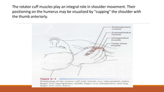
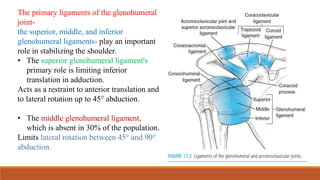
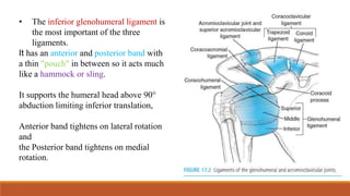
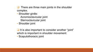
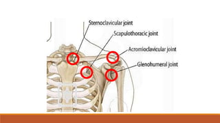
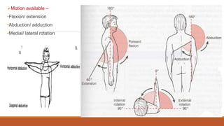
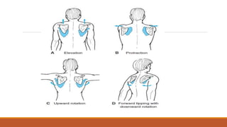
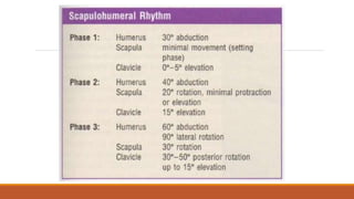
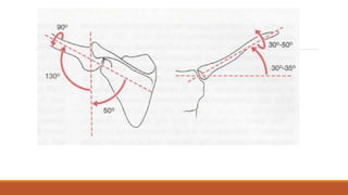
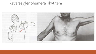
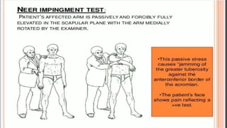
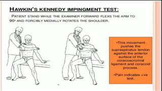
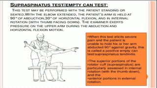
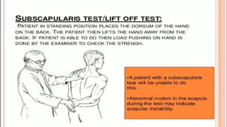
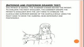
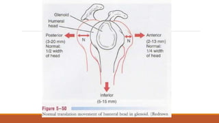
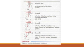
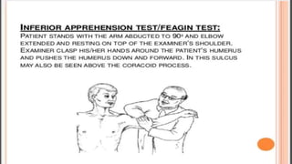
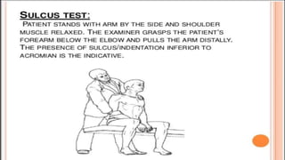
BY: SYED MASOOD.
MPT 2ND YEAR
ORTHO
Introduction to the Shoulder complex
Shoulder girdle + shoulder joint = shoulder complex
Shoulder girdle:
◦ Scapula, clavicle and articulations
Shoulder joint:
◦ Humeral head and glenoid fossa of scapula
 There are three main joints in the shoulder
complex.
• Shoulder girdle:
Acromioclavicular joint
Sternoclavicular joint
• Shoulder joint
 It is also important to consider another “joint”
which is important in shoulder movement:
• Scapulothoracic joint
Motion available –
•Flexion/ extension
•Abduction/ adduction
•Medial/ lateral rotation
Angle of inclination of
humerus –
•Frontal plane
•130-150 degree angle
created between axis of head
and shaft of humerus in
frontal plane
Angle of torsion-retroversion –
•Transverse plane
•Humeral head is rotated 30°
posteriorly
•Orients humeral head in
scapular plane for articulation
with glenoid fossa
•Increases stability
Glenohumeral joint
GH joint is a multiaxial, ball & socket, synovial joint.
Articulating surfaces: concave glenoid cavity and convex humeral head.
The glenoid in the resting position has a 5° superior tilt or inclination and a 7°
retroversion (slight medial rotation).
The structural relationship of the bony anatomy, ligaments, and glenoid labrum and
the adhesive and cohesive forces in the joint provide static stability.
Glenohumeral joit
Resting position 40-55° abduction with 30° horizontal adduction.
Close pack position Full abduction and lateral rotation
Capsular pattern Lateral rotation, abduction, medial rotation
The tendons of the rotator cuff blend
with ligaments and glenoid labrum so
when muscles contract they provide
dynamic stability by tightening the
static restraints.
The coordinated response of the
muscles of the cuff and tension in the
ligaments provide varying degrees of
support.
The rotator cuff muscles play an integral role in shoulder movement. Their
positioning on the humerus may be visualized by "cupping" the shoulder with
the thumb anteriorly.
The primary ligaments of the glenohumeral
joint-
the superior, middle, and inferior
glenohumeral ligaments- play an important
role in stabilizing the shoulder.
• The superior glenohumeral ligament's
primary role is limiting inferior
translation in adduction.
Acts as a restraint to anterior translation and
to lateral rotation up to 45° abduction.
• The middle glenohumeral ligament,
which is absent in 30% of the population.
Limits lateral rotation between 45° and 90°
abduction.
• The inferior glenohumeral ligament is
the most important of the three
ligaments.
It has an anterior and posterior band with
a thin "pouch" in between so it acts much
like a hammock or sling.
It supports the humeral head above 90°
abduction limiting inferior translation,
Anterior band tightens on lateral rotation
and
the Posterior band tightens on medial
rotation.
• The coracohumeral ligament primarily limits
inferior translation and helps limit lateral
rotation below 60° abduction.
This ligament is found in the rotator interval
thus the ligament "unites" the two tendons
anteriorly.
• The coracoacromial ligament forms an arch
over the humeral head acting as a block to
superior translation.
• The transverse humeral ligament forms a roof
over the bicipital groove to hold the long head
of biceps tendon within the groove.
Osteo and Arthrokinematics:-
Three degree of freedom.
•Flexion- posterior spining
•Extension- anterior spining
•Abduction- glide downwards and rolling upwards
•Adduction- glide upwards and rolling downwards
•Lateral rotation- posterior rolling and anterior glide
•Medial rotation- anterior rolling and posterior glide.
Acromioclavicular joint
Plane synovial joint. Augments the range of motion (ROM) of the humerus in the glenoid. Three
degree of freedom. Disc separates the joint surfaces. Fibrous capsule around the joint.
AcromioClavicular Joint
Resting position Arm by side
Close pack
position
90° abduction
Capsular
pattern
Pain at extremes of ROM, especially
horizontal adduction n full elevation
This joint depends on ligaments for its
strength.
The acromioclavicular ligaments
surround the joint and are commonly
the first ligaments injured when the
joint is stressed. To restrain posterior
translation and posterior axial rotation
at the AC joint.
The coracoclavicular ligament
(trapezoid, conoid) is the primary
support of the acromioclavicular joint.
If a step deformity occurs, this ligament
has been torn.
Sternoclavicular joint
Articulating surfaces : concave surface of medial clavicle and convex sternum.
Enables the humerus in the glenoid to move through a full 180° of abduction. It is
a saddle shaped synovial joint “with three degrees of freedom”.
There is a substantial disc between the two bony joint surfaces,
As concave medial clavicle is moving over convex sternum, according to concave-
convex rule, when concave surface is moving over convex surface then rolling
and glide will occur in same direction.
SternoClavicular Joint
Resting position Arm at side
Close pack position Full elevation and protraction
Capsular Pattern Pain at extremes of ROM, especially horizontal
adduction n full elevation
Protraction- anterior glide of clavicle and
anterior rotation
Retraction- posterior glide of clavicle and
posterior rotation
Elevation-upward rotation and inferior
glide
Depression- downward rotation and
superior glide
Rotation
The ligaments of the sternoclavicular joint include
1. the anterior and posterior sternoclavicular ligaments, which support the joint
anteriorly and posteriorly,
2. the interclavicular ligament, and
3. the costoclavicular ligament running from the clavicle to the first rib and its
costal cartilage. This is the main ligament maintaining the integrity of the
sternoclavicular joint.
Scapulothoracic joint
Since SC joint is not true joint, normal concave convex rule doesnot apply.
The muscles acting on the scapula help to control its movements. The medial
border of the scapula is not parallel with the spinous processes but is angled
about 3° away (top to bottom), and the scapula lies 20° to 30° forward relative
to the sagittal plane.
ScapuloThoracic joint
Resting position Arm by side
Close pack position no
Capsular pattern Not a true joint so does not have capsular pattern
Reverse glenohumeral rhythem
Painful arc in shoulder
Assessment
Patient history
1.What is the patient's age?
Many problems of the shoulder can be age related.
For example, rotator cuff degeneration usually occurs in patients who are
between 40 and 60 years of age.
Primary impingement due to degeneration and weakness is usually seen in
patients older than 35.
Secondary impingement due to instability caused by weakness in the scapular
or humeral control muscles is more common in people in their late 20s
especially those involved in vigorous overhead activities such as swimmers or
pitchers in baseball.
Calcium deposits may occur between the ages of 20 and 40.
Chondrosarcomas may be seen in those older than 30 years of age.
Frozen shoulder is seen in persons between the ages of 45 and 60 years.
2.Does the patient support the upper limb in a protected position or hesitate to
move it?
This action could mean that one of the joints of the shoulder complex is unstable or
that there is an acute problem in the shoulder.
3.If there was an injury, what exactly was the mechanism of injury?
Did the patient fall on an outstretched hand ("FOOSH" injury), which could indicate a
fracture or dislocation of the glenohumeral joint?
The spectrum of instability varies from gross or anatomical instability-
TUBS type (Traumatic onset, Unidirectional anterior with a Bankart lesion responding
to Surgery)
AMBRI type (Atraumatic cause, Multidirectional with Bilateral shoulder findings with
Rehabilitation as appropriate treatment and, rarely, Inferior capsular shift surgery).
Does the shoulder feel unstable or feel like it is "coming out" during movement?
4.Are there any movements or positions that cause the patient pain or symptoms? If
so, which ones?
5.What is the extent and behavior of the patient's pain?
For example, deep, boring, toothache-like pain in the neck or shoulder region, or both, may indicate
thoracic outlet syndrome or acute brachial plexus neuropathy.
Strains of the rotator cuff usually cause dull, toothache-like pain that is worse at night, whereas
acute calcific tendinitis usually causes a hot, burning type of pain.
Sprain of the first or second rib from direct trauma or sudden contraction of the scalene may mimic
an acute impingement or rotator cuff injury
6.Are there any activities that cause or increase the pain?
7.What is the patient unable to do functionally?
8.How long has the problem bothered the patient?
For example, idiopathic frozen shoulder goes through three stages: the condition becomes
progressively worse, plateaus, and then progressively improves, with each stage lasting 3 to 5
months.
9.Do any positions relieve the pain?
10.Is there any indication of muscle spasm, deformity, bruising, wasting,
paresthesia, or numbness?
11.Does the patient complain of a feeling of weakness and heaviness in the limb
after activity? Does the limb tire easily?
12.Is there any indication of nerve injury?
13.Which hand is dominant?
Often the dominant shoulder is lower than the nondominant shoulder and the ROM
may not be the same for both. Usually, the dominant shoulder shows greater
muscularity and often less ROM.
OBSERVATION
ANTERIOR VIEW:
the examiner should begin by ensuring that the head and neck are in the midline of the body
and observing their relation to the shoulders.
While observing the shoulder, the examiner should look for the possibility of a step deformity.
Such a deformity may be caused by an acromioclavicular dislocation, with the distal end of the
clavicle lying superior to the acromion process. Such a deformity seen at rest indicates both the
acromioclavicular and coracoclavicular ligaments have been torn.
Occasionally, swelling may be evident anterior to the acromioclavicular joint. This is called the
Fountain sign and indicates degeneration has caused communication between the
acromioclavicular joint and swollen subacromial bursa underneath.
If sulcus deformity occurs when traction is applied it may be caused by multidirectional
instability.
In most people, the dominant side is lower than the nondominant side. This difference may be
caused by the extra use of the dominant side, which results in stretching of the ligaments, joint
capsules, and muscle , allowing the arm to "sag" slightly.
POSTERIOR VIEW:
the examiner again notes bony and soft-tissue contours and body alignment.
For example, atrophy of the upper trapezius may indicate spinal accessory nerve palsy, whereas
atrophy of supraspinatus and/or infraspinatus may indicate supraspinous nerve palsy.
The spines of the scapulae, which begin medially at the level of the third (T3) thoracic vertebra,
should be at the same angle.
Lennie test. Sobush and associates developed a method for measuring the scapular position.
If the scapula is sitting lower than normal against the chest wall, the superior medial border of the
scapula may "washboard" over the ribs, causing a snapping or clunking sound (snapping scapula)
during abduction and adduction.
The inferior angles of the scapulae should be equidistant from the spine. The examiner should note
the possible presence of classic winging of the scapula medial border moves away from the posterior
chest wall.
Dynamic winging (i.e., winging with movement) may be caused by a lesion of the
long thoracic nerve affecting serratus anterior, trapezius palsy (spinal accessory
nerve), rhomboid weakness, multidirectional instability, voluntary action, or a
painful shoulder resulting in splinting of the glenohumeral joint, which in turn
causes reverse scapulohumeral rhythm.
Static winging (i.e., winging occurring at rest) is usually caused by a structural
deformity of the scapula, clavicle, spine, or ribs.
shoulder assessment (full)

shoulder assessment (full)

  • 1.
    BY: SYED MASOOD. MPT2ND YEAR ORTHO
  • 2.
    Introduction to theShoulder complex Shoulder girdle + shoulder joint = shoulder complex Shoulder girdle: ◦ Scapula, clavicle and articulations Shoulder joint: ◦ Humeral head and glenoid fossa of scapula
  • 3.
     There arethree main joints in the shoulder complex. • Shoulder girdle: Acromioclavicular joint Sternoclavicular joint • Shoulder joint  It is also important to consider another “joint” which is important in shoulder movement: • Scapulothoracic joint
  • 5.
    Motion available – •Flexion/extension •Abduction/ adduction •Medial/ lateral rotation
  • 6.
    Angle of inclinationof humerus – •Frontal plane •130-150 degree angle created between axis of head and shaft of humerus in frontal plane Angle of torsion-retroversion – •Transverse plane •Humeral head is rotated 30° posteriorly •Orients humeral head in scapular plane for articulation with glenoid fossa •Increases stability
  • 7.
    Glenohumeral joint GH jointis a multiaxial, ball & socket, synovial joint. Articulating surfaces: concave glenoid cavity and convex humeral head. The glenoid in the resting position has a 5° superior tilt or inclination and a 7° retroversion (slight medial rotation). The structural relationship of the bony anatomy, ligaments, and glenoid labrum and the adhesive and cohesive forces in the joint provide static stability. Glenohumeral joit Resting position 40-55° abduction with 30° horizontal adduction. Close pack position Full abduction and lateral rotation Capsular pattern Lateral rotation, abduction, medial rotation
  • 8.
    The tendons ofthe rotator cuff blend with ligaments and glenoid labrum so when muscles contract they provide dynamic stability by tightening the static restraints. The coordinated response of the muscles of the cuff and tension in the ligaments provide varying degrees of support.
  • 9.
    The rotator cuffmuscles play an integral role in shoulder movement. Their positioning on the humerus may be visualized by "cupping" the shoulder with the thumb anteriorly.
  • 10.
    The primary ligamentsof the glenohumeral joint- the superior, middle, and inferior glenohumeral ligaments- play an important role in stabilizing the shoulder. • The superior glenohumeral ligament's primary role is limiting inferior translation in adduction. Acts as a restraint to anterior translation and to lateral rotation up to 45° abduction. • The middle glenohumeral ligament, which is absent in 30% of the population. Limits lateral rotation between 45° and 90° abduction.
  • 11.
    • The inferiorglenohumeral ligament is the most important of the three ligaments. It has an anterior and posterior band with a thin "pouch" in between so it acts much like a hammock or sling. It supports the humeral head above 90° abduction limiting inferior translation, Anterior band tightens on lateral rotation and the Posterior band tightens on medial rotation.
  • 12.
    • The coracohumeralligament primarily limits inferior translation and helps limit lateral rotation below 60° abduction. This ligament is found in the rotator interval thus the ligament "unites" the two tendons anteriorly. • The coracoacromial ligament forms an arch over the humeral head acting as a block to superior translation. • The transverse humeral ligament forms a roof over the bicipital groove to hold the long head of biceps tendon within the groove.
  • 14.
    Osteo and Arthrokinematics:- Threedegree of freedom. •Flexion- posterior spining •Extension- anterior spining •Abduction- glide downwards and rolling upwards •Adduction- glide upwards and rolling downwards •Lateral rotation- posterior rolling and anterior glide •Medial rotation- anterior rolling and posterior glide.
  • 15.
    Acromioclavicular joint Plane synovialjoint. Augments the range of motion (ROM) of the humerus in the glenoid. Three degree of freedom. Disc separates the joint surfaces. Fibrous capsule around the joint. AcromioClavicular Joint Resting position Arm by side Close pack position 90° abduction Capsular pattern Pain at extremes of ROM, especially horizontal adduction n full elevation
  • 16.
    This joint dependson ligaments for its strength. The acromioclavicular ligaments surround the joint and are commonly the first ligaments injured when the joint is stressed. To restrain posterior translation and posterior axial rotation at the AC joint. The coracoclavicular ligament (trapezoid, conoid) is the primary support of the acromioclavicular joint. If a step deformity occurs, this ligament has been torn.
  • 17.
    Sternoclavicular joint Articulating surfaces: concave surface of medial clavicle and convex sternum. Enables the humerus in the glenoid to move through a full 180° of abduction. It is a saddle shaped synovial joint “with three degrees of freedom”. There is a substantial disc between the two bony joint surfaces, As concave medial clavicle is moving over convex sternum, according to concave- convex rule, when concave surface is moving over convex surface then rolling and glide will occur in same direction. SternoClavicular Joint Resting position Arm at side Close pack position Full elevation and protraction Capsular Pattern Pain at extremes of ROM, especially horizontal adduction n full elevation
  • 18.
    Protraction- anterior glideof clavicle and anterior rotation Retraction- posterior glide of clavicle and posterior rotation Elevation-upward rotation and inferior glide Depression- downward rotation and superior glide Rotation
  • 19.
    The ligaments ofthe sternoclavicular joint include 1. the anterior and posterior sternoclavicular ligaments, which support the joint anteriorly and posteriorly, 2. the interclavicular ligament, and 3. the costoclavicular ligament running from the clavicle to the first rib and its costal cartilage. This is the main ligament maintaining the integrity of the sternoclavicular joint.
  • 20.
    Scapulothoracic joint Since SCjoint is not true joint, normal concave convex rule doesnot apply. The muscles acting on the scapula help to control its movements. The medial border of the scapula is not parallel with the spinous processes but is angled about 3° away (top to bottom), and the scapula lies 20° to 30° forward relative to the sagittal plane. ScapuloThoracic joint Resting position Arm by side Close pack position no Capsular pattern Not a true joint so does not have capsular pattern
  • 24.
  • 26.
  • 32.
  • 33.
    Patient history 1.What isthe patient's age? Many problems of the shoulder can be age related. For example, rotator cuff degeneration usually occurs in patients who are between 40 and 60 years of age. Primary impingement due to degeneration and weakness is usually seen in patients older than 35. Secondary impingement due to instability caused by weakness in the scapular or humeral control muscles is more common in people in their late 20s especially those involved in vigorous overhead activities such as swimmers or pitchers in baseball. Calcium deposits may occur between the ages of 20 and 40. Chondrosarcomas may be seen in those older than 30 years of age. Frozen shoulder is seen in persons between the ages of 45 and 60 years.
  • 34.
    2.Does the patientsupport the upper limb in a protected position or hesitate to move it? This action could mean that one of the joints of the shoulder complex is unstable or that there is an acute problem in the shoulder. 3.If there was an injury, what exactly was the mechanism of injury? Did the patient fall on an outstretched hand ("FOOSH" injury), which could indicate a fracture or dislocation of the glenohumeral joint? The spectrum of instability varies from gross or anatomical instability- TUBS type (Traumatic onset, Unidirectional anterior with a Bankart lesion responding to Surgery) AMBRI type (Atraumatic cause, Multidirectional with Bilateral shoulder findings with Rehabilitation as appropriate treatment and, rarely, Inferior capsular shift surgery). Does the shoulder feel unstable or feel like it is "coming out" during movement? 4.Are there any movements or positions that cause the patient pain or symptoms? If so, which ones?
  • 35.
    5.What is theextent and behavior of the patient's pain? For example, deep, boring, toothache-like pain in the neck or shoulder region, or both, may indicate thoracic outlet syndrome or acute brachial plexus neuropathy. Strains of the rotator cuff usually cause dull, toothache-like pain that is worse at night, whereas acute calcific tendinitis usually causes a hot, burning type of pain. Sprain of the first or second rib from direct trauma or sudden contraction of the scalene may mimic an acute impingement or rotator cuff injury 6.Are there any activities that cause or increase the pain? 7.What is the patient unable to do functionally? 8.How long has the problem bothered the patient? For example, idiopathic frozen shoulder goes through three stages: the condition becomes progressively worse, plateaus, and then progressively improves, with each stage lasting 3 to 5 months.
  • 36.
    9.Do any positionsrelieve the pain? 10.Is there any indication of muscle spasm, deformity, bruising, wasting, paresthesia, or numbness? 11.Does the patient complain of a feeling of weakness and heaviness in the limb after activity? Does the limb tire easily? 12.Is there any indication of nerve injury? 13.Which hand is dominant? Often the dominant shoulder is lower than the nondominant shoulder and the ROM may not be the same for both. Usually, the dominant shoulder shows greater muscularity and often less ROM.
  • 37.
    OBSERVATION ANTERIOR VIEW: the examinershould begin by ensuring that the head and neck are in the midline of the body and observing their relation to the shoulders. While observing the shoulder, the examiner should look for the possibility of a step deformity. Such a deformity may be caused by an acromioclavicular dislocation, with the distal end of the clavicle lying superior to the acromion process. Such a deformity seen at rest indicates both the acromioclavicular and coracoclavicular ligaments have been torn. Occasionally, swelling may be evident anterior to the acromioclavicular joint. This is called the Fountain sign and indicates degeneration has caused communication between the acromioclavicular joint and swollen subacromial bursa underneath. If sulcus deformity occurs when traction is applied it may be caused by multidirectional instability. In most people, the dominant side is lower than the nondominant side. This difference may be caused by the extra use of the dominant side, which results in stretching of the ligaments, joint capsules, and muscle , allowing the arm to "sag" slightly.
  • 40.
    POSTERIOR VIEW: the examineragain notes bony and soft-tissue contours and body alignment. For example, atrophy of the upper trapezius may indicate spinal accessory nerve palsy, whereas atrophy of supraspinatus and/or infraspinatus may indicate supraspinous nerve palsy. The spines of the scapulae, which begin medially at the level of the third (T3) thoracic vertebra, should be at the same angle. Lennie test. Sobush and associates developed a method for measuring the scapular position. If the scapula is sitting lower than normal against the chest wall, the superior medial border of the scapula may "washboard" over the ribs, causing a snapping or clunking sound (snapping scapula) during abduction and adduction. The inferior angles of the scapulae should be equidistant from the spine. The examiner should note the possible presence of classic winging of the scapula medial border moves away from the posterior chest wall.
  • 42.
    Dynamic winging (i.e.,winging with movement) may be caused by a lesion of the long thoracic nerve affecting serratus anterior, trapezius palsy (spinal accessory nerve), rhomboid weakness, multidirectional instability, voluntary action, or a painful shoulder resulting in splinting of the glenohumeral joint, which in turn causes reverse scapulohumeral rhythm. Static winging (i.e., winging occurring at rest) is usually caused by a structural deformity of the scapula, clavicle, spine, or ribs.