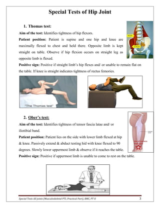
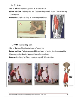
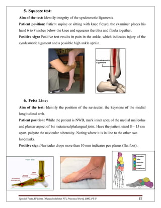
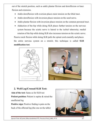
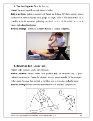
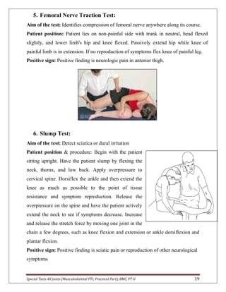
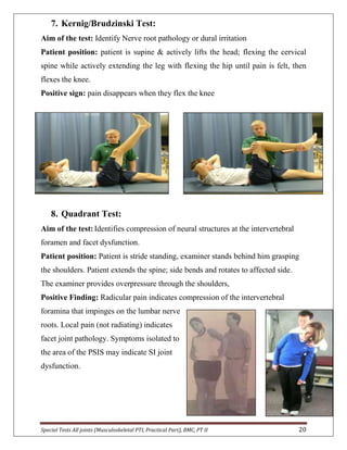
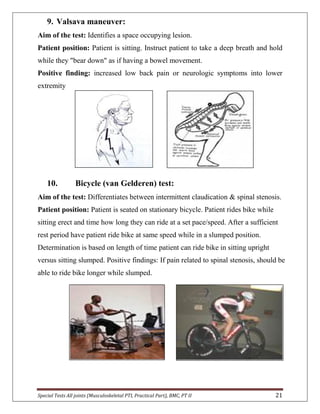
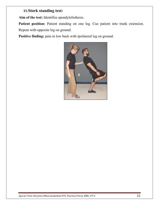
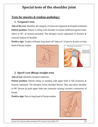
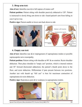
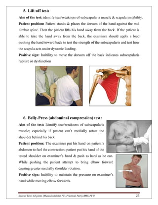
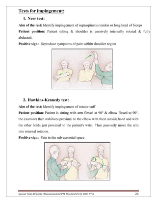
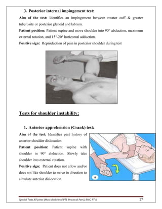
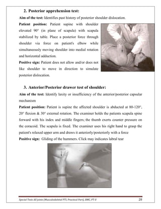
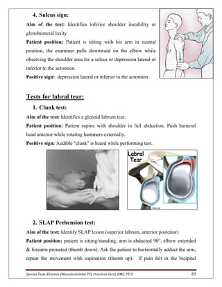
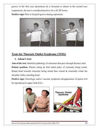
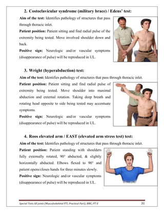
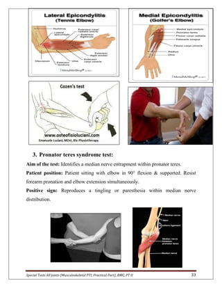
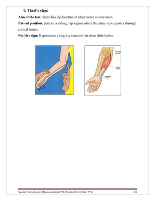
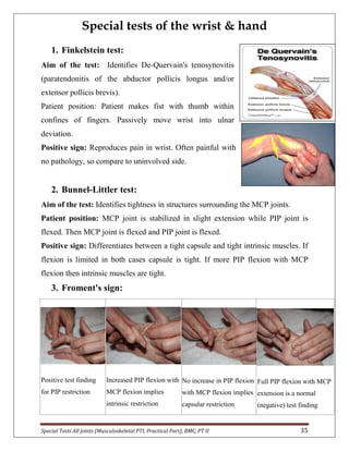
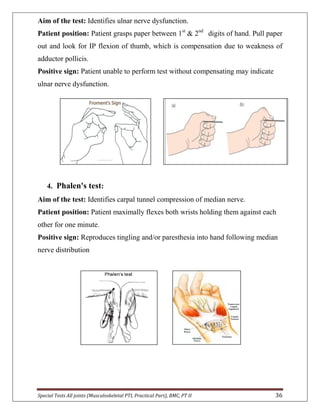
SlideShare a Scribd company logo
College of Physical Therapy
PT (II)
Practical Part of
Musculoskeletal Physical Therapy I
By:
Dr. Reem Sayed Dawood (Assist Prof of Physical Therapy)
Special Tests All joints (Musculoskeletal PTI, Practical Part), BMC, PT II 2
Contents:
No. Subject Page no.
1) Special tests of hip joint…………..…………………..……. 3
2) Special tests of knee joint…………..…….……….….……. 9
3) Special tests of ankle & foot ………....……..……………. 13
4) Special tests of lumbar spine joint…………...…………... 16
5) Special tests of shoulder joint…………..………...………. 23
6) Special tests of elbow joint…………..………...….………. 32
7) Special tests of wrist & hand…...…….…....………..…….. 35
8) Special tests of cervical spine joint……………..……….. 37
9) Students’ lab activities…………..………...….……….……. 41
10) References…………..…………………………..………..……. 42
Special Tests All joints (Musculoskeletal PTI, Practical Part), BMC, PT II 3
Special Tests of Hip Joint
1. Thomas test:
Aim of the test: Identifies tightness of hip flexors.
Patient position: Patient is supine and one hip and knee are
maximally flexed to chest and held there. Opposite limb is kept
straight on table. Observe if hip flexion occurs on straight leg as
opposite limb is flexed.
Positive sign: Positive if straight limb’s hip flexes and/ or unable to remain flat on
the table. If knee is straight indicates tightness of rectus femories.
2. Ober’s test:
Aim of the test: Identifies tightness of tensor fascia latae and/ or
iliotibial band.
Patient position: Patient lies on the side with lower limb flexed at hip
& knee. Passively extend & abduct testing hid with knee flexed to 90
degrees. Slowly lower uppermost limb & observe if it reaches the table.
Positive sign: Positive if uppermost limb is unable to come to rest on the table.
Special Tests All joints (Musculoskeletal PTI, Practical Part), BMC, PT II 4
3. Ely test:
Aim of the test: Identify tightness of rectus femoris.
Patient position: Patient prone and knee of testing limb is flexed. Observe the hip
of testing limb.
Positive sign: Positive if hip of the testing limb flexes.
4. 90-90 Hamstring test:
Aim of the test: Identifies tightness of hamstring.
Patient position: Patient supine and hip and knee of testing limb is supported in
90 degree flexion. Passively extend knee of testing limb.
Positive sign: Positive if knee is unable to reach full extension.
Special Tests All joints (Musculoskeletal PTI, Practical Part), BMC, PT II 5
5. Piriformis test:
Aim of the test: Identifies tightness of the piriformis muscle
(piriformis syndrome).
Patient position: Patient is supine and foot of tested leg is
placed lateral to the opposite limb’s knee. Testing hip is
adducted. Observe the position of testing knee relative to the
opposite knee.
Positive sign: Positive if testing knee is unable pass over resting knee and/ or
reproduction of pain in buttock and/ or along sciatic nerve distribution.
6. Patrick/FABER (flex, abd, external rot) test:
Aim of the test: Identify dysfunction of hip and sacroiliac joints such as mobility
restriction.
Patient position: Patient lies supine. Passively flex, abduct, and externally rotate
tested leg so that foot is resting above the opposite knee. Slowly testing leg down
towards the table surface.
Positive sign: Positive test when involved knee is unable to assume relaxed
position and/ or reproduction of painful symptoms.
Special Tests All joints (Musculoskeletal PTI, Practical Part), BMC, PT II 6
7. Standing flexion test:
Aim of the test: Identify dysfunction of sacroiliac joint.
Patient position: Patient stands with his feet apart,
therapist stands behind him with his thumbs placing on
PSIS, asked the patient to flex forward as much as he can,
therapist observe both PSIS normally both equally move.
Positive sign: Positive when one PSIS move further
cranially than the other, which is the affected side.
8. Standing flexion test:
Aim of the test: Identify dysfunction of sacroiliac joint.
Patient position: Patient is sitting with arms across the
chest, therapist stands behind him with his thumbs placing
on PSIS, asked the patient to flex forward & passes elbows
between the knees as if to touch the floor.
Positive sign: Positive when one PSIS move further
cranially than the other, which is the affected side.
9. Long sitting test:
Aim of the test: Identify anteriorly or posteriorly rotated innominate (based on
apparent leg length differences)
Patient position: Patient is supine, both hips & knees extend, therapist standing
with thumbs on patient’s medial malleoli relative to each other. Patient slowly
assumes the long-sitting position & malleolar position is reassessed.
Positive sign: When leg appears longer in supine but shorter in long sitting this
indicates anterior innominate rotation on affected side. Opposite is for posterior
innominate rotation.
Special Tests All joints (Musculoskeletal PTI, Practical Part), BMC, PT II 7
10. Trendelenberg’s sign:
Aim of the test: Identifies weakness of gluteus medius or unstable hip.
Patient position: Patient standing and asked to stand on one leg (flex
the opposite knee). Observe the pelvis on the stance leg.
Positive sign: Positive when contra-lateral pelvis drops when lower
limb support is removed.
Special Tests All joints (Musculoskeletal PTI, Practical Part), BMC, PT II 8
11. Leg length test.
Aim of the test: Identifies true leg length discrepancy.
Patient position: Patient supine and pelvis is balanced aligned with lower limbs
and trunk. Measure distance from ASlS to medial malleolus on each limb several
times for consistency and compare results.
Positive sign: A difference in lengths between two limbs is noted identifying a
true leg length discrepancy. This test will determine if the limb discrepancy is true
or functional. True discrepancy is caused by an anatomical difference in bone
lengths (either tibia or femur). Functional discrepancies are not anatomical in
origin and are the result of compensation due to abnormal position or posture such
as pronation of a foot or pelvic obliquity.
Special Tests All joints (Musculoskeletal PTI, Practical Part), BMC, PT II 9
Special tests of the knee joint
1. Collateral ligament instability tests (valgus & varus stress tests):
Aim of the test: Identifies collateral ligaments laxity.
Patient position: Patient is supine, the entire lower extremity is
supported & knee placed in 20-30° of flexion. Valgus force
placed through knee to test medial collateral ligament. Varus
force placed through knee to test lateral collateral ligament.
Positive sign: Primary finding is laxity, but pain may be noted.
2. Lachman stress test:
Aim of the test: Indicates integrity of anterior/posterior cruciate ligament (ACL/ PCL).
Patient position: Patient supine with testing knee flexed 20-30°. Stabilize femur
and passively try to glide tibia anterior (posterior).
Positive sign: finding is excessive anterior (posterior) glide of tibia.
Special Tests All joints (Musculoskeletal PTI, Practical Part), BMC, PT II 10
3. Anterior (posterior) drawer test:
Aim of the test: Indicates integrity of anterior (posterior) cruciate ligament.
Patient position: Patient supine and testing hip flexed to 45 degree and knee flexed to
90 degree. Passively glide tibia anteriorly (posteriorly) following the joint plane.
Positive sign: Positive finding is excessive anterior (posterior) glide.
4. McMurray test:
Aim of the test: Identifies meniscal tears.
Patient position: Patient supine with testing knee is in
maximal flexion. Passively internally rotate and extend the
knee. This tests lateral meniscus. Test the medial meniscus
with the same procedure except rotate the tibia into lateral
rotation.
Positive sign: Positive finding is reproduction of click and/ or pain in the knee joint.
Special Tests All joints (Musculoskeletal PTI, Practical Part), BMC, PT II 11
5. Apley test:
Aim of the test: Helps to differentiate between meniscal
tears and ligamentous lesions.
Patient position: Patient prone with testing knee flexed to
90 degrees. Stabilize patient’s thigh to table with your
knee. Passively distract the knee joint then slowly rotate
tibia internally and externally. Next step is to apply a
compressive load to knee joint and once again slowly
rotate tibia internally and externally.
Positive sign: Pain or decreased motion during compression indicates a meniscal
dysfunction. If pain or decreased motion occurs during the distraction then it is
most likely a Ligamentous dysfunction.
6. Clarke’s sign:
Aim of the test: Indicates Patellofemoral dysfunction.
Patient position: Patient supine with knee in extension resting on table. Push
posterior on superior pole of patella then ask patient to perform active contraction
of the quadriceps muscle.
Positive sign: Pain is produced in
knee as a result of the test.
Special Tests All joints (Musculoskeletal PTI, Practical Part), BMC, PT II 12
7. Patellar tap test (Ballotable patella):
Aim of the test: Indicates infrapatellar effusion.
Patient position: Patient supine with knee in extension resting on table. Apply a
soft tap over the central patella.
Positive sign: Positive finding is perception of the patella floating (dancing patella
sign).
8. Patellar apprehension test:
Aim of the test: indicate past history of patella dislocation.
Patient position: patient supine &patella is passively glided laterally
Positive sign: patient does not allow &/or doesn't like patella to move in lateral
direction to stimulate sublaxation/dislocation.
Special Tests All joints (Musculoskeletal PTI, Practical Part), BMC, PT II 13
Special tests of the ankle & foot joint
1. Anterior drawer test:
Aim of the test: Identifies Ligament instability (particularly anterior talofibular-
ATF ligament).
Patient position: Patient is supine with heel just off edge of table in 20° plantar
flexion. Stabilize lower leg and grasp foot. Pull foot (talus) anterior.
Positive sign: Positive finding if foot (talus) has excessive anterior glide and/ or
pain is noted.
2. Talar tilt (Kleiger):
Aim of the test: Identifies Ligament instability (particularly calcaneofibular
ligament)
Patient position: Patient side lying with knee slightly flexed and ankle in neutral.
Move foot into adduction testing calcaneofibular ligament and into abduction
testing deltoid ligament.
Positive sign: Positive finding if excessive adduction or abduction occurs and/ or
pain is noted.
Special Tests All joints (Musculoskeletal PTI, Practical Part), BMC, PT II 14
3. Thompson test:
Aim of the test: Evaluate the integrity of the Achilles tendon.
Patient position: Patient prone with foot off the edge of table. The examiner
squeezes the calf muscles; normally there is planter flexion.
Positive sign: No movement of foot while squeezing calf indicates positive finding.
4. Homan’s test
Aim of the test: Test for deep vein thrombosis (DVT)
Patient position: Patient supine or sitting with knee flexed, the examiner forcibly
dorsiflex the patient's ankle then palpate the calf muscle.
Positive sign: Pain in calf is a positive sign and should be referred
Special Tests All joints (Musculoskeletal PTI, Practical Part), BMC, PT II 15
5. Squeeze test:
Aim of the test: Identify integrity of the syndesmotic ligaments
Patient position: Patient supine or sitting with knee flexed, the examiner places his
hand 6 to 8 inches below the knee and squeezes the tibia and fibula together.
Positive sign: Positive test results in pain in the ankle, which indicates injury of the
syndesmotic ligament and a possible high ankle sprain.
6. Feiss Line:
Aim of the test: Identify the position of the navicular; the keystone of the medial
longitudinal arch.
Patient position: While the patient is NWB, mark inner apex of the medial malleolus
and plantar aspect of 1st metatarsalphalangeal joint. Have the patient stand 8 – 15 cm
apart, palpate the navicular tuberosity. Noting where it is in line to the other two
landmarks.
Positive sign: Navicular drops more than 10 mm indicates pes planus (flat foot).
Special Tests All joints (Musculoskeletal PTI, Practical Part), BMC, PT II 16
Special tests of the Lumbar Spine
1. Straight Leg Raising (SLR)/Lasegue Test:
Aim of the test: Identifies dysfunction of neurologic structures that supply lower
limb & also indicates the unilateral dysfunction of the sacroiliac joint.
Patient position: Patient is supine with legs resting on table. Passively flex hip of
one leg with knee extended until patient complains of shooting pain into lower
limb. Slowly lower limb until pain subsides then passively dorsiflex foot.
Positive sign: Positive finding is reproduction of pathologic neurologic symptoms
when foot is dorsiflexed.
More illustration of SLR test: ROM can demonstrate problems in different areas;
 0 – 30° equals hip pathology or severely inflamed nerve root.
 30 – 50° indicates sciatic nerve involvement
 50 – 70° is probable hamstring involvement
 70 – 90° when sacroiliac joint is stressed
Neural tension & mobilization for lower quadrant by SLR;
While patient is supine several variations may be done; ankle dorsiflexion, ankle
plantar flexion with inversion, hip adduction, hip medial rotation, and passive
neck flexion. The maneuver may also be performed long-sitting (slump-sitting
position—see below) and side-lying. These various positions of the lower
extremity and neck are used to differentiate tight or strained hamstrings from
possible sites of restriction or nerve mobility in the lumbosacral plexus and sciatic
nerve.
Once the position that places tension on the involved neurological tissue is found,
maintain the stretch position and then move one of the joints a few degrees in and
Special Tests All joints (Musculoskeletal PTI, Practical Part), BMC, PT II 17
out of the stretch position, such as ankle plantar flexion and dorsiflexion or knee
flexion and extension.
 Ankle dorsiflexion with eversion places more tension on the tibial tract.
 Ankle dorsiflexion with inversion places tension on the sural nerve.
 Ankle plantar flexion with inversion places tension on the common peroneal tract.
 Adduction of the hip while doing SLR places further tension on the nervous
system because the sciatic nerve is lateral to the ischial tuberosity; medial
rotation of the hip while doing SLR also increases tension on the sciatic nerve.
Passive neck flexion while doing SLR pulls the spinal cord cranially and places
the entire nervous system on a stretch; this technique is called SLR
modification test.
2. Well Leg/Crossed SLR Test:
Aim of the test: Same as for SLR test
Patient position: Patient is supine & raised the
unaffected leg
Positive sign: Positive finding is pain on the
back of the affected leg (the one on the table)
Special Tests All joints (Musculoskeletal PTI, Practical Part), BMC, PT II 18
3. Tension Sign for Sciatic Nerve:
Aim of the test: Identifies sciatic nerve irritation
Patient position: patient is supine with flexed hip & knee 90°; the examiner grasps
the heel with one hand & the other grasps the thigh. Knee is then extended as far as
possible with the examiner palpating the tibial portion of the sciatic nerve as it
passes behind popliteal space
Positive finding: Tenderness and reproduction of sciatica symptoms
4. Bowstring Test (Cram Test):
Aim of test: Indicates sciatic nerve tension.
Patient position: Patient supine with passive SLR on involved side. If pain
radiating the examiner flexes the subject’s knee to approximately 20° in attempt to
reduce pain. Pressure then applied to popliteal area to reproduce radicular pain.
Positive finding: Painful radicular reproduction with popliteal compression.
Special Tests All joints (Musculoskeletal PTI, Practical Part), BMC, PT II 19
5. Femoral Nerve Traction Test:
Aim of the test: Identifies compression of femoral nerve anywhere along its course.
Patient position: Patient lies on non-painful side with trunk in neutral, head flexed
slightly, and lower limb's hip and knee flexed. Passively extend hip while knee of
painful limb is in extension. If no reproduction of symptoms flex knee of painful leg.
Positive sign: Positive finding is neurologic pain in anterior thigh.
6. Slump Test:
Aim of the test: Detect sciatica or dural irritation
Patient position & procedure: Begin with the patient
sitting upright. Have the patient slump by flexing the
neck, thorax, and low back. Apply overpressure to
cervical spine. Dorsiflex the ankle and then extend the
knee as much as possible to the point of tissue
resistance and symptom reproduction. Release the
overpressure on the spine and have the patient actively
extend the neck to see if symptoms decrease. Increase
and release the stretch force by moving one joint in the
chain a few degrees, such as knee flexion and extension or ankle dorsiflexion and
plantar flexion.
Positive sign: Positive finding is sciatic pain or reproduction of other neurological
symptoms
Special Tests All joints (Musculoskeletal PTI, Practical Part), BMC, PT II 20
7. Kernig/Brudzinski Test:
Aim of the test: Identify Nerve root pathology or dural irritation
Patient position: patient is supine & actively lifts the head; flexing the cervical
spine while actively extending the leg with flexing the hip until pain is felt, then
flexes the knee.
Positive sign: pain disappears when they flex the knee
8. Quadrant Test:
Aim of the test: Identifies compression of neural structures at the intervertebral
foramen and facet dysfunction.
Patient position: Patient is stride standing, examiner stands behind him grasping
the shoulders. Patient extends the spine; side bends and rotates to affected side.
The examiner provides overpressure through the shoulders,
Positive Finding: Radicular pain indicates compression of the intervertebral
foramina that impinges on the lumbar nerve
roots. Local pain (not radiating) indicates
facet joint pathology. Symptoms isolated to
the area of the PSIS may indicate SI joint
dysfunction.
Special Tests All joints (Musculoskeletal PTI, Practical Part), BMC, PT II 21
9. Valsava maneuver:
Aim of the test: Identifies a space occupying lesion.
Patient position: Patient is sitting. Instruct patient to take a deep breath and hold
while they "bear down" as if having a bowel movement.
Positive finding: increased low back pain or neurologic symptoms into lower
extremity
10. Bicycle (van Gelderen) test:
Aim of the test: Differentiates between intermittent claudication & spinal stenosis.
Patient position: Patient is seated on stationary bicycle. Patient rides bike while
sitting erect and time how long they can ride at a set pace/speed. After a sufficient
rest period have patient ride bike at same speed while in a slumped position.
Determination is based on length of time patient can ride bike in sitting upright
versus sitting slumped. Positive findings: If pain related to spinal stenosis, should be
able to ride bike longer while slumped.
Special Tests All joints (Musculoskeletal PTI, Practical Part), BMC, PT II 22
11.Stork standing test:
Aim of the test: Identifies spondylolisthesis.
Patient position: Patient standing on one leg. Cue patient into trunk extension.
Repeat with opposite leg on ground.
Positive finding: pain in low back with ipsilateral leg on ground.
Special Tests All joints (Musculoskeletal PTI, Practical Part), BMC, PT II 23
Special tests of the shoulder joint
Tests for muscles & tendons pathology:
1. Yergason's test:
Aim of the test: Identifies the integrity of transverse ligament & bicipital tendonitis.
Patient position: Patient is sitting with shoulder in neutral stabilized against trunk,
elbow at 90°, & forearm pronated. The therapist resists supination of forearm &
external rotation of shoulder
Positive sign: Tendon of biceps long head will "pop out" of groove & pain on long
head of biceps tendon.
2. Speed’s test (Biceps straight arm)
Aim of test: Identifies bicipital tendonitis
Patient position: Patient sitting or standing with upper limb in full extension &
forearm supinated. The therapist resists shoulder flexion. May also place shoulder
in 90° flexion & push upper limb into extension causing eccentric contraction of
biceps
Positive sign: Pain in long head of biceps tendon.
Special Tests All joints (Musculoskeletal PTI, Practical Part), BMC, PT II 24
3. Drop arm test:
Aim of test: Identifies tear &/or full rupture of rotator cuff
Patient position: Patient sitting with shoulder passively abducted to 120°. Patient
is instructed to slowly bring arm down to side .Guard patient's arm from falling in
case it gives way.
Positive sign: Patient unable to lower arm back down to side
4. Empty can test:
Aim of test: Identifies tear &/or impingement of supraspinatus tendon or possible
suprascapular nerve neuropathy.
Patient position: Patient sitting with shoulder at 90° & no rotation. Resist shoulder
abduction. Then place shoulder in "empty can" position, which is internal rotation
and 30° forward (horizontal adduction), the patient’s thumb point down to the
floor, and resist abduction. Differentiate if pain present between two positions.
Another test with thumb up “full can” is best for maximum contraction of
supraspinatus & resist abduction.
Positive sign: Reproduces pain &/or weakness in supraspinatus tendon.
Special Tests All joints (Musculoskeletal PTI, Practical Part), BMC, PT II 25
5. Lift-off test:
Aim of the test: identify tear/weakness of subscapularis muscle & scapula instability.
Patient position: Patient stands & places the dorsum of the hand against the mid
lumbar spine. Then the patient lifts his hand away from the back. If the patient is
able to take the hand away from the back, the examiner should apply a load
pushing the hand toward back to test the strength of the subscapularis and test how
the scapula acts under dynamic loading.
Positive sign: Inability to move the dorsum off the back indicates subscapularis
rupture or dysfunction
6. Belly-Press (abdominal compression) test:
Aim of the test: Identify tear/weakness of subscapularis
muscle; especially if patient can’t medially rotate the
shoulder behind his back.
Patient position: The examiner put his hand on patient’s
abdomen to feel the contraction; patient put his hand of the
tested shoulder on examiner’s hand & push as hard as he can.
While pushing the patient attempt to bring elbow forward
causing greater medially shoulder rotation.
Positive sign: Inability to maintain the pressure on examiner’s
hand while moving elbow forwards.
Special Tests All joints (Musculoskeletal PTI, Practical Part), BMC, PT II 26
Tests for impingement:
1. Neer test:
Aim of the test: Identify impingement of supraspinatus tendon or long head of biceps
Patient position: Patient silting & shoulder is passively internally rotated & fully
abducted.
Positive sign: Reproduce symptoms of pain within shoulder region
2. Hawkins-Kennedy test:
Aim of the test: Identify impingement of rotator cuff
Patient position: Patient is sitting with arm flexed at 90° & elbow flexed to 90°,
the examiner then stabilizes proximal to the elbow with their outside hand and with
the other holds just proximal to the patient's wrist. Then passively move the arm
into internal rotation.
Positive sign: Pain in the sub-acromial space
Special Tests All joints (Musculoskeletal PTI, Practical Part), BMC, PT II 27
3. Posterior internal impingement test:
Aim of the test: Identifies an impingement between rotator cuff & greater
tuberosity or posterior glenoid and labrum.
Patient position: Patient supine and move shoulder into 90° abduction, maximum
external rotation, and 15°-20° horizontal adduction.
Positive sign: Reproduction of pain in posterior shoulder during test
Tests for shoulder instability:
1. Anterior apprehension (Crank) test:
Aim of the test: Identifies past history of
anterior shoulder dislocation
Patient position: Patient supine with
shoulder in 90° abduction. Slowly take
shoulder into external rotation.
Positive sign: Patient does not allow and/or
does not like shoulder to move in direction to
simulate anterior dislocation.
Special Tests All joints (Musculoskeletal PTI, Practical Part), BMC, PT II 28
2. Posterior apprehension test:
Aim of the test: Identifies past history of posterior shoulder dislocation.
Patient position: Patient supine with shoulder
elevated 90° (in plane of scapula) with scapula
stabilized by table. Place a posterior force through
shoulder via force on patient's elbow while
simultaneously moving shoulder into medial rotation
and horizontal adduction.
Positive sign: Patient does not allow and/or does not
like shoulder to move in direction to simulate
posterior dislocation.
3. Anterior/Posterior drawer test of shoulder:
Aim of the test: Identify laxity or insufficiency of the anterior/posterior capsular
mechanism
Patient position: Patient is supine the affected shoulder is abducted at 80-120°,
20° flexion & 30° external rotation. The examiner holds the patients scapula spine
forward with his index and middle fingers; the thumb exerts counter pressure on
the coracoid. The scapula is fixed. The examiner uses his right hand to grasp the
patient's relaxed upper arm and draws it anteriorly/posteriorly with a force
Positive sign: Gliding of the hummers. Click may indicates labral tear
Special Tests All joints (Musculoskeletal PTI, Practical Part), BMC, PT II 29
4. Sulcus sign:
Aim of the test: Identifies inferior shoulder instability or
glenohumeral laxity
Patient position: Patient is sitting with his arm in neutral
position, the examiner pulls downward on the elbow while
observing the shoulder area for a sulcus or depression lateral or
inferior to the acromion.
Positive sign: depression lateral or inferior to the acromion
Tests for labral tear:
1. Clunk test:
Aim of the test: Identifies a glenoid labrum tear.
Patient position: Patient supine with shoulder in full abduction. Push humeral
head anterior while rotating hummers externally.
Positive sign: Audible "clunk" is heard while performing test.
2. SLAP Prehension test:
Aim of the test: Identify SLAP lesion (superior labrum, anterior posterior)
Patient position: patient is sitting/standing, arm is abducted 90°, elbow extended
& forearm pronated (thumb down). Ask the patient to horizontally adduct the arm,
repeat the movement with supination (thumb up). If pain felt in the bicipital
Special Tests All joints (Musculoskeletal PTI, Practical Part), BMC, PT II 30
groove in the first case (pronation) & is lessened or absent in the second case
(supination), the test is considered positive for a SLAP lesion.
Positive sign: Pain in bicipital groove during supination.
Tests for Thoracic Outlet Syndrome (TOS):
1. Adson's test:
Aim of the test: Identifies pathology of structures that pass through thoracic inlet.
Patient position: Patient sitting & find radial pulse of extremity being tested.
Rotate head towards extremity being tested then extend & externally rotate the
shoulder while extending head.
Positive sign: Neurologic and/or vascular symptoms (disappearance of pulse) will
be reproduced in upper limb (UL).
Special Tests All joints (Musculoskeletal PTI, Practical Part), BMC, PT II 31
2. Costoclavicular syndrome (military brace) / Edens’ test:
Aim of the test: Identifies pathology of structures that pass
through thoracic inlet.
Patient position: Patient sitting and find radial pulse of the
extremity being tested. Move involved shoulder down and
back.
Positive sign: Neurologic and/or vascular symptoms
(disappearance of pulse) will be reproduced in UL.
3. Wright (hyperabduction) test:
Aim of the test: Identifies pathology of structures that pass through thoracic inlet.
Patient position: Patient sitting and find radial pulse of
extremity being tested. Move shoulder into maximal
abduction and external rotation. Taking deep breath and
rotating head opposite to side being tested may accentuate
symptoms.
Positive sign: Neurologic and/or vascular symptoms
(disappearance of pulse) will be reproduced in UL.
4. Roos elevated arm / EAST (elevated arm stress test) test:
Aim of the test: Identifies pathology of structures that pass through thoracic inlet.
Patient position: Patient standing with shoulders
fully externally rotated, 90° abducted, & slightly
horizontally abducted. Elbows flexed to 90° and
patient opens/closes hands for three minutes slowly.
Positive sign: Neurologic and/or vascular symptoms
(disappearance of pulse) will be reproduced in UL.
Special Tests All joints (Musculoskeletal PTI, Practical Part), BMC, PT II 32
Special tests of the elbow joint
1. Ligament instability tests (valgus & varus stress tests):
Aim of the test: Identifies collateral ligaments laxity or restriction.
Patient position: Patient is sitting or supine. Entire upper limb is supported &
stabilized and elbow placed in 20°-30° of flexion. Valgus force placed through
elbow tests ulnar collateral ligament. Varus force placed through elbow tests radial
collateral ligament
Positive sign: Primary finding is laxity, but pain may be noted as well.
2. Tests for epicondylitis:
Aim of the test: To identify lateral or medial epicodylitis
A. Lateral epicondylities/Tennis Elbow (Cozen) Test:
Patient position: Patient is sitting with elbow in 90° & supported, resist wrist
extension, wrist radial deviation & forearm pronation with fingers fully flexed
(fist) simultaneously.
B. Medial epicondylities/Golfer Elbow test
Patient position: Patient is sitting with elbow in 90° & supported, passively supinate
forearm, extend elbow & wrist.
Positive sign: Pain at Lateral epicondyle for tennis elbow & at medial epicondyle
for golfer elbow
Special Tests All joints (Musculoskeletal PTI, Practical Part), BMC, PT II 33
3. Pronator teres syndrome test:
Aim of the test: Identifies a median nerve entrapment within pronator teres.
Patient position: Patient sitting with elbow in 90° flexion & supported. Resist
forearm pronation and elbow extension simultaneously.
Positive sign: Reproduces a tingling or paresthesia within median nerve
distribution.
Special Tests All joints (Musculoskeletal PTI, Practical Part), BMC, PT II 34
4. Tinel's sign:
Aim of the test: Identifies dysfunction of ulnar nerve at olecranon.
Patient position: patient is sitting, tap region where the ulnar nerve passes through
cubital tunnel.
Positive sign: Reproduces a tingling sensation in ulnar distribution.
Special Tests All joints (Musculoskeletal PTI, Practical Part), BMC, PT II 35
Special tests of the wrist & hand
1. Finkelstein test:
Aim of the test: Identifies De-Quervain's tenosynovitis
(paratendonitis of the abductor pollicis longus and/or
extensor pollicis brevis).
Patient position: Patient makes fist with thumb within
confines of fingers. Passively move wrist into ulnar
deviation.
Positive sign: Reproduces pain in wrist. Often painful with
no pathology, so compare to uninvolved side.
2. Bunnel-Littler test:
Aim of the test: Identifies tightness in structures surrounding the MCP joints.
Patient position: MCP joint is stabilized in slight extension while PIP joint is
flexed. Then MCP joint is flexed and PIP joint is flexed.
Positive sign: Differentiates between a tight capsule and tight intrinsic muscles. If
flexion is limited in both cases capsule is tight. If more PIP flexion with MCP
flexion then intrinsic muscles are tight.
3. Froment's sign:
Positive test finding
for PIP restriction
Increased PIP flexion with
MCP flexion implies
intrinsic restriction
No increase in PIP flexion
with MCP flexion implies
capsular restriction
Full PIP flexion with MCP
extension is a normal
(negative) test finding
Special Tests All joints (Musculoskeletal PTI, Practical Part), BMC, PT II 36
Aim of the test: Identifies ulnar nerve dysfunction.
Patient position: Patient grasps paper between 1st
& 2nd
digits of hand. Pull paper
out and look for IP flexion of thumb, which is compensation due to weakness of
adductor pollicis.
Positive sign: Patient unable to perform test without compensating may indicate
ulnar nerve dysfunction.
4. Phalen's test:
Aim of the test: Identifies carpal tunnel compression of median nerve.
Patient position: Patient maximally flexes both wrists holding them against each
other for one minute.
Positive sign: Reproduces tingling and/or paresthesia into hand following median
nerve distribution
Special Tests All joints (Musculoskeletal PTI, Practical Part), BMC, PT II 37
Special tests of the cervical joint
1. Vertebral artery test:
Aim of the test: Identifies the integrity of
vertebrobasilar artery (vertebrobasilar insufficiency)
Patient position: Patient is supine; the examiner takes
the patient head & neck into extension, right & left
rotation, & side bending. Hold each position 10-30 sec
unless symptoms are evoked.
Positive sign: dizziness, visual disturbances, disorientation, blurred speech,
nausea, vomiting, etc.
2. Hautant's test:
Aim of the test: Differentiates vascular versus vestibular causes of dizziness/vertigo.
Patient position: Two steps to this test.
a) Patient sitting with shoulders at 90° flexion & palms up. Have patient close
their eyes and remain in this position for 30 sec. If arms lose their position
there may be a vestibular condition.
b) Patient sitting with shoulders at 90° flexion & palms up. Have patient close
their eyes & cue patient into head and neck extension with rotation right then
Special Tests All joints (Musculoskeletal PTI, Practical Part), BMC, PT II 38
left, remaining in each position for 30 sec. If arms lose their position the
condition may be vascular in nature.
Positive sign: Position/movement of arms determines positive finding.
3. Foraminal compression (Spurling's) test.
Aim of the test: Identifies dysfunction (typically compression) of cervical nerve root.
Patient position: Patient sitting with head side bent towards uninvolved side.
Apply pressure through head straight down. Repeat with head side bent towards
involved side. This test is performed in 3 stages; if symptoms are produced, don’t
proceed to next stage. First stage involves compression with the head in neutral,
second in extension, third in extension & rotation to unaffected side then to
affected side.
Positive sign: Positive finding is pain and/or paresthesia in dermatomal pattern for
involved nerve root.
Special Tests All joints (Musculoskeletal PTI, Practical Part), BMC, PT II 39
4. Distraction test:
Aim of the test: Indicates compression of neural structures at the intervertebral
foramen or facet joint dysfunction.
Patient position: Patient sitting or supine, examiner
places one hand under patient’s chin & other hand
around the occiput and the head is passively distracted.
Positive sign: Positive finding is a decrease in
symptoms in neck (facet condition) or a decrease in
upper limb pain (neurologic condition).
5. Shoulder abduction test (Bakody’s sign)
Aim of the test: Indicates compression of neural structures
within intervertebral foramen.
Patient position: Patient sitting and asked to place one hand on
top of their head. Repeat with opposite hand.
Positive sign: Positive finding is a decrease in symptoms into
upper limb. If increases, it implies increase pressure in the interscalene triangle.
6. Upper limb tension tests (ULTT):
ULTT are equivalent to SLR test in lumbar spine. They are tension tests designed
to put stress on the neurological structures of UL, on all tissues of UL.
Modification of the position of shoulder, elbow, wrist & fingers places greater
stress on special nerves (nerve bias). The final movement is sometimes referred to
as sensitizing test (e.g., neck side flexion in ULTT).
Aim of the test: Evaluation of peripheral nerve compression.
Patient position: see the following picture & table
Positive sign: Neurologic symptoms will be reproduced in upper extremity.
Special Tests All joints (Musculoskeletal PTI, Practical Part), BMC, PT II 40
Special Tests All joints (Musculoskeletal PTI, Practical Part), BMC, PT II 41
Lab activities:
In groups illustrate, discuss & explain the following:
1) Lumbosacral plexuses.
2) Dermatomes & myotomes of the lower limbs.
3) Pathway & the origin of each of peripheral nerves of the lower limbs.
4) Location the common sites of each nerve to be palpated.
5) Tension tests for lower limb nerves.
6) Brachial plexuses.
7) Dermatomes & myotomes of the upper limbs.
8) Pathway & the origin of each of peripheral nerves of the upper limbs.
9) Location the common sites of each nerve to be palpated.
10) Tension tests for upper limb nerves.
Special Tests All joints (Musculoskeletal PTI, Practical Part), BMC, PT II 42
References:
 Magee David J: Orthopedic Physical Assessment, 6e (Musculoskeletal
Rehabilitation), 2013. Elsevier Saunders.
 Dutton M: Orthopedic examination, evaluation, & intervention. 2004, Mc
Graw Hill.
 Gross et al: Musculoskeletal examination, 4th
ed, 2016. Wiley & Sons, Ltd

More Related Content

What's hot

Piriformis syndrome
Piriformis syndromePiriformis syndrome
Piriformis syndrome
Andy Coleman
 
Painful Arc Syndrome.pptx
Painful Arc Syndrome.pptxPainful Arc Syndrome.pptx
Painful Arc Syndrome.pptx
Prashant Badiger
 
Patellar tendinopathy
Patellar tendinopathyPatellar tendinopathy
Patellar tendinopathy
pratigya deuja
 
Rotator cuff injuries
Rotator cuff injuriesRotator cuff injuries
Rotator cuff injuries
pratigya deuja
 
Hammer toes
Hammer toesHammer toes
Hammer toes
Md. Nayeem Hasan
 
Frozen Shoulder Physiotherapy Management
Frozen Shoulder Physiotherapy ManagementFrozen Shoulder Physiotherapy Management
Frozen Shoulder Physiotherapy Management
Vishal Deep
 
Volksmann contracture
Volksmann contracture Volksmann contracture
Volksmann contracture
Kimberly Walsh
 
SLAP Lesion and.pdf
SLAP Lesion and.pdfSLAP Lesion and.pdf
SLAP Lesion and.pdf
eyobkaseye
 
Genu recurvatum
Genu recurvatumGenu recurvatum
Genu recurvatum
Shishir Bin
 
Flat foot and Cavus foot
 Flat foot and Cavus foot Flat foot and Cavus foot
Flat foot and Cavus foot
Dr Thouseef Abdul Majeed
 
Subacromail bursitis
Subacromail bursitisSubacromail bursitis
Subacromail bursitis
Career Point University
 
Bicipital tendonitis
Bicipital tendonitisBicipital tendonitis
Cervical Spondylosis
Cervical SpondylosisCervical Spondylosis
Cervical Spondylosis
Shazia Abdul Hamid Khalfe
 
Frozen shoulder
Frozen shoulderFrozen shoulder
Frozen shoulder
Rachita Hada
 
Congenital Dislocation of the Hip - PHYSIOTHERAPY
Congenital Dislocation of the Hip - PHYSIOTHERAPYCongenital Dislocation of the Hip - PHYSIOTHERAPY
Congenital Dislocation of the Hip - PHYSIOTHERAPY
UPASANA AGARWAL
 
Fibrositis
FibrositisFibrositis
Posture assessment cpd
Posture assessment cpdPosture assessment cpd
Posture assessment cpd
Jayasri Prasanna
 
Carpal Tunnel Syndrome
Carpal Tunnel SyndromeCarpal Tunnel Syndrome
Carpal Tunnel Syndrome
Hemant Aggarwal
 
Physiotherapy management of poliomyelitis
Physiotherapy management of poliomyelitisPhysiotherapy management of poliomyelitis
Physiotherapy management of poliomyelitis
Sayali Gujjewar
 
Ankle Sprains
Ankle SprainsAnkle Sprains
Ankle Sprains
Apeksha Besekar
 

What's hot (20)

Piriformis syndrome
Piriformis syndromePiriformis syndrome
Piriformis syndrome
 
Painful Arc Syndrome.pptx
Painful Arc Syndrome.pptxPainful Arc Syndrome.pptx
Painful Arc Syndrome.pptx
 
Patellar tendinopathy
Patellar tendinopathyPatellar tendinopathy
Patellar tendinopathy
 
Rotator cuff injuries
Rotator cuff injuriesRotator cuff injuries
Rotator cuff injuries
 
Hammer toes
Hammer toesHammer toes
Hammer toes
 
Frozen Shoulder Physiotherapy Management
Frozen Shoulder Physiotherapy ManagementFrozen Shoulder Physiotherapy Management
Frozen Shoulder Physiotherapy Management
 
Volksmann contracture
Volksmann contracture Volksmann contracture
Volksmann contracture
 
SLAP Lesion and.pdf
SLAP Lesion and.pdfSLAP Lesion and.pdf
SLAP Lesion and.pdf
 
Genu recurvatum
Genu recurvatumGenu recurvatum
Genu recurvatum
 
Flat foot and Cavus foot
 Flat foot and Cavus foot Flat foot and Cavus foot
Flat foot and Cavus foot
 
Subacromail bursitis
Subacromail bursitisSubacromail bursitis
Subacromail bursitis
 
Bicipital tendonitis
Bicipital tendonitisBicipital tendonitis
Bicipital tendonitis
 
Cervical Spondylosis
Cervical SpondylosisCervical Spondylosis
Cervical Spondylosis
 
Frozen shoulder
Frozen shoulderFrozen shoulder
Frozen shoulder
 
Congenital Dislocation of the Hip - PHYSIOTHERAPY
Congenital Dislocation of the Hip - PHYSIOTHERAPYCongenital Dislocation of the Hip - PHYSIOTHERAPY
Congenital Dislocation of the Hip - PHYSIOTHERAPY
 
Fibrositis
FibrositisFibrositis
Fibrositis
 
Posture assessment cpd
Posture assessment cpdPosture assessment cpd
Posture assessment cpd
 
Carpal Tunnel Syndrome
Carpal Tunnel SyndromeCarpal Tunnel Syndrome
Carpal Tunnel Syndrome
 
Physiotherapy management of poliomyelitis
Physiotherapy management of poliomyelitisPhysiotherapy management of poliomyelitis
Physiotherapy management of poliomyelitis
 
Ankle Sprains
Ankle SprainsAnkle Sprains
Ankle Sprains
 

Similar to Special_test_for_all_joints.pdf

Knee
KneeKnee
MMT for knee
MMT for kneeMMT for knee
MMT for knee
Ambreen Sadaf
 
Muscskel.lecture kneeand shoulder
Muscskel.lecture kneeand shoulderMuscskel.lecture kneeand shoulder
Muscskel.lecture kneeand shoulder
Hue University of Medicine and Pharmacy
 
SPECIAL TEST OF UL.pptx
SPECIAL TEST OF UL.pptxSPECIAL TEST OF UL.pptx
SPECIAL TEST OF UL.pptx
Shamima Akter Swapna
 
Physical examination signs
Physical examination signsPhysical examination signs
Physical examination signs
coolboy101pk
 
Knee Examination.pptx
Knee Examination.pptxKnee Examination.pptx
Knee Examination.pptx
MohammedAli666017
 
FRO.ppt
FRO.pptFRO.ppt
Adv Musc Exam Portfolio 09
Adv Musc Exam Portfolio 09Adv Musc Exam Portfolio 09
Adv Musc Exam Portfolio 09
Steven Sager
 
Ullswater Physio CPD
Ullswater Physio CPDUllswater Physio CPD
Ullswater Physio CPD
Mary McCance MSc MCSP HCPC
 
Mmt f0r hip
Mmt f0r hip Mmt f0r hip
Mmt f0r hip
Ambreen Sadaf
 
Knee Joint Assessment Tests and Mobilizations
Knee Joint Assessment Tests and MobilizationsKnee Joint Assessment Tests and Mobilizations
Knee Joint Assessment Tests and Mobilizations
M Sohail Raza
 
ch 3 Shoulder Elbow joint.pptx
ch 3 Shoulder Elbow joint.pptxch 3 Shoulder Elbow joint.pptx
ch 3 Shoulder Elbow joint.pptx
AhmedFaisal59561
 
Hip asseement
Hip asseementHip asseement
Elbow joint Assessment.pptx
Elbow joint  Assessment.pptxElbow joint  Assessment.pptx
Elbow joint Assessment.pptx
Rutuja Butala
 
Nicola Taddio Arezzo Nicolas Foundation 2011 Shoulder Cadaver Lab
Nicola Taddio Arezzo Nicolas Foundation 2011 Shoulder Cadaver Lab Nicola Taddio Arezzo Nicolas Foundation 2011 Shoulder Cadaver Lab
Nicola Taddio Arezzo Nicolas Foundation 2011 Shoulder Cadaver Lab
Nicola Taddio
 
The Examination of the Knee_040716.pptx
The Examination of the Knee_040716.pptxThe Examination of the Knee_040716.pptx
The Examination of the Knee_040716.pptx
deepaksampath5
 
lect 1 Anthropometry -.pdf
lect 1 Anthropometry -.pdflect 1 Anthropometry -.pdf
lect 1 Anthropometry -.pdf
ssuser650c771
 
_FIU - Thoracic and Lumbar Spine Special Tests and Pathologies (1) - نسخة.ppt
_FIU - Thoracic and Lumbar Spine Special Tests and Pathologies (1) - نسخة.ppt_FIU - Thoracic and Lumbar Spine Special Tests and Pathologies (1) - نسخة.ppt
_FIU - Thoracic and Lumbar Spine Special Tests and Pathologies (1) - نسخة.ppt
Radwa Talaat
 
IVMS ICM-Physical diagnosis- Extremeties and Back
IVMS ICM-Physical diagnosis-  Extremeties and BackIVMS ICM-Physical diagnosis-  Extremeties and Back
IVMS ICM-Physical diagnosis- Extremeties and Back
Imhotep Virtual Medical School
 
Musculoskeletal Case Study
Musculoskeletal Case StudyMusculoskeletal Case Study
Musculoskeletal Case Study
Joshua de Rooy
 

Similar to Special_test_for_all_joints.pdf (20)

Knee
KneeKnee
Knee
 
MMT for knee
MMT for kneeMMT for knee
MMT for knee
 
Muscskel.lecture kneeand shoulder
Muscskel.lecture kneeand shoulderMuscskel.lecture kneeand shoulder
Muscskel.lecture kneeand shoulder
 
SPECIAL TEST OF UL.pptx
SPECIAL TEST OF UL.pptxSPECIAL TEST OF UL.pptx
SPECIAL TEST OF UL.pptx
 
Physical examination signs
Physical examination signsPhysical examination signs
Physical examination signs
 
Knee Examination.pptx
Knee Examination.pptxKnee Examination.pptx
Knee Examination.pptx
 
FRO.ppt
FRO.pptFRO.ppt
FRO.ppt
 
Adv Musc Exam Portfolio 09
Adv Musc Exam Portfolio 09Adv Musc Exam Portfolio 09
Adv Musc Exam Portfolio 09
 
Ullswater Physio CPD
Ullswater Physio CPDUllswater Physio CPD
Ullswater Physio CPD
 
Mmt f0r hip
Mmt f0r hip Mmt f0r hip
Mmt f0r hip
 
Knee Joint Assessment Tests and Mobilizations
Knee Joint Assessment Tests and MobilizationsKnee Joint Assessment Tests and Mobilizations
Knee Joint Assessment Tests and Mobilizations
 
ch 3 Shoulder Elbow joint.pptx
ch 3 Shoulder Elbow joint.pptxch 3 Shoulder Elbow joint.pptx
ch 3 Shoulder Elbow joint.pptx
 
Hip asseement
Hip asseementHip asseement
Hip asseement
 
Elbow joint Assessment.pptx
Elbow joint  Assessment.pptxElbow joint  Assessment.pptx
Elbow joint Assessment.pptx
 
Nicola Taddio Arezzo Nicolas Foundation 2011 Shoulder Cadaver Lab
Nicola Taddio Arezzo Nicolas Foundation 2011 Shoulder Cadaver Lab Nicola Taddio Arezzo Nicolas Foundation 2011 Shoulder Cadaver Lab
Nicola Taddio Arezzo Nicolas Foundation 2011 Shoulder Cadaver Lab
 
The Examination of the Knee_040716.pptx
The Examination of the Knee_040716.pptxThe Examination of the Knee_040716.pptx
The Examination of the Knee_040716.pptx
 
lect 1 Anthropometry -.pdf
lect 1 Anthropometry -.pdflect 1 Anthropometry -.pdf
lect 1 Anthropometry -.pdf
 
_FIU - Thoracic and Lumbar Spine Special Tests and Pathologies (1) - نسخة.ppt
_FIU - Thoracic and Lumbar Spine Special Tests and Pathologies (1) - نسخة.ppt_FIU - Thoracic and Lumbar Spine Special Tests and Pathologies (1) - نسخة.ppt
_FIU - Thoracic and Lumbar Spine Special Tests and Pathologies (1) - نسخة.ppt
 
IVMS ICM-Physical diagnosis- Extremeties and Back
IVMS ICM-Physical diagnosis-  Extremeties and BackIVMS ICM-Physical diagnosis-  Extremeties and Back
IVMS ICM-Physical diagnosis- Extremeties and Back
 
Musculoskeletal Case Study
Musculoskeletal Case StudyMusculoskeletal Case Study
Musculoskeletal Case Study
 

Recently uploaded

Thyroid Gland- Gross Anatomy by Dr. Rabia Inam Gandapore.pptx
Thyroid Gland- Gross Anatomy by Dr. Rabia Inam Gandapore.pptxThyroid Gland- Gross Anatomy by Dr. Rabia Inam Gandapore.pptx
Thyroid Gland- Gross Anatomy by Dr. Rabia Inam Gandapore.pptx
Dr. Rabia Inam Gandapore
 
Ear and its clinical correlations By Dr. Rabia Inam Gandapore.pptx
Ear and its clinical correlations By Dr. Rabia Inam Gandapore.pptxEar and its clinical correlations By Dr. Rabia Inam Gandapore.pptx
Ear and its clinical correlations By Dr. Rabia Inam Gandapore.pptx
Dr. Rabia Inam Gandapore
 
Top Effective Soaps for Fungal Skin Infections in India
Top Effective Soaps for Fungal Skin Infections in IndiaTop Effective Soaps for Fungal Skin Infections in India
Top Effective Soaps for Fungal Skin Infections in India
SwisschemDerma
 
Role of Mukta Pishti in the Management of Hyperthyroidism
Role of Mukta Pishti in the Management of HyperthyroidismRole of Mukta Pishti in the Management of Hyperthyroidism
Role of Mukta Pishti in the Management of Hyperthyroidism
Dr. Jyothirmai Paindla
 
Phone Us ❤8107221448❤ #ℂall #gIRLS In Dehradun By Dehradun @ℂall @Girls Hotel...
Phone Us ❤8107221448❤ #ℂall #gIRLS In Dehradun By Dehradun @ℂall @Girls Hotel...Phone Us ❤8107221448❤ #ℂall #gIRLS In Dehradun By Dehradun @ℂall @Girls Hotel...
Phone Us ❤8107221448❤ #ℂall #gIRLS In Dehradun By Dehradun @ℂall @Girls Hotel...
chandankumarsmartiso
 
Does Over-Masturbation Contribute to Chronic Prostatitis.pptx
Does Over-Masturbation Contribute to Chronic Prostatitis.pptxDoes Over-Masturbation Contribute to Chronic Prostatitis.pptx
Does Over-Masturbation Contribute to Chronic Prostatitis.pptx
walterHu5
 
8 Surprising Reasons To Meditate 40 Minutes A Day That Can Change Your Life.pptx
8 Surprising Reasons To Meditate 40 Minutes A Day That Can Change Your Life.pptx8 Surprising Reasons To Meditate 40 Minutes A Day That Can Change Your Life.pptx
8 Surprising Reasons To Meditate 40 Minutes A Day That Can Change Your Life.pptx
Holistified Wellness
 
Adhd Medication Shortage Uk - trinexpharmacy.com
Adhd Medication Shortage Uk - trinexpharmacy.comAdhd Medication Shortage Uk - trinexpharmacy.com
Adhd Medication Shortage Uk - trinexpharmacy.com
reignlana06
 
NVBDCP.pptx Nation vector borne disease control program
NVBDCP.pptx Nation vector borne disease control programNVBDCP.pptx Nation vector borne disease control program
NVBDCP.pptx Nation vector borne disease control program
Sapna Thakur
 
share - Lions, tigers, AI and health misinformation, oh my!.pptx
share - Lions, tigers, AI and health misinformation, oh my!.pptxshare - Lions, tigers, AI and health misinformation, oh my!.pptx
share - Lions, tigers, AI and health misinformation, oh my!.pptx
Tina Purnat
 
ABDOMINAL TRAUMA in pediatrics part one.
ABDOMINAL TRAUMA in pediatrics part one.ABDOMINAL TRAUMA in pediatrics part one.
ABDOMINAL TRAUMA in pediatrics part one.
drhasanrajab
 
Vestibulocochlear Nerve by Dr. Rabia Inam Gandapore.pptx
Vestibulocochlear Nerve by Dr. Rabia Inam Gandapore.pptxVestibulocochlear Nerve by Dr. Rabia Inam Gandapore.pptx
Vestibulocochlear Nerve by Dr. Rabia Inam Gandapore.pptx
Dr. Rabia Inam Gandapore
 
Dehradun #ℂall #gIRLS Oyo Hotel 8107221448 #ℂall #gIRL in Dehradun
Dehradun #ℂall #gIRLS Oyo Hotel 8107221448 #ℂall #gIRL in DehradunDehradun #ℂall #gIRLS Oyo Hotel 8107221448 #ℂall #gIRL in Dehradun
Dehradun #ℂall #gIRLS Oyo Hotel 8107221448 #ℂall #gIRL in Dehradun
chandankumarsmartiso
 
Chapter 11 Nutrition and Chronic Diseases.pptx
Chapter 11 Nutrition and Chronic Diseases.pptxChapter 11 Nutrition and Chronic Diseases.pptx
Chapter 11 Nutrition and Chronic Diseases.pptx
Earlene McNair
 
CHEMOTHERAPY_RDP_CHAPTER 1_ANTI TB DRUGS.pdf
CHEMOTHERAPY_RDP_CHAPTER 1_ANTI TB DRUGS.pdfCHEMOTHERAPY_RDP_CHAPTER 1_ANTI TB DRUGS.pdf
CHEMOTHERAPY_RDP_CHAPTER 1_ANTI TB DRUGS.pdf
rishi2789
 
Journal Article Review on Rasamanikya
Journal Article Review on RasamanikyaJournal Article Review on Rasamanikya
Journal Article Review on Rasamanikya
Dr. Jyothirmai Paindla
 
Histololgy of Female Reproductive System.pptx
Histololgy of Female Reproductive System.pptxHistololgy of Female Reproductive System.pptx
Histololgy of Female Reproductive System.pptx
AyeshaZaid1
 
CHEMOTHERAPY_RDP_CHAPTER 2 _LEPROSY.pdf1
CHEMOTHERAPY_RDP_CHAPTER 2 _LEPROSY.pdf1CHEMOTHERAPY_RDP_CHAPTER 2 _LEPROSY.pdf1
CHEMOTHERAPY_RDP_CHAPTER 2 _LEPROSY.pdf1
rishi2789
 
Local Advanced Lung Cancer: Artificial Intelligence, Synergetics, Complex Sys...
Local Advanced Lung Cancer: Artificial Intelligence, Synergetics, Complex Sys...Local Advanced Lung Cancer: Artificial Intelligence, Synergetics, Complex Sys...
Local Advanced Lung Cancer: Artificial Intelligence, Synergetics, Complex Sys...
Oleg Kshivets
 
THERAPEUTIC ANTISENSE MOLECULES .pptx
THERAPEUTIC ANTISENSE MOLECULES    .pptxTHERAPEUTIC ANTISENSE MOLECULES    .pptx
THERAPEUTIC ANTISENSE MOLECULES .pptx
70KRISHPATEL
 

Recently uploaded (20)

Thyroid Gland- Gross Anatomy by Dr. Rabia Inam Gandapore.pptx
Thyroid Gland- Gross Anatomy by Dr. Rabia Inam Gandapore.pptxThyroid Gland- Gross Anatomy by Dr. Rabia Inam Gandapore.pptx
Thyroid Gland- Gross Anatomy by Dr. Rabia Inam Gandapore.pptx
 
Ear and its clinical correlations By Dr. Rabia Inam Gandapore.pptx
Ear and its clinical correlations By Dr. Rabia Inam Gandapore.pptxEar and its clinical correlations By Dr. Rabia Inam Gandapore.pptx
Ear and its clinical correlations By Dr. Rabia Inam Gandapore.pptx
 
Top Effective Soaps for Fungal Skin Infections in India
Top Effective Soaps for Fungal Skin Infections in IndiaTop Effective Soaps for Fungal Skin Infections in India
Top Effective Soaps for Fungal Skin Infections in India
 
Role of Mukta Pishti in the Management of Hyperthyroidism
Role of Mukta Pishti in the Management of HyperthyroidismRole of Mukta Pishti in the Management of Hyperthyroidism
Role of Mukta Pishti in the Management of Hyperthyroidism
 
Phone Us ❤8107221448❤ #ℂall #gIRLS In Dehradun By Dehradun @ℂall @Girls Hotel...
Phone Us ❤8107221448❤ #ℂall #gIRLS In Dehradun By Dehradun @ℂall @Girls Hotel...Phone Us ❤8107221448❤ #ℂall #gIRLS In Dehradun By Dehradun @ℂall @Girls Hotel...
Phone Us ❤8107221448❤ #ℂall #gIRLS In Dehradun By Dehradun @ℂall @Girls Hotel...
 
Does Over-Masturbation Contribute to Chronic Prostatitis.pptx
Does Over-Masturbation Contribute to Chronic Prostatitis.pptxDoes Over-Masturbation Contribute to Chronic Prostatitis.pptx
Does Over-Masturbation Contribute to Chronic Prostatitis.pptx
 
8 Surprising Reasons To Meditate 40 Minutes A Day That Can Change Your Life.pptx
8 Surprising Reasons To Meditate 40 Minutes A Day That Can Change Your Life.pptx8 Surprising Reasons To Meditate 40 Minutes A Day That Can Change Your Life.pptx
8 Surprising Reasons To Meditate 40 Minutes A Day That Can Change Your Life.pptx
 
Adhd Medication Shortage Uk - trinexpharmacy.com
Adhd Medication Shortage Uk - trinexpharmacy.comAdhd Medication Shortage Uk - trinexpharmacy.com
Adhd Medication Shortage Uk - trinexpharmacy.com
 
NVBDCP.pptx Nation vector borne disease control program
NVBDCP.pptx Nation vector borne disease control programNVBDCP.pptx Nation vector borne disease control program
NVBDCP.pptx Nation vector borne disease control program
 
share - Lions, tigers, AI and health misinformation, oh my!.pptx
share - Lions, tigers, AI and health misinformation, oh my!.pptxshare - Lions, tigers, AI and health misinformation, oh my!.pptx
share - Lions, tigers, AI and health misinformation, oh my!.pptx
 
ABDOMINAL TRAUMA in pediatrics part one.
ABDOMINAL TRAUMA in pediatrics part one.ABDOMINAL TRAUMA in pediatrics part one.
ABDOMINAL TRAUMA in pediatrics part one.
 
Vestibulocochlear Nerve by Dr. Rabia Inam Gandapore.pptx
Vestibulocochlear Nerve by Dr. Rabia Inam Gandapore.pptxVestibulocochlear Nerve by Dr. Rabia Inam Gandapore.pptx
Vestibulocochlear Nerve by Dr. Rabia Inam Gandapore.pptx
 
Dehradun #ℂall #gIRLS Oyo Hotel 8107221448 #ℂall #gIRL in Dehradun
Dehradun #ℂall #gIRLS Oyo Hotel 8107221448 #ℂall #gIRL in DehradunDehradun #ℂall #gIRLS Oyo Hotel 8107221448 #ℂall #gIRL in Dehradun
Dehradun #ℂall #gIRLS Oyo Hotel 8107221448 #ℂall #gIRL in Dehradun
 
Chapter 11 Nutrition and Chronic Diseases.pptx
Chapter 11 Nutrition and Chronic Diseases.pptxChapter 11 Nutrition and Chronic Diseases.pptx
Chapter 11 Nutrition and Chronic Diseases.pptx
 
CHEMOTHERAPY_RDP_CHAPTER 1_ANTI TB DRUGS.pdf
CHEMOTHERAPY_RDP_CHAPTER 1_ANTI TB DRUGS.pdfCHEMOTHERAPY_RDP_CHAPTER 1_ANTI TB DRUGS.pdf
CHEMOTHERAPY_RDP_CHAPTER 1_ANTI TB DRUGS.pdf
 
Journal Article Review on Rasamanikya
Journal Article Review on RasamanikyaJournal Article Review on Rasamanikya
Journal Article Review on Rasamanikya
 
Histololgy of Female Reproductive System.pptx
Histololgy of Female Reproductive System.pptxHistololgy of Female Reproductive System.pptx
Histololgy of Female Reproductive System.pptx
 
CHEMOTHERAPY_RDP_CHAPTER 2 _LEPROSY.pdf1
CHEMOTHERAPY_RDP_CHAPTER 2 _LEPROSY.pdf1CHEMOTHERAPY_RDP_CHAPTER 2 _LEPROSY.pdf1
CHEMOTHERAPY_RDP_CHAPTER 2 _LEPROSY.pdf1
 
Local Advanced Lung Cancer: Artificial Intelligence, Synergetics, Complex Sys...
Local Advanced Lung Cancer: Artificial Intelligence, Synergetics, Complex Sys...Local Advanced Lung Cancer: Artificial Intelligence, Synergetics, Complex Sys...
Local Advanced Lung Cancer: Artificial Intelligence, Synergetics, Complex Sys...
 
THERAPEUTIC ANTISENSE MOLECULES .pptx
THERAPEUTIC ANTISENSE MOLECULES    .pptxTHERAPEUTIC ANTISENSE MOLECULES    .pptx
THERAPEUTIC ANTISENSE MOLECULES .pptx
 

Special_test_for_all_joints.pdf

  • 1. College of Physical Therapy PT (II) Practical Part of Musculoskeletal Physical Therapy I By: Dr. Reem Sayed Dawood (Assist Prof of Physical Therapy)
  • 2. Special Tests All joints (Musculoskeletal PTI, Practical Part), BMC, PT II 2 Contents: No. Subject Page no. 1) Special tests of hip joint…………..…………………..……. 3 2) Special tests of knee joint…………..…….……….….……. 9 3) Special tests of ankle & foot ………....……..……………. 13 4) Special tests of lumbar spine joint…………...…………... 16 5) Special tests of shoulder joint…………..………...………. 23 6) Special tests of elbow joint…………..………...….………. 32 7) Special tests of wrist & hand…...…….…....………..…….. 35 8) Special tests of cervical spine joint……………..……….. 37 9) Students’ lab activities…………..………...….……….……. 41 10) References…………..…………………………..………..……. 42
  • 3. Special Tests All joints (Musculoskeletal PTI, Practical Part), BMC, PT II 3 Special Tests of Hip Joint 1. Thomas test: Aim of the test: Identifies tightness of hip flexors. Patient position: Patient is supine and one hip and knee are maximally flexed to chest and held there. Opposite limb is kept straight on table. Observe if hip flexion occurs on straight leg as opposite limb is flexed. Positive sign: Positive if straight limb’s hip flexes and/ or unable to remain flat on the table. If knee is straight indicates tightness of rectus femories. 2. Ober’s test: Aim of the test: Identifies tightness of tensor fascia latae and/ or iliotibial band. Patient position: Patient lies on the side with lower limb flexed at hip & knee. Passively extend & abduct testing hid with knee flexed to 90 degrees. Slowly lower uppermost limb & observe if it reaches the table. Positive sign: Positive if uppermost limb is unable to come to rest on the table.
  • 4. Special Tests All joints (Musculoskeletal PTI, Practical Part), BMC, PT II 4 3. Ely test: Aim of the test: Identify tightness of rectus femoris. Patient position: Patient prone and knee of testing limb is flexed. Observe the hip of testing limb. Positive sign: Positive if hip of the testing limb flexes. 4. 90-90 Hamstring test: Aim of the test: Identifies tightness of hamstring. Patient position: Patient supine and hip and knee of testing limb is supported in 90 degree flexion. Passively extend knee of testing limb. Positive sign: Positive if knee is unable to reach full extension.
  • 5. Special Tests All joints (Musculoskeletal PTI, Practical Part), BMC, PT II 5 5. Piriformis test: Aim of the test: Identifies tightness of the piriformis muscle (piriformis syndrome). Patient position: Patient is supine and foot of tested leg is placed lateral to the opposite limb’s knee. Testing hip is adducted. Observe the position of testing knee relative to the opposite knee. Positive sign: Positive if testing knee is unable pass over resting knee and/ or reproduction of pain in buttock and/ or along sciatic nerve distribution. 6. Patrick/FABER (flex, abd, external rot) test: Aim of the test: Identify dysfunction of hip and sacroiliac joints such as mobility restriction. Patient position: Patient lies supine. Passively flex, abduct, and externally rotate tested leg so that foot is resting above the opposite knee. Slowly testing leg down towards the table surface. Positive sign: Positive test when involved knee is unable to assume relaxed position and/ or reproduction of painful symptoms.
  • 6. Special Tests All joints (Musculoskeletal PTI, Practical Part), BMC, PT II 6 7. Standing flexion test: Aim of the test: Identify dysfunction of sacroiliac joint. Patient position: Patient stands with his feet apart, therapist stands behind him with his thumbs placing on PSIS, asked the patient to flex forward as much as he can, therapist observe both PSIS normally both equally move. Positive sign: Positive when one PSIS move further cranially than the other, which is the affected side. 8. Standing flexion test: Aim of the test: Identify dysfunction of sacroiliac joint. Patient position: Patient is sitting with arms across the chest, therapist stands behind him with his thumbs placing on PSIS, asked the patient to flex forward & passes elbows between the knees as if to touch the floor. Positive sign: Positive when one PSIS move further cranially than the other, which is the affected side. 9. Long sitting test: Aim of the test: Identify anteriorly or posteriorly rotated innominate (based on apparent leg length differences) Patient position: Patient is supine, both hips & knees extend, therapist standing with thumbs on patient’s medial malleoli relative to each other. Patient slowly assumes the long-sitting position & malleolar position is reassessed. Positive sign: When leg appears longer in supine but shorter in long sitting this indicates anterior innominate rotation on affected side. Opposite is for posterior innominate rotation.
  • 7. Special Tests All joints (Musculoskeletal PTI, Practical Part), BMC, PT II 7 10. Trendelenberg’s sign: Aim of the test: Identifies weakness of gluteus medius or unstable hip. Patient position: Patient standing and asked to stand on one leg (flex the opposite knee). Observe the pelvis on the stance leg. Positive sign: Positive when contra-lateral pelvis drops when lower limb support is removed.
  • 8. Special Tests All joints (Musculoskeletal PTI, Practical Part), BMC, PT II 8 11. Leg length test. Aim of the test: Identifies true leg length discrepancy. Patient position: Patient supine and pelvis is balanced aligned with lower limbs and trunk. Measure distance from ASlS to medial malleolus on each limb several times for consistency and compare results. Positive sign: A difference in lengths between two limbs is noted identifying a true leg length discrepancy. This test will determine if the limb discrepancy is true or functional. True discrepancy is caused by an anatomical difference in bone lengths (either tibia or femur). Functional discrepancies are not anatomical in origin and are the result of compensation due to abnormal position or posture such as pronation of a foot or pelvic obliquity.
  • 9. Special Tests All joints (Musculoskeletal PTI, Practical Part), BMC, PT II 9 Special tests of the knee joint 1. Collateral ligament instability tests (valgus & varus stress tests): Aim of the test: Identifies collateral ligaments laxity. Patient position: Patient is supine, the entire lower extremity is supported & knee placed in 20-30° of flexion. Valgus force placed through knee to test medial collateral ligament. Varus force placed through knee to test lateral collateral ligament. Positive sign: Primary finding is laxity, but pain may be noted. 2. Lachman stress test: Aim of the test: Indicates integrity of anterior/posterior cruciate ligament (ACL/ PCL). Patient position: Patient supine with testing knee flexed 20-30°. Stabilize femur and passively try to glide tibia anterior (posterior). Positive sign: finding is excessive anterior (posterior) glide of tibia.
  • 10. Special Tests All joints (Musculoskeletal PTI, Practical Part), BMC, PT II 10 3. Anterior (posterior) drawer test: Aim of the test: Indicates integrity of anterior (posterior) cruciate ligament. Patient position: Patient supine and testing hip flexed to 45 degree and knee flexed to 90 degree. Passively glide tibia anteriorly (posteriorly) following the joint plane. Positive sign: Positive finding is excessive anterior (posterior) glide. 4. McMurray test: Aim of the test: Identifies meniscal tears. Patient position: Patient supine with testing knee is in maximal flexion. Passively internally rotate and extend the knee. This tests lateral meniscus. Test the medial meniscus with the same procedure except rotate the tibia into lateral rotation. Positive sign: Positive finding is reproduction of click and/ or pain in the knee joint.
  • 11. Special Tests All joints (Musculoskeletal PTI, Practical Part), BMC, PT II 11 5. Apley test: Aim of the test: Helps to differentiate between meniscal tears and ligamentous lesions. Patient position: Patient prone with testing knee flexed to 90 degrees. Stabilize patient’s thigh to table with your knee. Passively distract the knee joint then slowly rotate tibia internally and externally. Next step is to apply a compressive load to knee joint and once again slowly rotate tibia internally and externally. Positive sign: Pain or decreased motion during compression indicates a meniscal dysfunction. If pain or decreased motion occurs during the distraction then it is most likely a Ligamentous dysfunction. 6. Clarke’s sign: Aim of the test: Indicates Patellofemoral dysfunction. Patient position: Patient supine with knee in extension resting on table. Push posterior on superior pole of patella then ask patient to perform active contraction of the quadriceps muscle. Positive sign: Pain is produced in knee as a result of the test.
  • 12. Special Tests All joints (Musculoskeletal PTI, Practical Part), BMC, PT II 12 7. Patellar tap test (Ballotable patella): Aim of the test: Indicates infrapatellar effusion. Patient position: Patient supine with knee in extension resting on table. Apply a soft tap over the central patella. Positive sign: Positive finding is perception of the patella floating (dancing patella sign). 8. Patellar apprehension test: Aim of the test: indicate past history of patella dislocation. Patient position: patient supine &patella is passively glided laterally Positive sign: patient does not allow &/or doesn't like patella to move in lateral direction to stimulate sublaxation/dislocation.
  • 13. Special Tests All joints (Musculoskeletal PTI, Practical Part), BMC, PT II 13 Special tests of the ankle & foot joint 1. Anterior drawer test: Aim of the test: Identifies Ligament instability (particularly anterior talofibular- ATF ligament). Patient position: Patient is supine with heel just off edge of table in 20° plantar flexion. Stabilize lower leg and grasp foot. Pull foot (talus) anterior. Positive sign: Positive finding if foot (talus) has excessive anterior glide and/ or pain is noted. 2. Talar tilt (Kleiger): Aim of the test: Identifies Ligament instability (particularly calcaneofibular ligament) Patient position: Patient side lying with knee slightly flexed and ankle in neutral. Move foot into adduction testing calcaneofibular ligament and into abduction testing deltoid ligament. Positive sign: Positive finding if excessive adduction or abduction occurs and/ or pain is noted.
  • 14. Special Tests All joints (Musculoskeletal PTI, Practical Part), BMC, PT II 14 3. Thompson test: Aim of the test: Evaluate the integrity of the Achilles tendon. Patient position: Patient prone with foot off the edge of table. The examiner squeezes the calf muscles; normally there is planter flexion. Positive sign: No movement of foot while squeezing calf indicates positive finding. 4. Homan’s test Aim of the test: Test for deep vein thrombosis (DVT) Patient position: Patient supine or sitting with knee flexed, the examiner forcibly dorsiflex the patient's ankle then palpate the calf muscle. Positive sign: Pain in calf is a positive sign and should be referred
  • 15. Special Tests All joints (Musculoskeletal PTI, Practical Part), BMC, PT II 15 5. Squeeze test: Aim of the test: Identify integrity of the syndesmotic ligaments Patient position: Patient supine or sitting with knee flexed, the examiner places his hand 6 to 8 inches below the knee and squeezes the tibia and fibula together. Positive sign: Positive test results in pain in the ankle, which indicates injury of the syndesmotic ligament and a possible high ankle sprain. 6. Feiss Line: Aim of the test: Identify the position of the navicular; the keystone of the medial longitudinal arch. Patient position: While the patient is NWB, mark inner apex of the medial malleolus and plantar aspect of 1st metatarsalphalangeal joint. Have the patient stand 8 – 15 cm apart, palpate the navicular tuberosity. Noting where it is in line to the other two landmarks. Positive sign: Navicular drops more than 10 mm indicates pes planus (flat foot).
  • 16. Special Tests All joints (Musculoskeletal PTI, Practical Part), BMC, PT II 16 Special tests of the Lumbar Spine 1. Straight Leg Raising (SLR)/Lasegue Test: Aim of the test: Identifies dysfunction of neurologic structures that supply lower limb & also indicates the unilateral dysfunction of the sacroiliac joint. Patient position: Patient is supine with legs resting on table. Passively flex hip of one leg with knee extended until patient complains of shooting pain into lower limb. Slowly lower limb until pain subsides then passively dorsiflex foot. Positive sign: Positive finding is reproduction of pathologic neurologic symptoms when foot is dorsiflexed. More illustration of SLR test: ROM can demonstrate problems in different areas;  0 – 30° equals hip pathology or severely inflamed nerve root.  30 – 50° indicates sciatic nerve involvement  50 – 70° is probable hamstring involvement  70 – 90° when sacroiliac joint is stressed Neural tension & mobilization for lower quadrant by SLR; While patient is supine several variations may be done; ankle dorsiflexion, ankle plantar flexion with inversion, hip adduction, hip medial rotation, and passive neck flexion. The maneuver may also be performed long-sitting (slump-sitting position—see below) and side-lying. These various positions of the lower extremity and neck are used to differentiate tight or strained hamstrings from possible sites of restriction or nerve mobility in the lumbosacral plexus and sciatic nerve. Once the position that places tension on the involved neurological tissue is found, maintain the stretch position and then move one of the joints a few degrees in and
  • 17. Special Tests All joints (Musculoskeletal PTI, Practical Part), BMC, PT II 17 out of the stretch position, such as ankle plantar flexion and dorsiflexion or knee flexion and extension.  Ankle dorsiflexion with eversion places more tension on the tibial tract.  Ankle dorsiflexion with inversion places tension on the sural nerve.  Ankle plantar flexion with inversion places tension on the common peroneal tract.  Adduction of the hip while doing SLR places further tension on the nervous system because the sciatic nerve is lateral to the ischial tuberosity; medial rotation of the hip while doing SLR also increases tension on the sciatic nerve. Passive neck flexion while doing SLR pulls the spinal cord cranially and places the entire nervous system on a stretch; this technique is called SLR modification test. 2. Well Leg/Crossed SLR Test: Aim of the test: Same as for SLR test Patient position: Patient is supine & raised the unaffected leg Positive sign: Positive finding is pain on the back of the affected leg (the one on the table)
  • 18. Special Tests All joints (Musculoskeletal PTI, Practical Part), BMC, PT II 18 3. Tension Sign for Sciatic Nerve: Aim of the test: Identifies sciatic nerve irritation Patient position: patient is supine with flexed hip & knee 90°; the examiner grasps the heel with one hand & the other grasps the thigh. Knee is then extended as far as possible with the examiner palpating the tibial portion of the sciatic nerve as it passes behind popliteal space Positive finding: Tenderness and reproduction of sciatica symptoms 4. Bowstring Test (Cram Test): Aim of test: Indicates sciatic nerve tension. Patient position: Patient supine with passive SLR on involved side. If pain radiating the examiner flexes the subject’s knee to approximately 20° in attempt to reduce pain. Pressure then applied to popliteal area to reproduce radicular pain. Positive finding: Painful radicular reproduction with popliteal compression.
  • 19. Special Tests All joints (Musculoskeletal PTI, Practical Part), BMC, PT II 19 5. Femoral Nerve Traction Test: Aim of the test: Identifies compression of femoral nerve anywhere along its course. Patient position: Patient lies on non-painful side with trunk in neutral, head flexed slightly, and lower limb's hip and knee flexed. Passively extend hip while knee of painful limb is in extension. If no reproduction of symptoms flex knee of painful leg. Positive sign: Positive finding is neurologic pain in anterior thigh. 6. Slump Test: Aim of the test: Detect sciatica or dural irritation Patient position & procedure: Begin with the patient sitting upright. Have the patient slump by flexing the neck, thorax, and low back. Apply overpressure to cervical spine. Dorsiflex the ankle and then extend the knee as much as possible to the point of tissue resistance and symptom reproduction. Release the overpressure on the spine and have the patient actively extend the neck to see if symptoms decrease. Increase and release the stretch force by moving one joint in the chain a few degrees, such as knee flexion and extension or ankle dorsiflexion and plantar flexion. Positive sign: Positive finding is sciatic pain or reproduction of other neurological symptoms
  • 20. Special Tests All joints (Musculoskeletal PTI, Practical Part), BMC, PT II 20 7. Kernig/Brudzinski Test: Aim of the test: Identify Nerve root pathology or dural irritation Patient position: patient is supine & actively lifts the head; flexing the cervical spine while actively extending the leg with flexing the hip until pain is felt, then flexes the knee. Positive sign: pain disappears when they flex the knee 8. Quadrant Test: Aim of the test: Identifies compression of neural structures at the intervertebral foramen and facet dysfunction. Patient position: Patient is stride standing, examiner stands behind him grasping the shoulders. Patient extends the spine; side bends and rotates to affected side. The examiner provides overpressure through the shoulders, Positive Finding: Radicular pain indicates compression of the intervertebral foramina that impinges on the lumbar nerve roots. Local pain (not radiating) indicates facet joint pathology. Symptoms isolated to the area of the PSIS may indicate SI joint dysfunction.
  • 21. Special Tests All joints (Musculoskeletal PTI, Practical Part), BMC, PT II 21 9. Valsava maneuver: Aim of the test: Identifies a space occupying lesion. Patient position: Patient is sitting. Instruct patient to take a deep breath and hold while they "bear down" as if having a bowel movement. Positive finding: increased low back pain or neurologic symptoms into lower extremity 10. Bicycle (van Gelderen) test: Aim of the test: Differentiates between intermittent claudication & spinal stenosis. Patient position: Patient is seated on stationary bicycle. Patient rides bike while sitting erect and time how long they can ride at a set pace/speed. After a sufficient rest period have patient ride bike at same speed while in a slumped position. Determination is based on length of time patient can ride bike in sitting upright versus sitting slumped. Positive findings: If pain related to spinal stenosis, should be able to ride bike longer while slumped.
  • 22. Special Tests All joints (Musculoskeletal PTI, Practical Part), BMC, PT II 22 11.Stork standing test: Aim of the test: Identifies spondylolisthesis. Patient position: Patient standing on one leg. Cue patient into trunk extension. Repeat with opposite leg on ground. Positive finding: pain in low back with ipsilateral leg on ground.
  • 23. Special Tests All joints (Musculoskeletal PTI, Practical Part), BMC, PT II 23 Special tests of the shoulder joint Tests for muscles & tendons pathology: 1. Yergason's test: Aim of the test: Identifies the integrity of transverse ligament & bicipital tendonitis. Patient position: Patient is sitting with shoulder in neutral stabilized against trunk, elbow at 90°, & forearm pronated. The therapist resists supination of forearm & external rotation of shoulder Positive sign: Tendon of biceps long head will "pop out" of groove & pain on long head of biceps tendon. 2. Speed’s test (Biceps straight arm) Aim of test: Identifies bicipital tendonitis Patient position: Patient sitting or standing with upper limb in full extension & forearm supinated. The therapist resists shoulder flexion. May also place shoulder in 90° flexion & push upper limb into extension causing eccentric contraction of biceps Positive sign: Pain in long head of biceps tendon.
  • 24. Special Tests All joints (Musculoskeletal PTI, Practical Part), BMC, PT II 24 3. Drop arm test: Aim of test: Identifies tear &/or full rupture of rotator cuff Patient position: Patient sitting with shoulder passively abducted to 120°. Patient is instructed to slowly bring arm down to side .Guard patient's arm from falling in case it gives way. Positive sign: Patient unable to lower arm back down to side 4. Empty can test: Aim of test: Identifies tear &/or impingement of supraspinatus tendon or possible suprascapular nerve neuropathy. Patient position: Patient sitting with shoulder at 90° & no rotation. Resist shoulder abduction. Then place shoulder in "empty can" position, which is internal rotation and 30° forward (horizontal adduction), the patient’s thumb point down to the floor, and resist abduction. Differentiate if pain present between two positions. Another test with thumb up “full can” is best for maximum contraction of supraspinatus & resist abduction. Positive sign: Reproduces pain &/or weakness in supraspinatus tendon.
  • 25. Special Tests All joints (Musculoskeletal PTI, Practical Part), BMC, PT II 25 5. Lift-off test: Aim of the test: identify tear/weakness of subscapularis muscle & scapula instability. Patient position: Patient stands & places the dorsum of the hand against the mid lumbar spine. Then the patient lifts his hand away from the back. If the patient is able to take the hand away from the back, the examiner should apply a load pushing the hand toward back to test the strength of the subscapularis and test how the scapula acts under dynamic loading. Positive sign: Inability to move the dorsum off the back indicates subscapularis rupture or dysfunction 6. Belly-Press (abdominal compression) test: Aim of the test: Identify tear/weakness of subscapularis muscle; especially if patient can’t medially rotate the shoulder behind his back. Patient position: The examiner put his hand on patient’s abdomen to feel the contraction; patient put his hand of the tested shoulder on examiner’s hand & push as hard as he can. While pushing the patient attempt to bring elbow forward causing greater medially shoulder rotation. Positive sign: Inability to maintain the pressure on examiner’s hand while moving elbow forwards.
  • 26. Special Tests All joints (Musculoskeletal PTI, Practical Part), BMC, PT II 26 Tests for impingement: 1. Neer test: Aim of the test: Identify impingement of supraspinatus tendon or long head of biceps Patient position: Patient silting & shoulder is passively internally rotated & fully abducted. Positive sign: Reproduce symptoms of pain within shoulder region 2. Hawkins-Kennedy test: Aim of the test: Identify impingement of rotator cuff Patient position: Patient is sitting with arm flexed at 90° & elbow flexed to 90°, the examiner then stabilizes proximal to the elbow with their outside hand and with the other holds just proximal to the patient's wrist. Then passively move the arm into internal rotation. Positive sign: Pain in the sub-acromial space
  • 27. Special Tests All joints (Musculoskeletal PTI, Practical Part), BMC, PT II 27 3. Posterior internal impingement test: Aim of the test: Identifies an impingement between rotator cuff & greater tuberosity or posterior glenoid and labrum. Patient position: Patient supine and move shoulder into 90° abduction, maximum external rotation, and 15°-20° horizontal adduction. Positive sign: Reproduction of pain in posterior shoulder during test Tests for shoulder instability: 1. Anterior apprehension (Crank) test: Aim of the test: Identifies past history of anterior shoulder dislocation Patient position: Patient supine with shoulder in 90° abduction. Slowly take shoulder into external rotation. Positive sign: Patient does not allow and/or does not like shoulder to move in direction to simulate anterior dislocation.
  • 28. Special Tests All joints (Musculoskeletal PTI, Practical Part), BMC, PT II 28 2. Posterior apprehension test: Aim of the test: Identifies past history of posterior shoulder dislocation. Patient position: Patient supine with shoulder elevated 90° (in plane of scapula) with scapula stabilized by table. Place a posterior force through shoulder via force on patient's elbow while simultaneously moving shoulder into medial rotation and horizontal adduction. Positive sign: Patient does not allow and/or does not like shoulder to move in direction to simulate posterior dislocation. 3. Anterior/Posterior drawer test of shoulder: Aim of the test: Identify laxity or insufficiency of the anterior/posterior capsular mechanism Patient position: Patient is supine the affected shoulder is abducted at 80-120°, 20° flexion & 30° external rotation. The examiner holds the patients scapula spine forward with his index and middle fingers; the thumb exerts counter pressure on the coracoid. The scapula is fixed. The examiner uses his right hand to grasp the patient's relaxed upper arm and draws it anteriorly/posteriorly with a force Positive sign: Gliding of the hummers. Click may indicates labral tear
  • 29. Special Tests All joints (Musculoskeletal PTI, Practical Part), BMC, PT II 29 4. Sulcus sign: Aim of the test: Identifies inferior shoulder instability or glenohumeral laxity Patient position: Patient is sitting with his arm in neutral position, the examiner pulls downward on the elbow while observing the shoulder area for a sulcus or depression lateral or inferior to the acromion. Positive sign: depression lateral or inferior to the acromion Tests for labral tear: 1. Clunk test: Aim of the test: Identifies a glenoid labrum tear. Patient position: Patient supine with shoulder in full abduction. Push humeral head anterior while rotating hummers externally. Positive sign: Audible "clunk" is heard while performing test. 2. SLAP Prehension test: Aim of the test: Identify SLAP lesion (superior labrum, anterior posterior) Patient position: patient is sitting/standing, arm is abducted 90°, elbow extended & forearm pronated (thumb down). Ask the patient to horizontally adduct the arm, repeat the movement with supination (thumb up). If pain felt in the bicipital
  • 30. Special Tests All joints (Musculoskeletal PTI, Practical Part), BMC, PT II 30 groove in the first case (pronation) & is lessened or absent in the second case (supination), the test is considered positive for a SLAP lesion. Positive sign: Pain in bicipital groove during supination. Tests for Thoracic Outlet Syndrome (TOS): 1. Adson's test: Aim of the test: Identifies pathology of structures that pass through thoracic inlet. Patient position: Patient sitting & find radial pulse of extremity being tested. Rotate head towards extremity being tested then extend & externally rotate the shoulder while extending head. Positive sign: Neurologic and/or vascular symptoms (disappearance of pulse) will be reproduced in upper limb (UL).
  • 31. Special Tests All joints (Musculoskeletal PTI, Practical Part), BMC, PT II 31 2. Costoclavicular syndrome (military brace) / Edens’ test: Aim of the test: Identifies pathology of structures that pass through thoracic inlet. Patient position: Patient sitting and find radial pulse of the extremity being tested. Move involved shoulder down and back. Positive sign: Neurologic and/or vascular symptoms (disappearance of pulse) will be reproduced in UL. 3. Wright (hyperabduction) test: Aim of the test: Identifies pathology of structures that pass through thoracic inlet. Patient position: Patient sitting and find radial pulse of extremity being tested. Move shoulder into maximal abduction and external rotation. Taking deep breath and rotating head opposite to side being tested may accentuate symptoms. Positive sign: Neurologic and/or vascular symptoms (disappearance of pulse) will be reproduced in UL. 4. Roos elevated arm / EAST (elevated arm stress test) test: Aim of the test: Identifies pathology of structures that pass through thoracic inlet. Patient position: Patient standing with shoulders fully externally rotated, 90° abducted, & slightly horizontally abducted. Elbows flexed to 90° and patient opens/closes hands for three minutes slowly. Positive sign: Neurologic and/or vascular symptoms (disappearance of pulse) will be reproduced in UL.
  • 32. Special Tests All joints (Musculoskeletal PTI, Practical Part), BMC, PT II 32 Special tests of the elbow joint 1. Ligament instability tests (valgus & varus stress tests): Aim of the test: Identifies collateral ligaments laxity or restriction. Patient position: Patient is sitting or supine. Entire upper limb is supported & stabilized and elbow placed in 20°-30° of flexion. Valgus force placed through elbow tests ulnar collateral ligament. Varus force placed through elbow tests radial collateral ligament Positive sign: Primary finding is laxity, but pain may be noted as well. 2. Tests for epicondylitis: Aim of the test: To identify lateral or medial epicodylitis A. Lateral epicondylities/Tennis Elbow (Cozen) Test: Patient position: Patient is sitting with elbow in 90° & supported, resist wrist extension, wrist radial deviation & forearm pronation with fingers fully flexed (fist) simultaneously. B. Medial epicondylities/Golfer Elbow test Patient position: Patient is sitting with elbow in 90° & supported, passively supinate forearm, extend elbow & wrist. Positive sign: Pain at Lateral epicondyle for tennis elbow & at medial epicondyle for golfer elbow
  • 33. Special Tests All joints (Musculoskeletal PTI, Practical Part), BMC, PT II 33 3. Pronator teres syndrome test: Aim of the test: Identifies a median nerve entrapment within pronator teres. Patient position: Patient sitting with elbow in 90° flexion & supported. Resist forearm pronation and elbow extension simultaneously. Positive sign: Reproduces a tingling or paresthesia within median nerve distribution.
  • 34. Special Tests All joints (Musculoskeletal PTI, Practical Part), BMC, PT II 34 4. Tinel's sign: Aim of the test: Identifies dysfunction of ulnar nerve at olecranon. Patient position: patient is sitting, tap region where the ulnar nerve passes through cubital tunnel. Positive sign: Reproduces a tingling sensation in ulnar distribution.
  • 35. Special Tests All joints (Musculoskeletal PTI, Practical Part), BMC, PT II 35 Special tests of the wrist & hand 1. Finkelstein test: Aim of the test: Identifies De-Quervain's tenosynovitis (paratendonitis of the abductor pollicis longus and/or extensor pollicis brevis). Patient position: Patient makes fist with thumb within confines of fingers. Passively move wrist into ulnar deviation. Positive sign: Reproduces pain in wrist. Often painful with no pathology, so compare to uninvolved side. 2. Bunnel-Littler test: Aim of the test: Identifies tightness in structures surrounding the MCP joints. Patient position: MCP joint is stabilized in slight extension while PIP joint is flexed. Then MCP joint is flexed and PIP joint is flexed. Positive sign: Differentiates between a tight capsule and tight intrinsic muscles. If flexion is limited in both cases capsule is tight. If more PIP flexion with MCP flexion then intrinsic muscles are tight. 3. Froment's sign: Positive test finding for PIP restriction Increased PIP flexion with MCP flexion implies intrinsic restriction No increase in PIP flexion with MCP flexion implies capsular restriction Full PIP flexion with MCP extension is a normal (negative) test finding
  • 36. Special Tests All joints (Musculoskeletal PTI, Practical Part), BMC, PT II 36 Aim of the test: Identifies ulnar nerve dysfunction. Patient position: Patient grasps paper between 1st & 2nd digits of hand. Pull paper out and look for IP flexion of thumb, which is compensation due to weakness of adductor pollicis. Positive sign: Patient unable to perform test without compensating may indicate ulnar nerve dysfunction. 4. Phalen's test: Aim of the test: Identifies carpal tunnel compression of median nerve. Patient position: Patient maximally flexes both wrists holding them against each other for one minute. Positive sign: Reproduces tingling and/or paresthesia into hand following median nerve distribution
  • 37. Special Tests All joints (Musculoskeletal PTI, Practical Part), BMC, PT II 37 Special tests of the cervical joint 1. Vertebral artery test: Aim of the test: Identifies the integrity of vertebrobasilar artery (vertebrobasilar insufficiency) Patient position: Patient is supine; the examiner takes the patient head & neck into extension, right & left rotation, & side bending. Hold each position 10-30 sec unless symptoms are evoked. Positive sign: dizziness, visual disturbances, disorientation, blurred speech, nausea, vomiting, etc. 2. Hautant's test: Aim of the test: Differentiates vascular versus vestibular causes of dizziness/vertigo. Patient position: Two steps to this test. a) Patient sitting with shoulders at 90° flexion & palms up. Have patient close their eyes and remain in this position for 30 sec. If arms lose their position there may be a vestibular condition. b) Patient sitting with shoulders at 90° flexion & palms up. Have patient close their eyes & cue patient into head and neck extension with rotation right then
  • 38. Special Tests All joints (Musculoskeletal PTI, Practical Part), BMC, PT II 38 left, remaining in each position for 30 sec. If arms lose their position the condition may be vascular in nature. Positive sign: Position/movement of arms determines positive finding. 3. Foraminal compression (Spurling's) test. Aim of the test: Identifies dysfunction (typically compression) of cervical nerve root. Patient position: Patient sitting with head side bent towards uninvolved side. Apply pressure through head straight down. Repeat with head side bent towards involved side. This test is performed in 3 stages; if symptoms are produced, don’t proceed to next stage. First stage involves compression with the head in neutral, second in extension, third in extension & rotation to unaffected side then to affected side. Positive sign: Positive finding is pain and/or paresthesia in dermatomal pattern for involved nerve root.
  • 39. Special Tests All joints (Musculoskeletal PTI, Practical Part), BMC, PT II 39 4. Distraction test: Aim of the test: Indicates compression of neural structures at the intervertebral foramen or facet joint dysfunction. Patient position: Patient sitting or supine, examiner places one hand under patient’s chin & other hand around the occiput and the head is passively distracted. Positive sign: Positive finding is a decrease in symptoms in neck (facet condition) or a decrease in upper limb pain (neurologic condition). 5. Shoulder abduction test (Bakody’s sign) Aim of the test: Indicates compression of neural structures within intervertebral foramen. Patient position: Patient sitting and asked to place one hand on top of their head. Repeat with opposite hand. Positive sign: Positive finding is a decrease in symptoms into upper limb. If increases, it implies increase pressure in the interscalene triangle. 6. Upper limb tension tests (ULTT): ULTT are equivalent to SLR test in lumbar spine. They are tension tests designed to put stress on the neurological structures of UL, on all tissues of UL. Modification of the position of shoulder, elbow, wrist & fingers places greater stress on special nerves (nerve bias). The final movement is sometimes referred to as sensitizing test (e.g., neck side flexion in ULTT). Aim of the test: Evaluation of peripheral nerve compression. Patient position: see the following picture & table Positive sign: Neurologic symptoms will be reproduced in upper extremity.
  • 40. Special Tests All joints (Musculoskeletal PTI, Practical Part), BMC, PT II 40
  • 41. Special Tests All joints (Musculoskeletal PTI, Practical Part), BMC, PT II 41 Lab activities: In groups illustrate, discuss & explain the following: 1) Lumbosacral plexuses. 2) Dermatomes & myotomes of the lower limbs. 3) Pathway & the origin of each of peripheral nerves of the lower limbs. 4) Location the common sites of each nerve to be palpated. 5) Tension tests for lower limb nerves. 6) Brachial plexuses. 7) Dermatomes & myotomes of the upper limbs. 8) Pathway & the origin of each of peripheral nerves of the upper limbs. 9) Location the common sites of each nerve to be palpated. 10) Tension tests for upper limb nerves.
  • 42. Special Tests All joints (Musculoskeletal PTI, Practical Part), BMC, PT II 42 References:  Magee David J: Orthopedic Physical Assessment, 6e (Musculoskeletal Rehabilitation), 2013. Elsevier Saunders.  Dutton M: Orthopedic examination, evaluation, & intervention. 2004, Mc Graw Hill.  Gross et al: Musculoskeletal examination, 4th ed, 2016. Wiley & Sons, Ltd