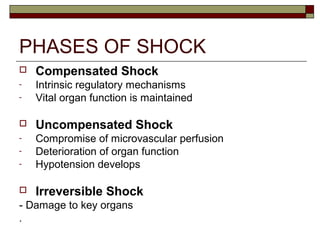
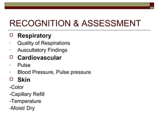
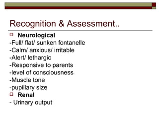
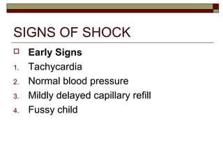
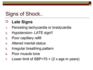
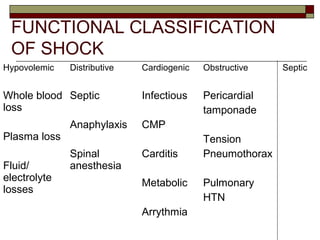
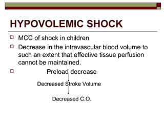
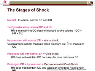
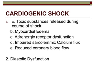
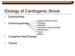
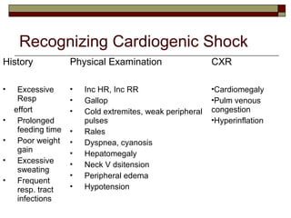
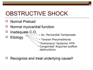
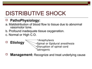
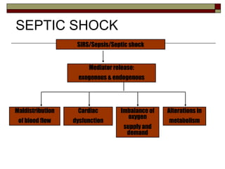
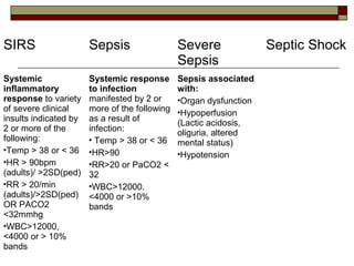
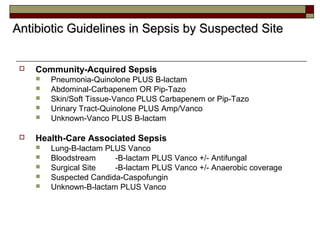
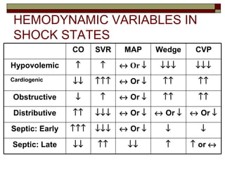
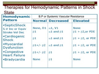
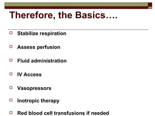
SHOCK IN CHILDREN
Sanju Susan Samuel
Fellow-Pediatric Critical Care Medicine
OBJECTIVES
 Definition
 Physiology
 Classification of Shock
 Common Etiologies
 Recognition and Assessment
 Management
DEFINITION
 An acute, complex state of circulatory
dysfunction that results in failure to deliver
sufficient amount of oxygen and nutrients to
meet tissue metabolic demands.
 Therefore, basically DO2 < VO2.
 If prolonged and left untreated- Can lead to
multiple organ failure and eventually death.
igure 1. FACTORS AFFECTING OXYGEN DELIVERY
DO2
CaO2
CO
SV
HR
Oxygenation
Hgb
A-a gradient
DPG
Acid-Base Balance
Blockers
Competitors
Temperature
Drugs
Conduction System
Ventricular
Compliance
EDV
ESV Contractility
CVP
Venous Volume
Venous Tone
Afterload Blockers
Temperature Competitors
Drugs Autonomic Tone
Metabolic Milieu
Ions
Acid Base
Temperature
Drugs
Toxins
Influenced By
Influenced By
Influenced By
Influenced By
What is needed to maintain
Perfusion??
 PUMP- Heart
 PIPES- Vessels
 FLUID- Blood
 Pump Failure
 Pipe Failure
 Loss of Volume
How can Perfusion fail??
Causes of Inadequate Perfusion
 Inadequate Pump
 Inadequate preload
 Poor contractility
 Excessive Afterload
 Inadequate HR
 Inadequate Fluid Volume
 Hypovolemia
 Inadequate Container
 Excessive Dilatation
 Inadequate systemic vascular resistance
So-what happens??
Anaerobic MetabolismAnaerobic MetabolismAnaerobic Metabolism
GLUCOSE METABOLISM
2 LACTIC ACID
2 ATP
HEAT (32 kcal)
Aerobic MetabolismAerobic MetabolismAerobic Metabolism
6 O2
GLUCOSE
METABOLISM
6 CO2
6 H2O
36 ATP
HEAT (417 kcal)
PHASES OF SHOCK
 Compensated Shock
- Intrinsic regulatory mechanisms
- Vital organ function is maintained
 Uncompensated Shock
- Compromise of microvascular perfusion
- Deterioration of organ function
- Hypotension develops
 Irreversible Shock
- Damage to key organs
.
RECOGNITION & ASSESSMENT
 Respiratory
- Quality of Respirations
- Auscultatory Findings
 Cardiovascular
- Pulse
- Blood Pressure, Pulse pressure
 Skin
-Color
-Capillary Refill
-Temperature
-Moist/ Dry
Recognition & Assessment..
 Neurological
-Full/ flat/ sunken fontanelle
-Calm/ anxious/ irritable
-Alert/ lethargic
-Responsive to parents
-level of consciousness
-Muscle tone
-pupillary size
 Renal
- Urinary output
SIGNS OF SHOCK
 Early Signs
1. Tachycardia
2. Normal blood pressure
3. Mildly delayed capillary refill
4. Fussy child
Signs of Shock..
 Late Signs
1. Persisting tachycardia or bradycardia
2. Hypotension- LATE sign!!
3. Poor capillary refill
4. Altered mental status
5. Irregular breathing pattern
6. Poor muscle tone
7. Lower limit of SBP=70 + (2 x age in years)
FUNCTIONAL CLASSIFICATION
OF SHOCK
Hypovolemic Distributive Cardiogenic Obstructive Septic
Whole blood
loss
Plasma loss
Fluid/
electrolyte
losses
Septic
Anaphylaxis
Spinal
anesthesia
Infectious
CMP
Carditis
Metabolic
Arrythmia
Pericardial
tamponade
Tension
Pneumothorax
Pulmonary
HTN
HYPOVOLEMIC SHOCK
 MCC of shock in children
 Decrease in the intravascular blood volume to
such an extent that effective tissue perfusion
cannot be maintained.
 Preload decrease
Decreased Stroke Volume
Decreased C.O.
Management of Hypovolemic
Shock
 Establishment of adequate oxygenation and
ventilation
 O2- ALWAYS the first drug administered.
 Adequate IV or IO
 Early correction of hypovolemia
-Crystalloids: Readily available, safe, least expensive
-First bolus 20cc/kg- ASAP
-Continuous monitoring of vitals
-Monitoring of CVP: Maintain > 10mmHg
-Identify causes of ongoing losses
- Blood available: if hemorrhagic shock.
Solution makeup
Osmol Glucose Na+ Cl- K+ Ca+ Lactate
 5% D/W 278 50g/l 0 0 0 0 0
 10%D/W 556 100g/l 0 0 0 0 0
 .45% NS 154 0 77 77 0 0 0
 .9% NS 308 0 154 154 0 0 0
 LR 274 0 130 109 4 1.5 28
Na, Cl, K, Ca, and lactate are measured in mmol/liter.
The Stages of Shock
Normal Eucardia, normal BP and CR
Tachycardia alone, normal BP and CR
HR is maintaining CO despite reduced stroke volume (CO =
HR x SV)
Hypotension with normal CR = Warm shock
Vascular tone cannot maintain blood pressure but HR maintains
CO
Prolonged CR with normal BP = Cold shock
HR does not maintain CO but vascular tone maintains BP
Prolonged CR + hypotension = Decompensated Cold Shock
HR does not maintain CO and vascular tone does not maintain
(Carcillo et al., Pediatrics 2009)(Slides are courtesy of Dr. Carcillo)
CARDIOGENIC SHOCK
1. a. Toxic substances released during
course of shock.
b. Myocardial Edema
c. Adrenergic receptor dysfunction
d. Impaired sarcolemmic Calcium flux
e. Reduced coronary blood flow
2. Diastolic Dysfunction
Pathophysiology
LV able to eject less volume of bld/ beat
Dec. Stroke Volume
Increased Venous Return
Increased EDV
Increased LV diastolic filling pressure
Backflow from LV to lungs
Dec.C.O
Increased O2
extraction by
tissues
Arterial O2
desaturation
Etiology of Cardiogenic Shock
 Dysrrhythmias
 Cardiomyopathies
 Congenital Heart Disease
 Trauma
Hypoxic-Ischemic event
Infectious
Metabolic
Connective Tissue Disorder
NM disorders
Toxins
Others
Recognizing Cardiogenic Shock
History Physical Examination CXR
 Excessive
Resp
effort
 Prolonged
feeding time
 Poor weight
gain
 Excessive
sweating
 Frequent
resp. tract
infections
 Inc HR, Inc RR
 Gallop
 Cold extremites, weak peripheral
pulses
 Rales
 Dyspnea, cyanosis
 Hepatomegaly
 Neck V dsitension
 Peripheral edema
 Hypotension
Cardiomegaly
Pulm venous
congestion
Hyperinflation
Managing Cardiogenic Shock…
Minimize Myocardial
O2 demands
Maximize Myocardial
Performance
Exclude & Explore
Intubation
Maintain normothermia
Provide sedation
Correct anemia
Correct Dysrhythmias
Optimize Preload
Improve Contractility
Reduce Afterload
Exclude traumatic or
CHD
Explore surgical options
OBSTRUCTIVE SHOCK
 Normal Preload
 Normal myocardial function
 Inadequate C.O.
 Etiology
 Recognize and treat underlying cause!!
Tension Pneumothorax
Pulmonary/ Systemic HTN
Congenital/ Acquired outflow
obstructions
Ac. Pericardial Tamponade
DISTRIBUTIVE SHOCK
 PathoPhysiology:
a. Maldistribution of blood flow to tissue due to abnormal
vasomotor tone.
b. Profound inadequate tissue oxygenation.
c. Normal or High C.O.
 Etiology
 Management: Recognize and treat underlying cause
Anaphylaxis
Spinal or Epidural anesthesia
Disruption of spinal cord
Iatrogenic
SIRS/Sepsis/Septic shock
Mediator release:
exogenous & endogenous
Maldistribution
of blood flow
Cardiac
dysfunction
Imbalance of
oxygen
supply and
demand
Alterations in
metabolism
SEPTIC SHOCK
ACUTE ORGAN
DYSFUNCTION
(Severe Sepsis)
DEATH
SEPSIS
SIRS Sepsis Severe
Sepsis
Septic Shock
Systemic
inflammatory
response to variety
of severe clinical
insults indicated by
2 or more of the
following:
Temp > 38 or < 36
HR > 90bpm
(adults)/ >2SD(ped)
RR > 20/min
(adults)/>2SD(ped)
OR PACO2
<32mmhg
WBC>12000,
<4000 or > 10%
bands
Systemic response
to infection
manifested by 2 or
more of the following
as a result of
infection:
 Temp > 38 or < 36
HR>90
RR>20 or PaCO2 <
32
WBC>12000.
<4000 or >10%
bands
Sepsis associated
with:
Organ dysfunction
Hypoperfusion
(Lactic acidosis,
oliguria, altered
mental status)
Hypotension
Warm Shock Cold Shock Fluid-Refractory/
Dopamine resistant
Catecholamine
Resistant
Refractory Shock
Early,
compensated
Clinical Signs
-Inc.HR
-Warm
extremities,
bounding pulses
Physiologic
Parameters
-Wide PP
-Inc. C.O.
-Inc. MvO2
-Dec.SVR
Lab Data
-Hypocardia
-Inc. Lactate
-Inc.Glucose
Late, Uncompensated
Clinical Signs
-Cold, clammy extremities
-Rapid, thready pulses
-Shallow breathing
Physiologic Parameters
-Narrow PP
-Dec.CVP, C.O
-Dec. MvO2 sat
-Inc. SVR
-Oliguria
-Capillary Leak
Lab Data
-Metab. Acidosis
-Hypoxia
-Coagulopathy
-Hypoglycemia
Persistance of shock
despite > 60cc/kg
fluid resuscitation
Persistance of shock
despite Dopamine at
>10mcg/kg/mn
Persistance of shock
despite administration of
direct acting
catecholamines
Epinephrine/
Nor-Epinephrine
Persistance of shock
despite:
-Goal direct inotropic/
pressor therapy
-Use of vasodilators
-Maintenance of
metabolic and
hormonal homeostasis
Early Goal directed therapy in treatment of sepsis and septic shock- Rivers et al., NEJM, Nov 2001
 Community-Acquired Sepsis
 Pneumonia-Quinolone PLUS B-lactam
 Abdominal-Carbapenem OR Pip-Tazo
 Skin/Soft Tissue-Vanco PLUS Carbapenem or Pip-Tazo
 Urinary Tract-Quinolone PLUS Amp/Vanco
 Unknown-Vanco PLUS B-lactam
 Health-Care Associated Sepsis
 Lung-B-lactam PLUS Vanco
 Bloodstream -B-lactam PLUS Vanco +/- Antifungal
 Surgical Site -B-lactam PLUS Vanco +/- Anaerobic coverage
 Suspected Candida-Caspofungin
 Unknown-B-lactam PLUS Vanco
Antibiotic Guidelines in Sepsis by Suspected SiteAntibiotic Guidelines in Sepsis by Suspected Site
HEMODYNAMIC VARIABLES IN
SHOCK STATES
↑ or ↔↑↓↓↑↑↓↓Septic: Late
↓↓↔ Or ↓↓↓↓↑↑↑Septic: Early
↔ Or ↓↔ Or ↓↔ Or ↓↓↓↓↑↑Distributive
↑↑↑↑↔ Or ↓↑↓Obstructive
↑↑↑↑↔ Or ↓↑↑↑↓↓Cardiogenic
↓↓↓↓↓↓↔ Or ↓↑↑Hypovolemic
CVPWedgeMAPSVRCO
B.P or Systemic Vascular Resistance
Therapies for Hemodynamic Patterns in Shock
State
Therefore, the Basics….
 Stabilize respiration
 Assess perfusion
 Fluid administration
 IV Access
 Vasopressors
 Inotropic therapy
 Red blood cell transfusions if needed
References
 Pediatric Critical Care: Fuhrman, Zimmerman
 Surviving Sepsis Guidelines
 E-medicine
 Uptodate online
 SCCM website

Shock in children-revppt

  • 1.
    SHOCK IN CHILDREN SanjuSusan Samuel Fellow-Pediatric Critical Care Medicine
  • 2.
    OBJECTIVES  Definition  Physiology Classification of Shock  Common Etiologies  Recognition and Assessment  Management
  • 3.
    DEFINITION  An acute,complex state of circulatory dysfunction that results in failure to deliver sufficient amount of oxygen and nutrients to meet tissue metabolic demands.  Therefore, basically DO2 < VO2.  If prolonged and left untreated- Can lead to multiple organ failure and eventually death.
  • 4.
    igure 1. FACTORSAFFECTING OXYGEN DELIVERY DO2 CaO2 CO SV HR Oxygenation Hgb A-a gradient DPG Acid-Base Balance Blockers Competitors Temperature Drugs Conduction System Ventricular Compliance EDV ESV Contractility CVP Venous Volume Venous Tone Afterload Blockers Temperature Competitors Drugs Autonomic Tone Metabolic Milieu Ions Acid Base Temperature Drugs Toxins Influenced By Influenced By Influenced By Influenced By
  • 5.
    What is neededto maintain Perfusion??  PUMP- Heart  PIPES- Vessels  FLUID- Blood  Pump Failure  Pipe Failure  Loss of Volume How can Perfusion fail??
  • 6.
    Causes of InadequatePerfusion  Inadequate Pump  Inadequate preload  Poor contractility  Excessive Afterload  Inadequate HR  Inadequate Fluid Volume  Hypovolemia  Inadequate Container  Excessive Dilatation  Inadequate systemic vascular resistance
  • 7.
    So-what happens?? Anaerobic MetabolismAnaerobicMetabolismAnaerobic Metabolism GLUCOSE METABOLISM 2 LACTIC ACID 2 ATP HEAT (32 kcal) Aerobic MetabolismAerobic MetabolismAerobic Metabolism 6 O2 GLUCOSE METABOLISM 6 CO2 6 H2O 36 ATP HEAT (417 kcal)
  • 8.
    PHASES OF SHOCK Compensated Shock - Intrinsic regulatory mechanisms - Vital organ function is maintained  Uncompensated Shock - Compromise of microvascular perfusion - Deterioration of organ function - Hypotension develops  Irreversible Shock - Damage to key organs .
  • 9.
    RECOGNITION & ASSESSMENT Respiratory - Quality of Respirations - Auscultatory Findings  Cardiovascular - Pulse - Blood Pressure, Pulse pressure  Skin -Color -Capillary Refill -Temperature -Moist/ Dry
  • 10.
    Recognition & Assessment.. Neurological -Full/ flat/ sunken fontanelle -Calm/ anxious/ irritable -Alert/ lethargic -Responsive to parents -level of consciousness -Muscle tone -pupillary size  Renal - Urinary output
  • 11.
    SIGNS OF SHOCK Early Signs 1. Tachycardia 2. Normal blood pressure 3. Mildly delayed capillary refill 4. Fussy child
  • 12.
    Signs of Shock.. Late Signs 1. Persisting tachycardia or bradycardia 2. Hypotension- LATE sign!! 3. Poor capillary refill 4. Altered mental status 5. Irregular breathing pattern 6. Poor muscle tone 7. Lower limit of SBP=70 + (2 x age in years)
  • 14.
    FUNCTIONAL CLASSIFICATION OF SHOCK HypovolemicDistributive Cardiogenic Obstructive Septic Whole blood loss Plasma loss Fluid/ electrolyte losses Septic Anaphylaxis Spinal anesthesia Infectious CMP Carditis Metabolic Arrythmia Pericardial tamponade Tension Pneumothorax Pulmonary HTN
  • 15.
    HYPOVOLEMIC SHOCK  MCCof shock in children  Decrease in the intravascular blood volume to such an extent that effective tissue perfusion cannot be maintained.  Preload decrease Decreased Stroke Volume Decreased C.O.
  • 16.
    Management of Hypovolemic Shock Establishment of adequate oxygenation and ventilation  O2- ALWAYS the first drug administered.  Adequate IV or IO  Early correction of hypovolemia -Crystalloids: Readily available, safe, least expensive -First bolus 20cc/kg- ASAP -Continuous monitoring of vitals -Monitoring of CVP: Maintain > 10mmHg -Identify causes of ongoing losses - Blood available: if hemorrhagic shock.
  • 17.
    Solution makeup Osmol GlucoseNa+ Cl- K+ Ca+ Lactate  5% D/W 278 50g/l 0 0 0 0 0  10%D/W 556 100g/l 0 0 0 0 0  .45% NS 154 0 77 77 0 0 0  .9% NS 308 0 154 154 0 0 0  LR 274 0 130 109 4 1.5 28 Na, Cl, K, Ca, and lactate are measured in mmol/liter.
  • 18.
    The Stages ofShock Normal Eucardia, normal BP and CR Tachycardia alone, normal BP and CR HR is maintaining CO despite reduced stroke volume (CO = HR x SV) Hypotension with normal CR = Warm shock Vascular tone cannot maintain blood pressure but HR maintains CO Prolonged CR with normal BP = Cold shock HR does not maintain CO but vascular tone maintains BP Prolonged CR + hypotension = Decompensated Cold Shock HR does not maintain CO and vascular tone does not maintain (Carcillo et al., Pediatrics 2009)(Slides are courtesy of Dr. Carcillo)
  • 19.
    CARDIOGENIC SHOCK 1. a.Toxic substances released during course of shock. b. Myocardial Edema c. Adrenergic receptor dysfunction d. Impaired sarcolemmic Calcium flux e. Reduced coronary blood flow 2. Diastolic Dysfunction
  • 20.
    Pathophysiology LV able toeject less volume of bld/ beat Dec. Stroke Volume Increased Venous Return Increased EDV Increased LV diastolic filling pressure Backflow from LV to lungs Dec.C.O Increased O2 extraction by tissues Arterial O2 desaturation
  • 21.
    Etiology of CardiogenicShock  Dysrrhythmias  Cardiomyopathies  Congenital Heart Disease  Trauma Hypoxic-Ischemic event Infectious Metabolic Connective Tissue Disorder NM disorders Toxins Others
  • 22.
    Recognizing Cardiogenic Shock HistoryPhysical Examination CXR  Excessive Resp effort  Prolonged feeding time  Poor weight gain  Excessive sweating  Frequent resp. tract infections  Inc HR, Inc RR  Gallop  Cold extremites, weak peripheral pulses  Rales  Dyspnea, cyanosis  Hepatomegaly  Neck V dsitension  Peripheral edema  Hypotension Cardiomegaly Pulm venous congestion Hyperinflation
  • 23.
    Managing Cardiogenic Shock… MinimizeMyocardial O2 demands Maximize Myocardial Performance Exclude & Explore Intubation Maintain normothermia Provide sedation Correct anemia Correct Dysrhythmias Optimize Preload Improve Contractility Reduce Afterload Exclude traumatic or CHD Explore surgical options
  • 24.
    OBSTRUCTIVE SHOCK  NormalPreload  Normal myocardial function  Inadequate C.O.  Etiology  Recognize and treat underlying cause!! Tension Pneumothorax Pulmonary/ Systemic HTN Congenital/ Acquired outflow obstructions Ac. Pericardial Tamponade
  • 25.
    DISTRIBUTIVE SHOCK  PathoPhysiology: a.Maldistribution of blood flow to tissue due to abnormal vasomotor tone. b. Profound inadequate tissue oxygenation. c. Normal or High C.O.  Etiology  Management: Recognize and treat underlying cause Anaphylaxis Spinal or Epidural anesthesia Disruption of spinal cord Iatrogenic
  • 26.
    SIRS/Sepsis/Septic shock Mediator release: exogenous& endogenous Maldistribution of blood flow Cardiac dysfunction Imbalance of oxygen supply and demand Alterations in metabolism SEPTIC SHOCK
  • 27.
  • 28.
    SIRS Sepsis Severe Sepsis SepticShock Systemic inflammatory response to variety of severe clinical insults indicated by 2 or more of the following: Temp > 38 or < 36 HR > 90bpm (adults)/ >2SD(ped) RR > 20/min (adults)/>2SD(ped) OR PACO2 <32mmhg WBC>12000, <4000 or > 10% bands Systemic response to infection manifested by 2 or more of the following as a result of infection:  Temp > 38 or < 36 HR>90 RR>20 or PaCO2 < 32 WBC>12000. <4000 or >10% bands Sepsis associated with: Organ dysfunction Hypoperfusion (Lactic acidosis, oliguria, altered mental status) Hypotension
  • 29.
    Warm Shock ColdShock Fluid-Refractory/ Dopamine resistant Catecholamine Resistant Refractory Shock Early, compensated Clinical Signs -Inc.HR -Warm extremities, bounding pulses Physiologic Parameters -Wide PP -Inc. C.O. -Inc. MvO2 -Dec.SVR Lab Data -Hypocardia -Inc. Lactate -Inc.Glucose Late, Uncompensated Clinical Signs -Cold, clammy extremities -Rapid, thready pulses -Shallow breathing Physiologic Parameters -Narrow PP -Dec.CVP, C.O -Dec. MvO2 sat -Inc. SVR -Oliguria -Capillary Leak Lab Data -Metab. Acidosis -Hypoxia -Coagulopathy -Hypoglycemia Persistance of shock despite > 60cc/kg fluid resuscitation Persistance of shock despite Dopamine at >10mcg/kg/mn Persistance of shock despite administration of direct acting catecholamines Epinephrine/ Nor-Epinephrine Persistance of shock despite: -Goal direct inotropic/ pressor therapy -Use of vasodilators -Maintenance of metabolic and hormonal homeostasis
  • 31.
    Early Goal directedtherapy in treatment of sepsis and septic shock- Rivers et al., NEJM, Nov 2001
  • 32.
     Community-Acquired Sepsis Pneumonia-Quinolone PLUS B-lactam  Abdominal-Carbapenem OR Pip-Tazo  Skin/Soft Tissue-Vanco PLUS Carbapenem or Pip-Tazo  Urinary Tract-Quinolone PLUS Amp/Vanco  Unknown-Vanco PLUS B-lactam  Health-Care Associated Sepsis  Lung-B-lactam PLUS Vanco  Bloodstream -B-lactam PLUS Vanco +/- Antifungal  Surgical Site -B-lactam PLUS Vanco +/- Anaerobic coverage  Suspected Candida-Caspofungin  Unknown-B-lactam PLUS Vanco Antibiotic Guidelines in Sepsis by Suspected SiteAntibiotic Guidelines in Sepsis by Suspected Site
  • 33.
    HEMODYNAMIC VARIABLES IN SHOCKSTATES ↑ or ↔↑↓↓↑↑↓↓Septic: Late ↓↓↔ Or ↓↓↓↓↑↑↑Septic: Early ↔ Or ↓↔ Or ↓↔ Or ↓↓↓↓↑↑Distributive ↑↑↑↑↔ Or ↓↑↓Obstructive ↑↑↑↑↔ Or ↓↑↑↑↓↓Cardiogenic ↓↓↓↓↓↓↔ Or ↓↑↑Hypovolemic CVPWedgeMAPSVRCO
  • 34.
    B.P or SystemicVascular Resistance Therapies for Hemodynamic Patterns in Shock State
  • 35.
    Therefore, the Basics…. Stabilize respiration  Assess perfusion  Fluid administration  IV Access  Vasopressors  Inotropic therapy  Red blood cell transfusions if needed
  • 36.
    References  Pediatric CriticalCare: Fuhrman, Zimmerman  Surviving Sepsis Guidelines  E-medicine  Uptodate online  SCCM website

Editor's Notes

  • #16 Activa
  • #20 Diastolic dysfunction-Impaired myocardial relaxation changes the press-vol raio during diastole and increases ventricular pressure at any vol. This lack of relaxation is hemodynamically unfavorable because Inc LV diast pressure is transmitted to the lung anf results in pulm edema and dyspnea. Elev LV diastolic press also dec myocardial perfusion pressure and can lead to subendocardial iscehmia. Can have normal &amp;lt;V systolic function.
  • #24 Optimize Preload- Salt and water restriction, Fluid challenges, diuretics/venodilators for congestion Improve Contractility- Provide O2, guarantee ventilation, correct acidosis and other metaboilc derangements, inotriopic drugs Reduce Afterload- Provide sedation, pain relief, correct hypothermia, vasodilators