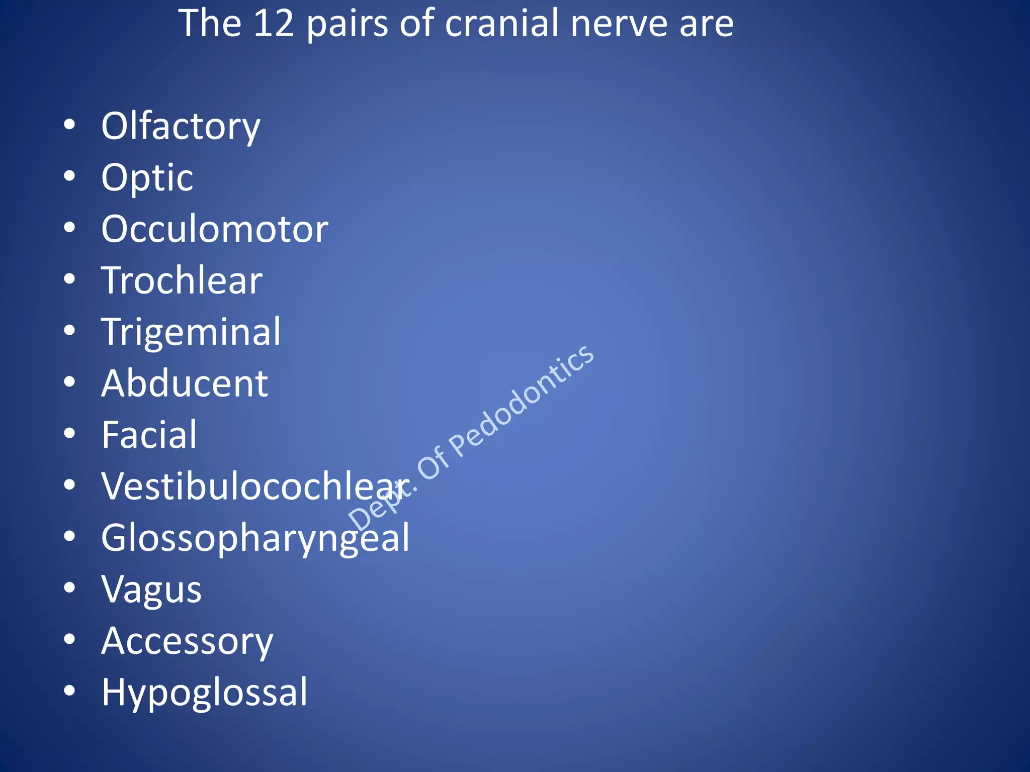
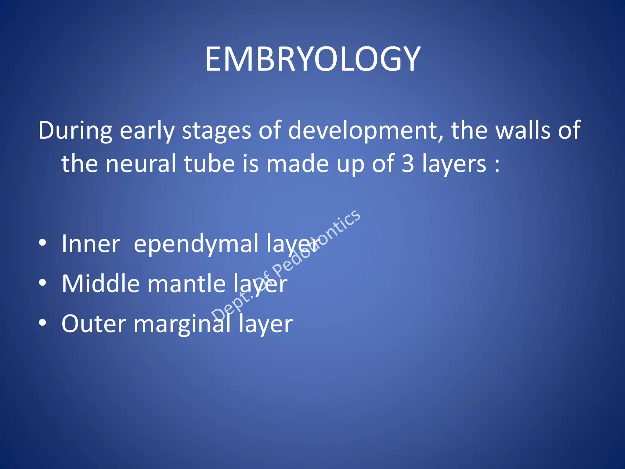
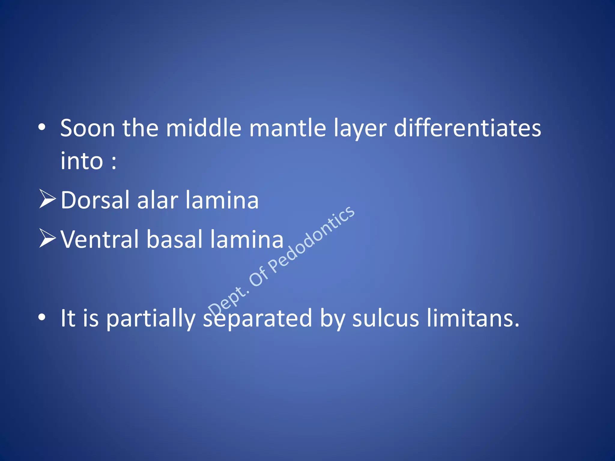
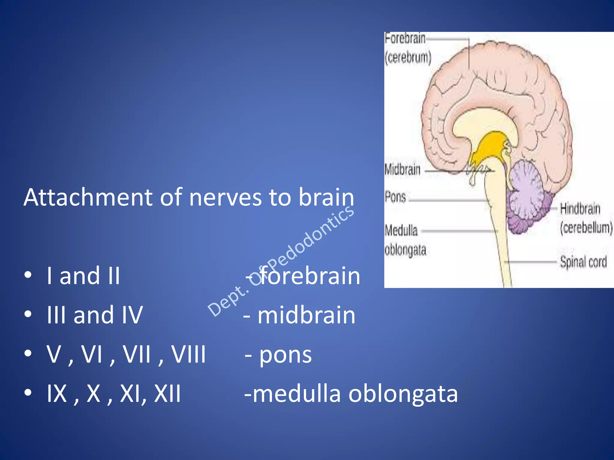
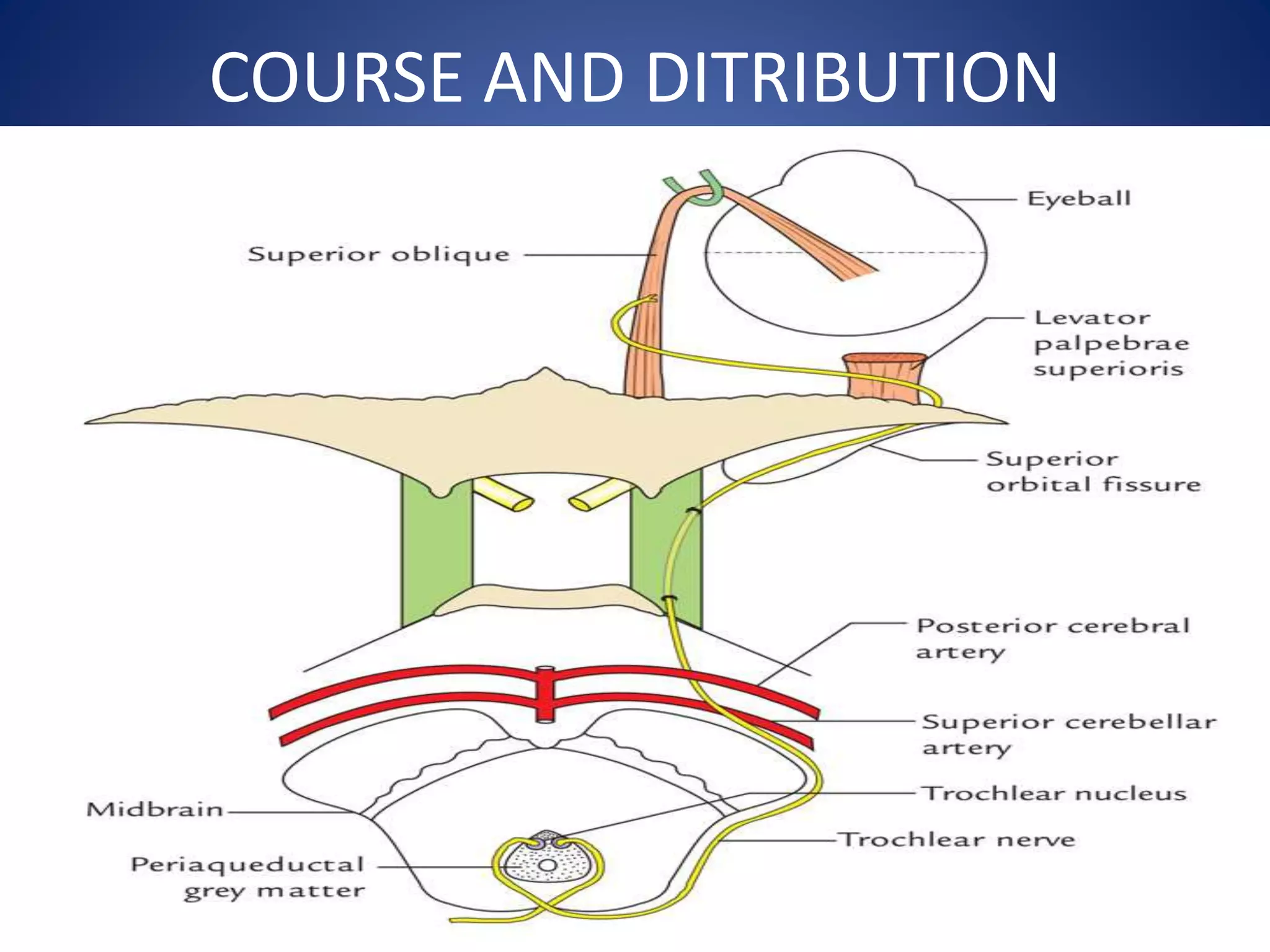
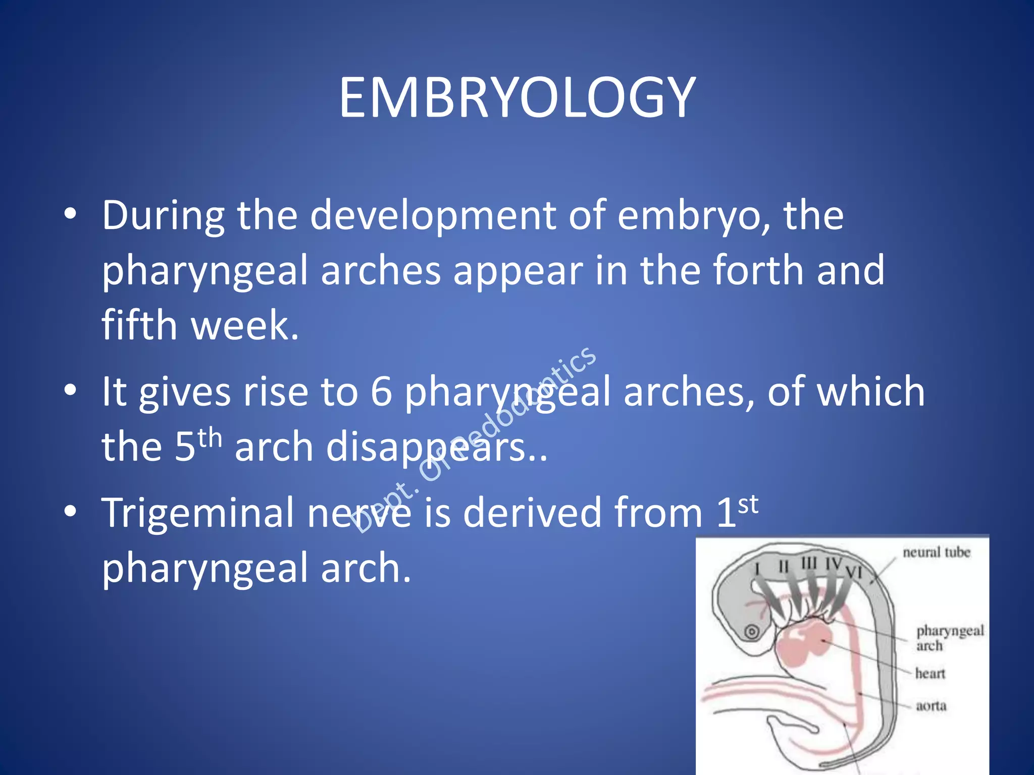
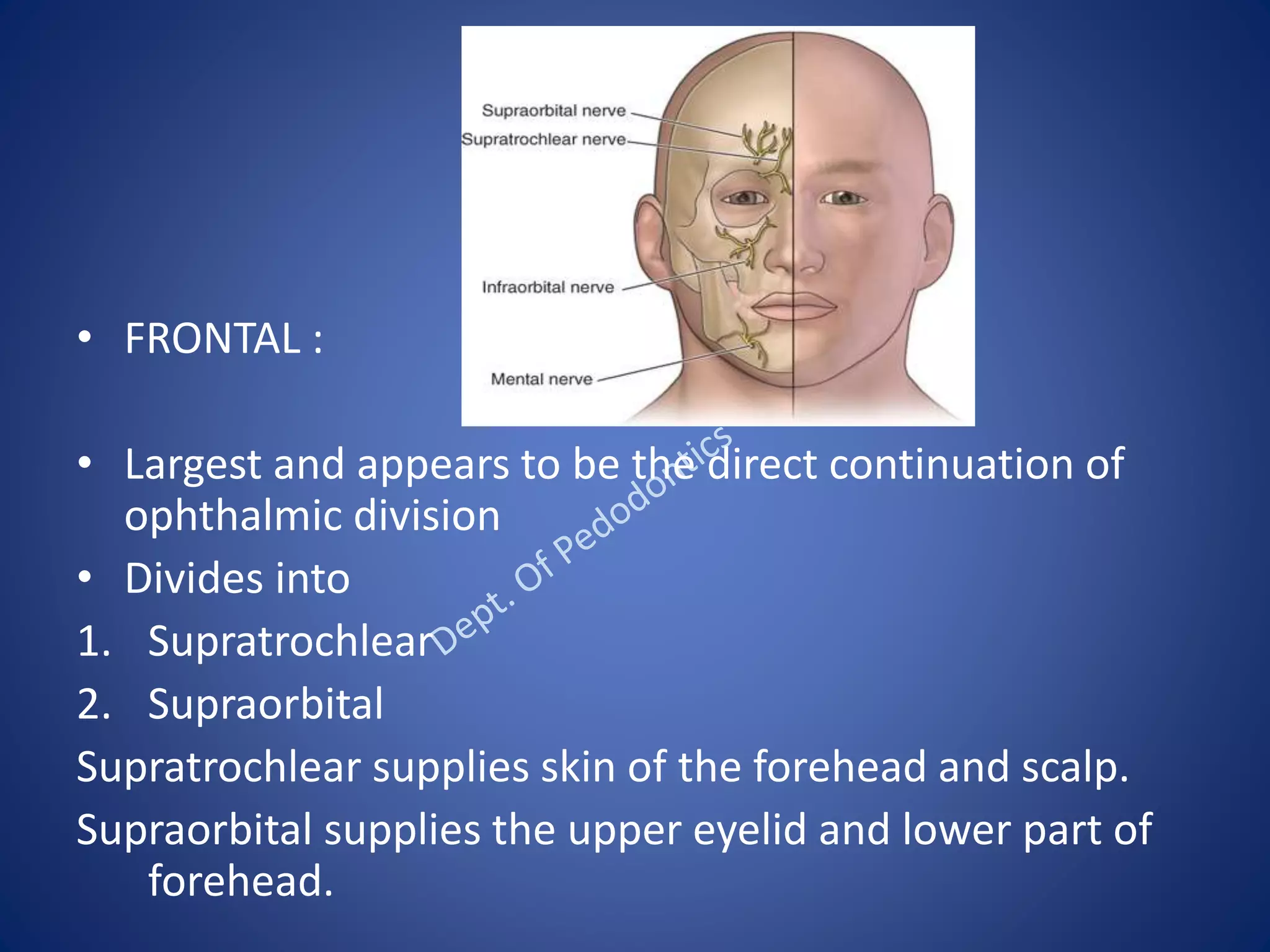
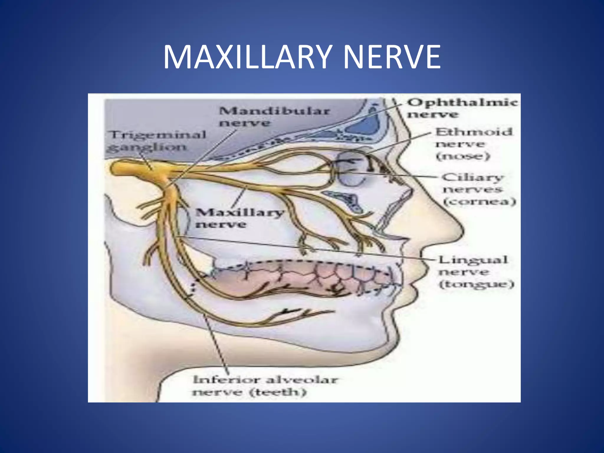
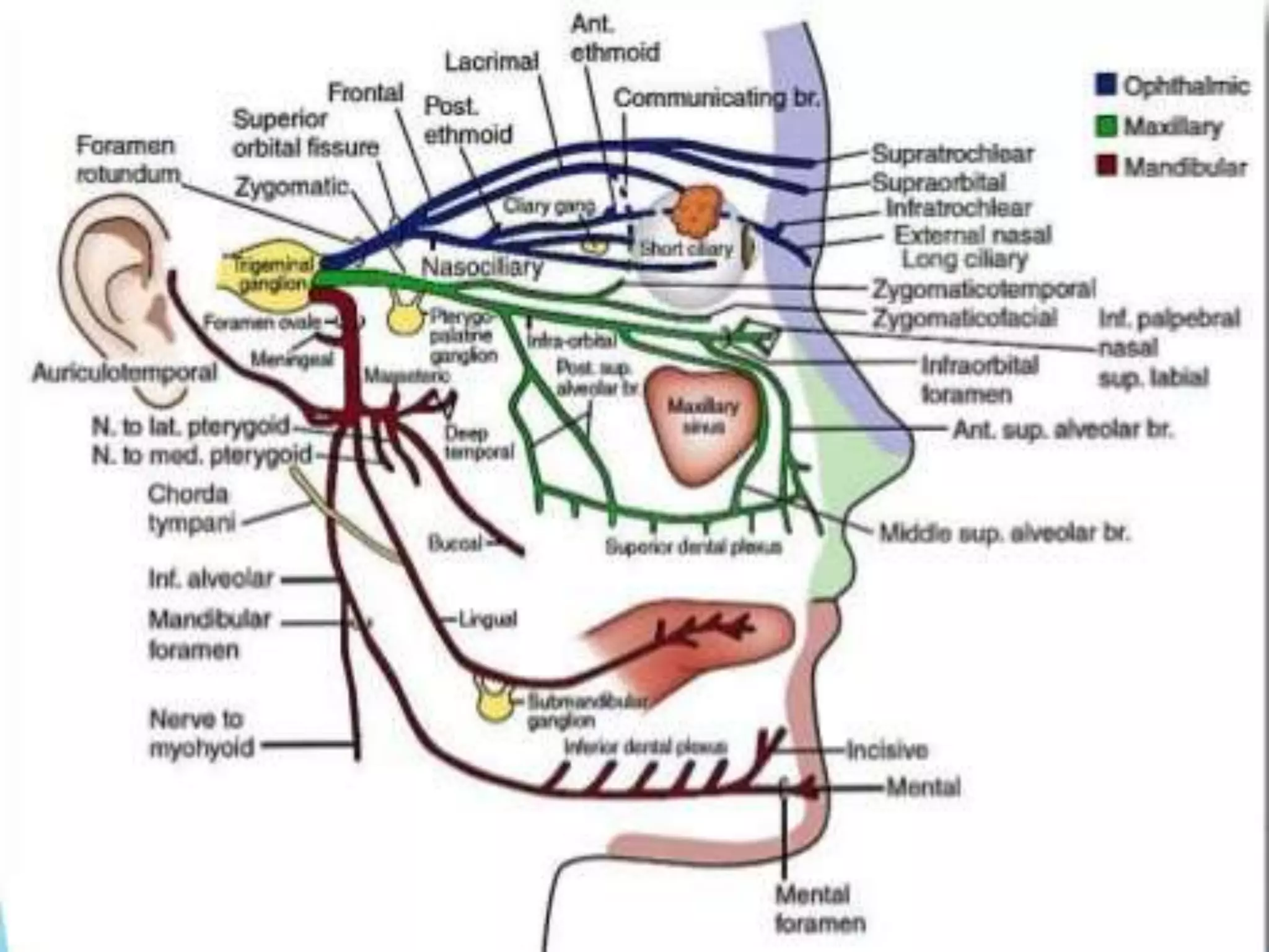
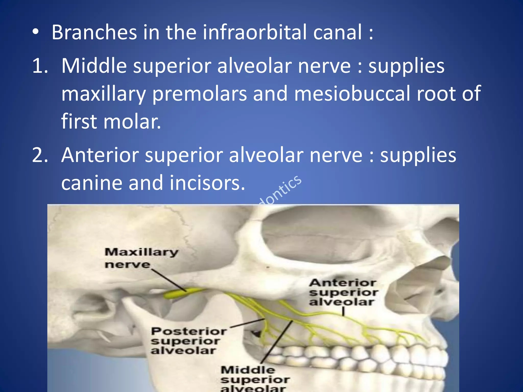
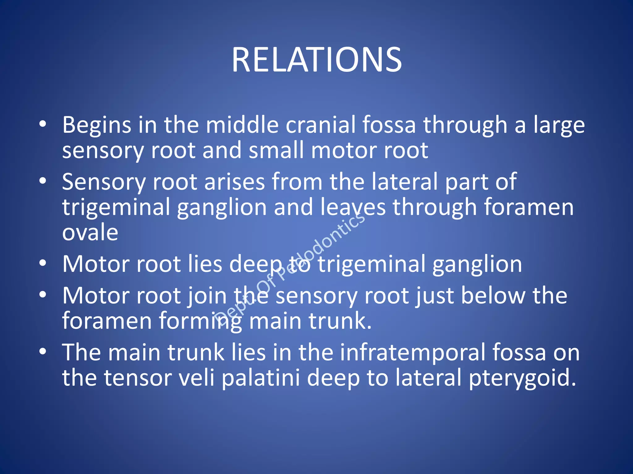
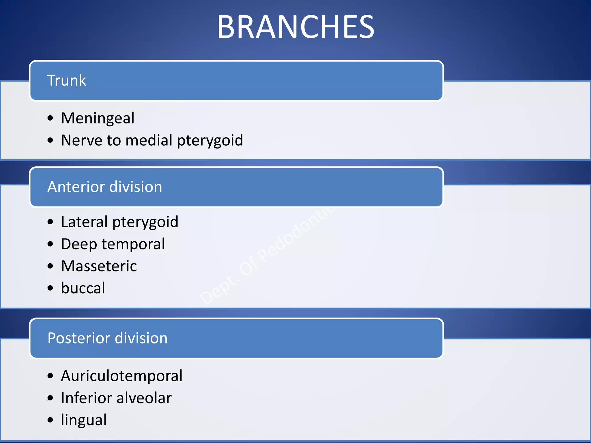
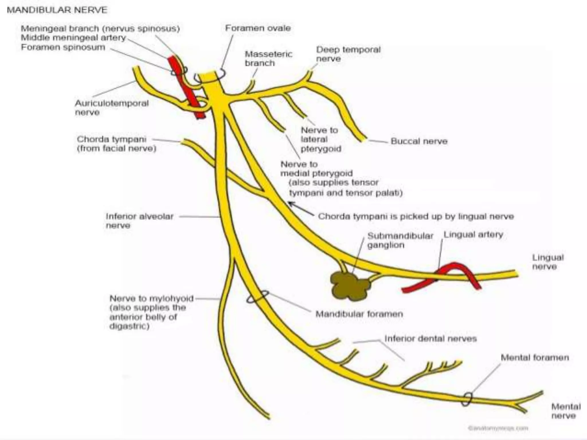
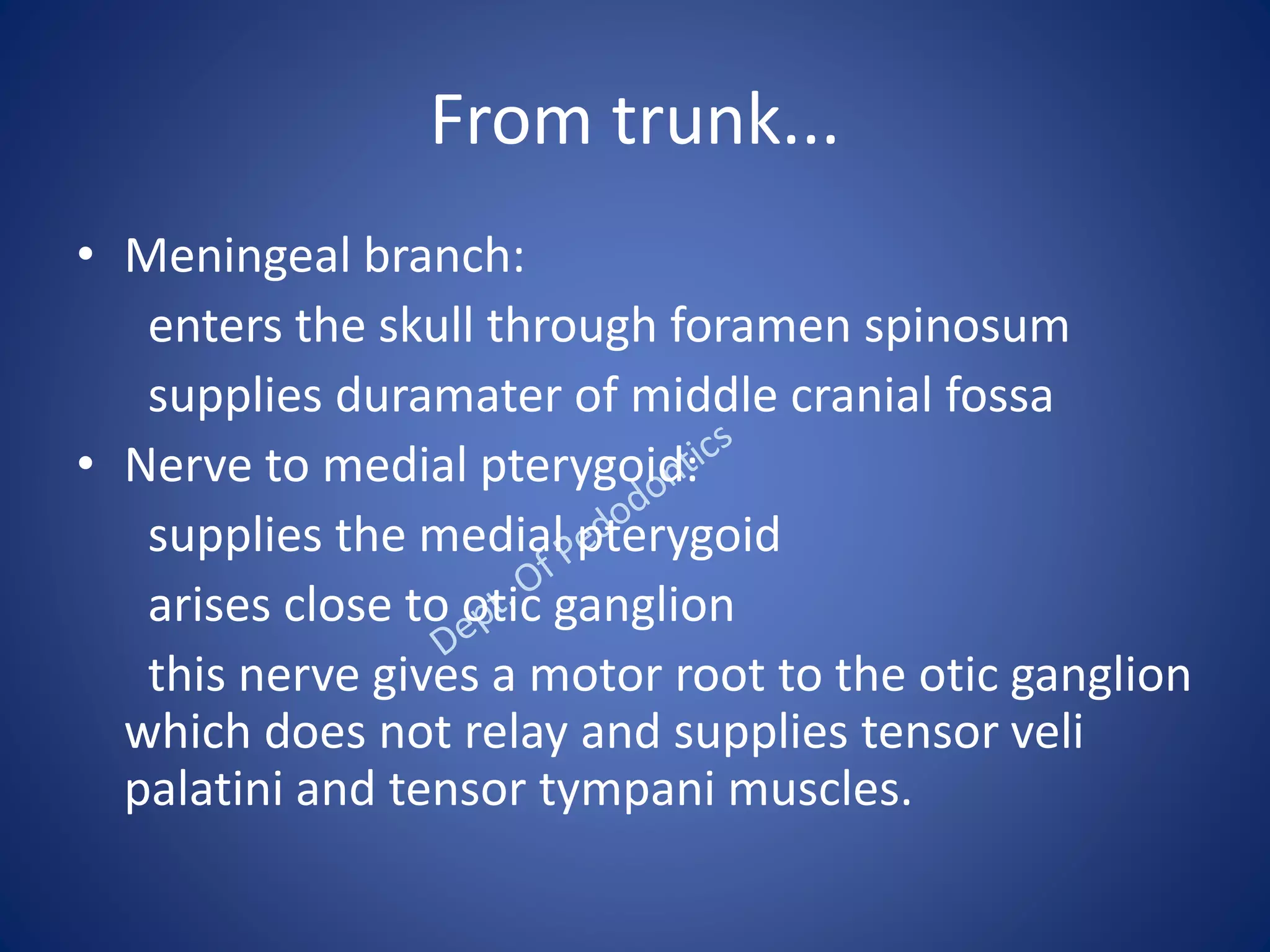
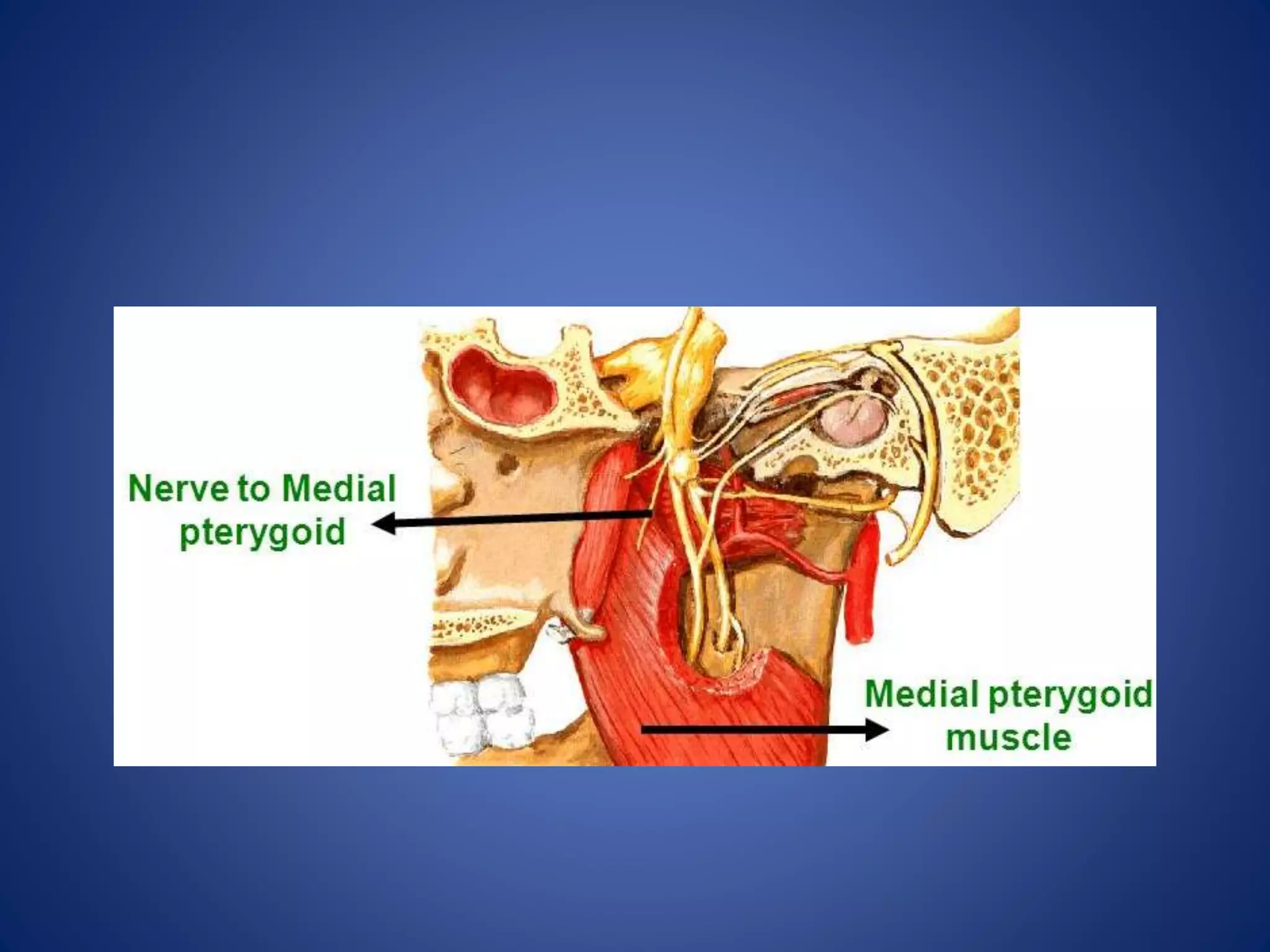
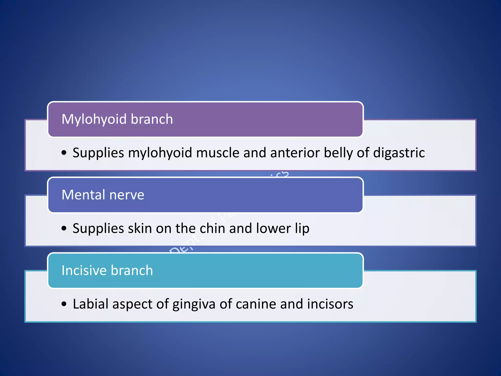
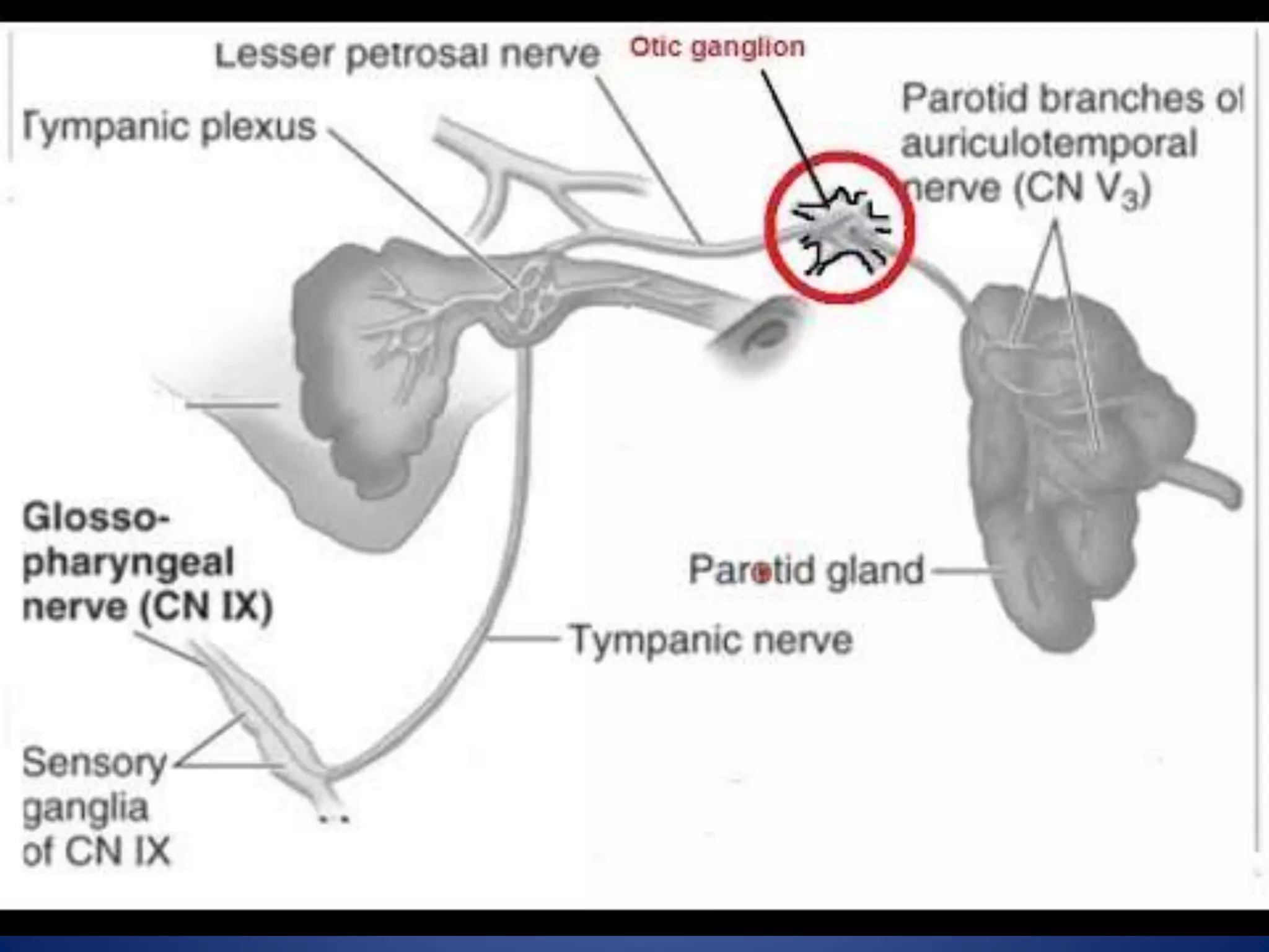
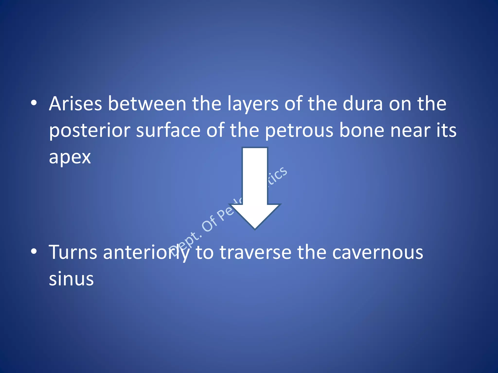
CRANIAL NERVES
Dr Suraj Nair
Senior Lecturer
Dept. Of Pedodontics
Malabar dental college
INTRODUCTION
• There are 12 pairs of cranial nerves
• A cranial nerve is made up of single or
multiple function called modality
• Each modality is sensory or motor in nature.
The 12 pairs of cranial nerve are
• Olfactory
• Optic
• Occulomotor
• Trochlear
• Trigeminal
• Abducent
• Facial
• Vestibulocochlear
• Glossopharyngeal
• Vagus
• Accessory
• Hypoglossal
EMBRYOLOGY
During early stages of development, the walls of
the neural tube is made up of 3 layers :
• Inner ependymal layer
• Middle mantle layer
• Outer marginal layer
• Soon the middle mantle layer differentiates
into :
Dorsal alar lamina
Ventral basal lamina
• It is partially separated by sulcus limitans.
Attachment of nerves to brain
• I and II - forebrain
• III and IV - midbrain
• V , VI , VII , VIII - pons
• IX , X , XI, XII -medulla oblongata
OLFACTORY NERVE
• It is sensory nerve
• Carries impulses of smell
• ORIGIN : olfactory epithelium
OLFACTORY CELL
Olfactory cells are 16-20 million in number
Olfactory cell present in the nasal mucosa serves
as first order neuron in olfactory pathway.
OLFACTORY NERVE
• Olfactory nerves are 20 in number
• Represent central processes of olfactory cells
• They pass through cribriform plate of ethmoid
and club with cells of olfactory bulb.
OLFACTION AND CODING
• In the olfactory epithelium, odorants bind to
specific receptors on the primary olfactory
neurons. This leads to the activation of a G
protein ,which activates adenylyl cyclase ,
which in turn produces cyclic adenosine
monophophate from ATP and activates an ion
channel that is permeable to cations Na+ and
Ca2+. This leads to a shift in membrane
potential leading to depolarization and
production of action potential.
CLINICAL ANATOMY
• ANOSMIA : Loss of olfactory fibres with
ageing.
• Head injury causes damage to olfactory bulbs
resulting in anosmia and CNS rhinorrhea.
• Abscess of frontal lobe of brain compresses
olfactory bulb resulting in anosmia.
OPTIC NERVE
FIELD OF VISION
It includes 4 fields :
• Upper temporal
• Lower temporal
• Upper nasal
• Lower nasal
RETINA
• Cells of the retina represents first order
neurons
• They receive impulses from the rods and
cones present in the retina
• Temporal field is seen by the nasal hemiretina
and vice versa.
RETINA
VISUAL PATHWAY
PUPILLARY LIGHT REFLEX
ACCOMMODATION REFLEX
CONJUNCTIVAL REFLEX
CLINICAL ANATOMY
• Lesions in retina leads to scotoma that is
certain points may become blind spots.
• Loss of vision in one half of visual field –
hemianopia.
• Optic nerve damage results in complete
blindness of that eye.
OCCULOMOTOR NERVE
• Distributed to extraocular and intraocular mucles.
• It is motor nerve
• ORIGIN : anterior surface of midbrain
• The nuclei associated is oculomotor nucleus in
the grey matter of midbrain.
COURSE AND DISTRIBUTION
CLINICAL ANATOMY
• Lesions lead to drooping of the eyelid due to
paralysis of levator palpebrae superioris
muscle
• Lateral squint
• Diplopia
• Ptosis
• Loss of accommodation
• Dilatation of the pupil
GOOD MORNING
TROCHLEAR NERVE
• 4 th cranial nerve
• Supplies only the superior oblique muscle of
the eyeball.
• Nuclei associated with this nerve is trochlear
nucleus
• The trochlear nucleus is situated in the
ventromedial part of the central grey matter
of midbrain at the level of inferior colliculus.
COURSE
COURSE AND DITRIBUTION
CLINICAL ANATOMY
• When trochlear nerve is damaged diplopia
occurs.
• Paralysis of the trochlear nerve results in :
defective depression of the adducted eye
and also diplopia.
TRIGEMINAL NERVE
EMBRYOLOGY
• During the development of embryo, the
pharyngeal arches appear in the forth and
fifth week.
• It gives rise to 6 pharyngeal arches, of which
the 5th arch disappears..
• Trigeminal nerve is derived from 1st
pharyngeal arch.
INTRODUCTION
• The largest cranial nerve
• It is a mixed nerve (sensory and motor)
• Sensory to – the skin of the face
mucosa of the cranial viscera
• Motor to – mucles of mastication
anterior belly of digatric
mylohyoid
tensor veli palatini
and tensor tympani
SENSORY NUCLEI
• MESENCEPHALIC NUCLEUS
 extends from pons till midbrain
 receives proprioceptive impulses from muscles
of mastication, temperomandibular joint and
teeth
• SUPERIOR SENSORY NUCLEUS
 fibers carrying touch and pressure relay in this
nucleus
• SPINAL NUCLEUS
 it takes pain and temperature sensations
from most of the face area
MOTOR NUCLEUS
• Innervates mucles of mastication , tensor veli
palatini and tensor tympani
• Located in the pons
• Derived from from first branchial arch
COURSE AND DISTRIBUTION
• Both sensory and motor root are attached
ventrally to junction of pons and middle
cerebellar peduncle with motor root lying
ventromedially to the sensory root.
• Pass anteriorly in middle cranial fossa to lie
below tentorium cerebelli in cavum
trigeminale, here motor root lies inferior to
sensory root
• Sensory root connected to postromedial
concave border of the trigeminal ganglion.
• Convex antrolateral margin of the ganglion
gives attachment to the 3 divisions of the
trigeminal nerve.
• Motor root turns further inferior with sensory
component to emerge out of foramen ovale as
Mandibular nerve.
• Ophthalmic and Maxillary division emerges
through superior orbital fissure and foramen
rotundum respectively.
GANGLION
• SEMILUNAR GANGLION
• GASSERIAN GANGLION
• Contains cell bodies of pseudounipolar
neuron.
• Lies in a bony fossa at apex of the petrous
temporal bone on floor of middle cranial fossa
just lateral to posterior part of lateral wall of
the cavernous sinus.
• Coverings : covered by dural pouch that is the
Meckels cave
• Cave is lined by piamater and arachnoid
• Thus the ganglion is bathed in CSF
• ARTERIAL SUPPLY : Ganglionic branches of
Internal carotid artery , Middle meningeal
artery and Accessory meningeal artery.
RELATIONS
• MEDIALLY : internal carotid artery and the
posterior part of cavernous sinus
• LATERALLY : middle meningeal artery
• SUPERIORLY : parahippocampal gyrus
• INFERIORLY : motor root of trigeminal nerve ,
greater petrosal nerve , apex of petrous
temporal bone and foramen lacerum
DIVISIONS
• OPHTHALMIC NERVE
• MAXILLARY NERVE
• MANDIBULAR NERVE
OPHTHALMIC NERVE
• Smallest division
• Sensory only
• Supplies eyeball, conjuctiva , lacrimal gland ,
mucosa of nose and paranasal sinus , skin of
forehead , eyelid .
COURSE
• BRANCHES
Frontal
Naociliary
Lacrimal
LACRIMAL NERVE:
• Smallest
• Supplies lacrimal gland and the conjuctiva
• It pierces the orbital septum and ends in the
skin of the upper eyelid
• FRONTAL :
• Largest and appears to be the direct continuation of
ophthalmic division
• Divides into
1. Supratrochlear
2. Supraorbital
Supratrochlear supplies skin of the forehead and scalp.
Supraorbital supplies the upper eyelid and lower part of
forehead.
• NASOCILLIARY :
• Intermediate in size
• Its branches are divided as following:
1. Branches in the orbit
2. Branches in the nasal cavity
3. Branches on the face
• Branches in the orbit:
1. Long root of ciliary ganglion :eyeball
2. Long ciliary nerve : iris and cornea
3. Posterior ethmoidal nerve : mucous
membrane lining of the posterior ethmoidal
and sphenoidal paranasal air cells.
4. Anterior ethmoidal nerve: anterior ethmoid
and frontal paranasal air cells.
• Branches in the nasal cavity :
Branches arising here supply the mucous
membrane of the nasal cavity.
• Terminal branches on the face :
They supply skin of both eyelid , lacrimal sac and
skin on the bridge of the nose.
MAXILLARY NERVE
• Sensory division
• 2nd division of trigeminal nerve
• Supplies derivatives of maxillary process and
frontonasal process
•Meningeal branches
In cranial cavity
•2 ganglionic branches
Pterygopalatine fossa
•Posterior superior alveolar
•Zygomatic nerve
In infra temporal fosa
•Anterior superior alveolar
•Middlesuperior alveolar
In infraorbital canal
•Labial
•Nasal
•palpebral
In face
• Branches given off on the cranium :
1. Meningeal branch : duramater of the anterior
and middle cranial fossae.
• Branches in the pterygopalatine fossa :
1. Ganglionic branch : contains secretomotor fibres
to the lacrimal gland .They provide sensory
fibres to the orbital periosteum and mucous
membrane of the nose , palate and pharynx.
In infratemporal fossa
1 . Zygomatic nerve : skin over the zygomatic
bone. It divides into zygomaticofacial and
zygomaticotemporal.
2 . Posterior superior alveolar nerve : supply 3
molar teeth except mesiobuccal root of first
molar.
• Branches in the infraorbital canal :
1. Middle superior alveolar nerve : supplies
maxillary premolars and mesiobuccal root of
first molar.
2. Anterior superior alveolar nerve : supplies
canine and incisors.
• Branches given on the face:
1. Palpebral branches : skin over the lower
eyelid and lateral angle ofthe eye.
2. Nasal branches : skin of the face and tip of
the nasal septum.
3. Superior labial branches : supplies cheek and
upper lip.
MANDIBULAR NERVE
• Largest
• Mixed nerve
• Nerve of the first branchial arch
RELATIONS
• Begins in the middle cranial fossa through a large
sensory root and small motor root
• Sensory root arises from the lateral part of
trigeminal ganglion and leaves through foramen
ovale
• Motor root lies deep to trigeminal ganglion
• Motor root join the sensory root just below the
foramen forming main trunk.
• The main trunk lies in the infratemporal fossa on
the tensor veli palatini deep to lateral pterygoid.
BRANCHES
• Meningeal
• Nerve to medial pterygoid
Trunk
• Lateral pterygoid
• Deep temporal
• Masseteric
• buccal
Anterior division
• Auriculotemporal
• Inferior alveolar
• lingual
Posterior division
From trunk...
• Meningeal branch:
enters the skull through foramen spinosum
supplies duramater of middle cranial fossa
• Nerve to medial pterygoid:
supplies the medial pterygoid
arises close to otic ganglion
this nerve gives a motor root to the otic ganglion
which does not relay and supplies tensor veli
palatini and tensor tympani muscles.
From the anterior division...
• Buccal nerve :
 only sensory branch of anterior division
 supplies skin of cheek and mucous
membrane of buccinator.
 Also supplies labial aspect of gingiva of
molars and premolars
• Deep temporal nerve :
Supplies temporalis muscle
• Nerve to lateral pterygoid :
Supplies lateral pterygoid
• Masseteric nerve
 supplies masseter and TMj
Emerges at the upper border of lateral pterygoid
From poterior division..
Auriculotemporal nerve :
• Auricular part supplies skin of the
tragus,upper pinna,external acoustic meatus
• Temporal part supplies skin of the temple
• Auriculotemporal nerve also supplies parotid
gland and TMJ
LINGUAL NERVE :
• Sensory to anterior 2/3rd of tongue.
• Supplies floor of oral cavity.
• The fibres of chorda tympani which is
secretomotor to submandibular and
sublingual salivary glands is also distributed
through lingual nerve.
• Begins from 1cm below the skull
• About 2 cm below the skull it is joined by
chorda tympani
• Lies in contact with mandible
• Medial to 3rd molar
INFERIOR ALVEOLAR NERVE :
• Largest terminal nerve
• It enters the mandibular foramen and runs in
the mandibular canal
• It is accompanied by inferior alveolar artery.
• Supplies mylohyoid muscle and anterior belly of digastric
Mylohyoid branch
• Supplies skin on the chin and lower lip
Mental nerve
• Labial aspect of gingiva of canine and incisors
Incisive branch
OTIC GANGLION
• Small, oval shaped, flattened ganglion
• Situated immediately below the foramen
ovale
• Lies on the medial surface of the mandibular
nerve and surrounds the origin of the nerve to
medial pterygoid.
CLINICAL ANATOMY
• IN INJURY TO :
OPHTHALMIC NERVE : There is loss of corneal
blink reflex.
MAXILLARY NERVE : There is loss of sneeze
reflex.
MANDIBULAR NERVE : There is loss of jaw
jerk reflex.
• TRIGEMINAL NEURALGIA :
Pain along the distribution of the nerve
Caused due to local lesion or unknown cause
Sharp shooting pain which lasts for few
seconds
Has trigger zones
Maxillary nerve is most frequently involved
• The Trigeminal ganglion harbours the Herpes
zoster virus causing shingles in the distribution
of nerve.
• Flaccid paralysis of muscles of mastication in
injury of mandibular nerve leading to
decrease strength for biting.
Mandibular nerve
• Reffered pain : in cases of cancer of
tongue,pain radiates to ear and to temporal
fossa, over the ditribution of auriculotemporal
nerve.
• Lingual nerve lies in contact with mandible,
medial to 3rd molar,in case of extraction of it
care must be taken not to injure nerve.
• In extraction of mandibular teeth , inferior
alveolar nerve needs to be anaesthetised .
The drug is given before it enters the
mandibular canal
• Inferior alveolar nerve as it travels through the
mandibular canal can be damaged by the
fracture of the mandible .This can be assessed
by testing sensation over the chin.
ABDUCENT NERVE
• 6th cranial nerve
• Supplies the lateral rectus muscle of the
eyeball.
• Nuclei associated with this nerve is the
abducent nucleus.
• Nucleus is situated in the upper part of the
floor of fourth ventricle in the lower pons.
COURSE
• Passes upward and anterolaterally in the
subarachnoid space of posterior cranial fossa
• Pierces the arachnoid and dura lateral to the
dorum sellae (part of the sphenoid bone)
• Arises between the layers of the dura on the
posterior surface of the petrous bone near its
apex
• Turns anteriorly to traverse the cavernous
sinus
• Enters the orbit through the superior orbital
fissure within the annular tendon to supply
the lateral rectus muscle
BLOOD SUPPLY
• The majority of the abducens nerves were
supplied by the anterolateral arteries , and
only some of them by the anterior inferior
cerebellar artery or the pontomedullary
artery.
CLINICAL ANATOMY
• Sixth nerve paralysis is one of the commonest
false localizing signs in cases with raised
intracranial pressure.
• Diplopia occurs
• Causes failure of abduction of the affected eye
THANK YOU

Seminar cranial nerves

  • 1.
    CRANIAL NERVES Dr SurajNair Senior Lecturer Dept. Of Pedodontics Malabar dental college
  • 2.
    INTRODUCTION • There are12 pairs of cranial nerves • A cranial nerve is made up of single or multiple function called modality • Each modality is sensory or motor in nature.
  • 3.
    The 12 pairsof cranial nerve are • Olfactory • Optic • Occulomotor • Trochlear • Trigeminal • Abducent • Facial • Vestibulocochlear • Glossopharyngeal • Vagus • Accessory • Hypoglossal
  • 4.
    EMBRYOLOGY During early stagesof development, the walls of the neural tube is made up of 3 layers : • Inner ependymal layer • Middle mantle layer • Outer marginal layer
  • 5.
    • Soon themiddle mantle layer differentiates into : Dorsal alar lamina Ventral basal lamina • It is partially separated by sulcus limitans.
  • 6.
    Attachment of nervesto brain • I and II - forebrain • III and IV - midbrain • V , VI , VII , VIII - pons • IX , X , XI, XII -medulla oblongata
  • 7.
  • 8.
    • It issensory nerve • Carries impulses of smell • ORIGIN : olfactory epithelium
  • 9.
    OLFACTORY CELL Olfactory cellsare 16-20 million in number Olfactory cell present in the nasal mucosa serves as first order neuron in olfactory pathway.
  • 10.
    OLFACTORY NERVE • Olfactorynerves are 20 in number • Represent central processes of olfactory cells • They pass through cribriform plate of ethmoid and club with cells of olfactory bulb.
  • 12.
    OLFACTION AND CODING •In the olfactory epithelium, odorants bind to specific receptors on the primary olfactory neurons. This leads to the activation of a G protein ,which activates adenylyl cyclase , which in turn produces cyclic adenosine monophophate from ATP and activates an ion channel that is permeable to cations Na+ and Ca2+. This leads to a shift in membrane potential leading to depolarization and production of action potential.
  • 14.
    CLINICAL ANATOMY • ANOSMIA: Loss of olfactory fibres with ageing. • Head injury causes damage to olfactory bulbs resulting in anosmia and CNS rhinorrhea. • Abscess of frontal lobe of brain compresses olfactory bulb resulting in anosmia.
  • 16.
  • 17.
    FIELD OF VISION Itincludes 4 fields : • Upper temporal • Lower temporal • Upper nasal • Lower nasal
  • 18.
    RETINA • Cells ofthe retina represents first order neurons • They receive impulses from the rods and cones present in the retina • Temporal field is seen by the nasal hemiretina and vice versa.
  • 19.
  • 20.
  • 21.
  • 22.
  • 23.
  • 24.
    CLINICAL ANATOMY • Lesionsin retina leads to scotoma that is certain points may become blind spots. • Loss of vision in one half of visual field – hemianopia. • Optic nerve damage results in complete blindness of that eye.
  • 26.
  • 27.
    • Distributed toextraocular and intraocular mucles. • It is motor nerve • ORIGIN : anterior surface of midbrain • The nuclei associated is oculomotor nucleus in the grey matter of midbrain.
  • 28.
  • 29.
    CLINICAL ANATOMY • Lesionslead to drooping of the eyelid due to paralysis of levator palpebrae superioris muscle • Lateral squint • Diplopia • Ptosis • Loss of accommodation • Dilatation of the pupil
  • 30.
  • 31.
  • 32.
    • 4 thcranial nerve • Supplies only the superior oblique muscle of the eyeball. • Nuclei associated with this nerve is trochlear nucleus • The trochlear nucleus is situated in the ventromedial part of the central grey matter of midbrain at the level of inferior colliculus.
  • 33.
  • 34.
  • 35.
    CLINICAL ANATOMY • Whentrochlear nerve is damaged diplopia occurs. • Paralysis of the trochlear nerve results in : defective depression of the adducted eye and also diplopia.
  • 36.
  • 37.
    EMBRYOLOGY • During thedevelopment of embryo, the pharyngeal arches appear in the forth and fifth week. • It gives rise to 6 pharyngeal arches, of which the 5th arch disappears.. • Trigeminal nerve is derived from 1st pharyngeal arch.
  • 38.
    INTRODUCTION • The largestcranial nerve • It is a mixed nerve (sensory and motor) • Sensory to – the skin of the face mucosa of the cranial viscera • Motor to – mucles of mastication anterior belly of digatric mylohyoid tensor veli palatini and tensor tympani
  • 39.
    SENSORY NUCLEI • MESENCEPHALICNUCLEUS  extends from pons till midbrain  receives proprioceptive impulses from muscles of mastication, temperomandibular joint and teeth • SUPERIOR SENSORY NUCLEUS  fibers carrying touch and pressure relay in this nucleus
  • 40.
    • SPINAL NUCLEUS it takes pain and temperature sensations from most of the face area
  • 41.
    MOTOR NUCLEUS • Innervatesmucles of mastication , tensor veli palatini and tensor tympani • Located in the pons • Derived from from first branchial arch
  • 43.
    COURSE AND DISTRIBUTION •Both sensory and motor root are attached ventrally to junction of pons and middle cerebellar peduncle with motor root lying ventromedially to the sensory root. • Pass anteriorly in middle cranial fossa to lie below tentorium cerebelli in cavum trigeminale, here motor root lies inferior to sensory root
  • 44.
    • Sensory rootconnected to postromedial concave border of the trigeminal ganglion. • Convex antrolateral margin of the ganglion gives attachment to the 3 divisions of the trigeminal nerve.
  • 45.
    • Motor rootturns further inferior with sensory component to emerge out of foramen ovale as Mandibular nerve. • Ophthalmic and Maxillary division emerges through superior orbital fissure and foramen rotundum respectively.
  • 46.
    GANGLION • SEMILUNAR GANGLION •GASSERIAN GANGLION • Contains cell bodies of pseudounipolar neuron. • Lies in a bony fossa at apex of the petrous temporal bone on floor of middle cranial fossa just lateral to posterior part of lateral wall of the cavernous sinus.
  • 47.
    • Coverings :covered by dural pouch that is the Meckels cave • Cave is lined by piamater and arachnoid • Thus the ganglion is bathed in CSF • ARTERIAL SUPPLY : Ganglionic branches of Internal carotid artery , Middle meningeal artery and Accessory meningeal artery.
  • 49.
    RELATIONS • MEDIALLY :internal carotid artery and the posterior part of cavernous sinus • LATERALLY : middle meningeal artery • SUPERIORLY : parahippocampal gyrus • INFERIORLY : motor root of trigeminal nerve , greater petrosal nerve , apex of petrous temporal bone and foramen lacerum
  • 50.
    DIVISIONS • OPHTHALMIC NERVE •MAXILLARY NERVE • MANDIBULAR NERVE
  • 51.
    OPHTHALMIC NERVE • Smallestdivision • Sensory only • Supplies eyeball, conjuctiva , lacrimal gland , mucosa of nose and paranasal sinus , skin of forehead , eyelid .
  • 52.
  • 53.
  • 55.
    LACRIMAL NERVE: • Smallest •Supplies lacrimal gland and the conjuctiva • It pierces the orbital septum and ends in the skin of the upper eyelid
  • 56.
    • FRONTAL : •Largest and appears to be the direct continuation of ophthalmic division • Divides into 1. Supratrochlear 2. Supraorbital Supratrochlear supplies skin of the forehead and scalp. Supraorbital supplies the upper eyelid and lower part of forehead.
  • 57.
    • NASOCILLIARY : •Intermediate in size • Its branches are divided as following: 1. Branches in the orbit 2. Branches in the nasal cavity 3. Branches on the face
  • 58.
    • Branches inthe orbit: 1. Long root of ciliary ganglion :eyeball 2. Long ciliary nerve : iris and cornea 3. Posterior ethmoidal nerve : mucous membrane lining of the posterior ethmoidal and sphenoidal paranasal air cells. 4. Anterior ethmoidal nerve: anterior ethmoid and frontal paranasal air cells.
  • 59.
    • Branches inthe nasal cavity : Branches arising here supply the mucous membrane of the nasal cavity. • Terminal branches on the face : They supply skin of both eyelid , lacrimal sac and skin on the bridge of the nose.
  • 60.
  • 61.
    • Sensory division •2nd division of trigeminal nerve • Supplies derivatives of maxillary process and frontonasal process
  • 63.
    •Meningeal branches In cranialcavity •2 ganglionic branches Pterygopalatine fossa •Posterior superior alveolar •Zygomatic nerve In infra temporal fosa •Anterior superior alveolar •Middlesuperior alveolar In infraorbital canal •Labial •Nasal •palpebral In face
  • 65.
    • Branches givenoff on the cranium : 1. Meningeal branch : duramater of the anterior and middle cranial fossae. • Branches in the pterygopalatine fossa : 1. Ganglionic branch : contains secretomotor fibres to the lacrimal gland .They provide sensory fibres to the orbital periosteum and mucous membrane of the nose , palate and pharynx.
  • 66.
    In infratemporal fossa 1. Zygomatic nerve : skin over the zygomatic bone. It divides into zygomaticofacial and zygomaticotemporal. 2 . Posterior superior alveolar nerve : supply 3 molar teeth except mesiobuccal root of first molar.
  • 67.
    • Branches inthe infraorbital canal : 1. Middle superior alveolar nerve : supplies maxillary premolars and mesiobuccal root of first molar. 2. Anterior superior alveolar nerve : supplies canine and incisors.
  • 68.
    • Branches givenon the face: 1. Palpebral branches : skin over the lower eyelid and lateral angle ofthe eye. 2. Nasal branches : skin of the face and tip of the nasal septum. 3. Superior labial branches : supplies cheek and upper lip.
  • 69.
    MANDIBULAR NERVE • Largest •Mixed nerve • Nerve of the first branchial arch
  • 70.
    RELATIONS • Begins inthe middle cranial fossa through a large sensory root and small motor root • Sensory root arises from the lateral part of trigeminal ganglion and leaves through foramen ovale • Motor root lies deep to trigeminal ganglion • Motor root join the sensory root just below the foramen forming main trunk. • The main trunk lies in the infratemporal fossa on the tensor veli palatini deep to lateral pterygoid.
  • 71.
    BRANCHES • Meningeal • Nerveto medial pterygoid Trunk • Lateral pterygoid • Deep temporal • Masseteric • buccal Anterior division • Auriculotemporal • Inferior alveolar • lingual Posterior division
  • 73.
    From trunk... • Meningealbranch: enters the skull through foramen spinosum supplies duramater of middle cranial fossa • Nerve to medial pterygoid: supplies the medial pterygoid arises close to otic ganglion this nerve gives a motor root to the otic ganglion which does not relay and supplies tensor veli palatini and tensor tympani muscles.
  • 75.
    From the anteriordivision... • Buccal nerve :  only sensory branch of anterior division  supplies skin of cheek and mucous membrane of buccinator.  Also supplies labial aspect of gingiva of molars and premolars
  • 76.
    • Deep temporalnerve : Supplies temporalis muscle • Nerve to lateral pterygoid : Supplies lateral pterygoid • Masseteric nerve  supplies masseter and TMj Emerges at the upper border of lateral pterygoid
  • 78.
    From poterior division.. Auriculotemporalnerve : • Auricular part supplies skin of the tragus,upper pinna,external acoustic meatus • Temporal part supplies skin of the temple • Auriculotemporal nerve also supplies parotid gland and TMJ
  • 79.
    LINGUAL NERVE : •Sensory to anterior 2/3rd of tongue. • Supplies floor of oral cavity. • The fibres of chorda tympani which is secretomotor to submandibular and sublingual salivary glands is also distributed through lingual nerve.
  • 80.
    • Begins from1cm below the skull • About 2 cm below the skull it is joined by chorda tympani • Lies in contact with mandible • Medial to 3rd molar
  • 81.
    INFERIOR ALVEOLAR NERVE: • Largest terminal nerve • It enters the mandibular foramen and runs in the mandibular canal • It is accompanied by inferior alveolar artery.
  • 82.
    • Supplies mylohyoidmuscle and anterior belly of digastric Mylohyoid branch • Supplies skin on the chin and lower lip Mental nerve • Labial aspect of gingiva of canine and incisors Incisive branch
  • 83.
    OTIC GANGLION • Small,oval shaped, flattened ganglion • Situated immediately below the foramen ovale • Lies on the medial surface of the mandibular nerve and surrounds the origin of the nerve to medial pterygoid.
  • 85.
    CLINICAL ANATOMY • ININJURY TO : OPHTHALMIC NERVE : There is loss of corneal blink reflex. MAXILLARY NERVE : There is loss of sneeze reflex. MANDIBULAR NERVE : There is loss of jaw jerk reflex.
  • 86.
    • TRIGEMINAL NEURALGIA: Pain along the distribution of the nerve Caused due to local lesion or unknown cause Sharp shooting pain which lasts for few seconds Has trigger zones Maxillary nerve is most frequently involved
  • 87.
    • The Trigeminalganglion harbours the Herpes zoster virus causing shingles in the distribution of nerve. • Flaccid paralysis of muscles of mastication in injury of mandibular nerve leading to decrease strength for biting.
  • 88.
    Mandibular nerve • Refferedpain : in cases of cancer of tongue,pain radiates to ear and to temporal fossa, over the ditribution of auriculotemporal nerve. • Lingual nerve lies in contact with mandible, medial to 3rd molar,in case of extraction of it care must be taken not to injure nerve.
  • 89.
    • In extractionof mandibular teeth , inferior alveolar nerve needs to be anaesthetised . The drug is given before it enters the mandibular canal • Inferior alveolar nerve as it travels through the mandibular canal can be damaged by the fracture of the mandible .This can be assessed by testing sensation over the chin.
  • 90.
    ABDUCENT NERVE • 6thcranial nerve • Supplies the lateral rectus muscle of the eyeball. • Nuclei associated with this nerve is the abducent nucleus. • Nucleus is situated in the upper part of the floor of fourth ventricle in the lower pons.
  • 92.
    COURSE • Passes upwardand anterolaterally in the subarachnoid space of posterior cranial fossa • Pierces the arachnoid and dura lateral to the dorum sellae (part of the sphenoid bone)
  • 93.
    • Arises betweenthe layers of the dura on the posterior surface of the petrous bone near its apex • Turns anteriorly to traverse the cavernous sinus
  • 94.
    • Enters theorbit through the superior orbital fissure within the annular tendon to supply the lateral rectus muscle
  • 95.
    BLOOD SUPPLY • Themajority of the abducens nerves were supplied by the anterolateral arteries , and only some of them by the anterior inferior cerebellar artery or the pontomedullary artery.
  • 96.
    CLINICAL ANATOMY • Sixthnerve paralysis is one of the commonest false localizing signs in cases with raised intracranial pressure. • Diplopia occurs • Causes failure of abduction of the affected eye
  • 97.