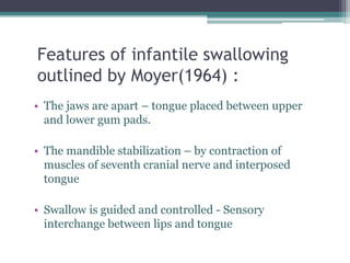
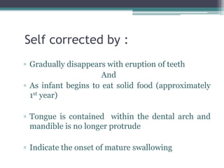
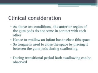
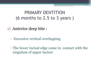
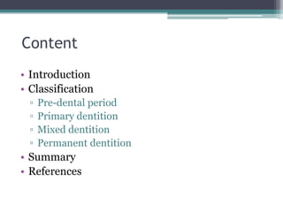
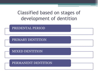
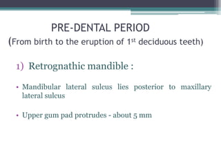
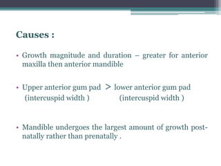
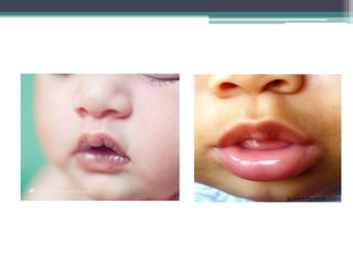
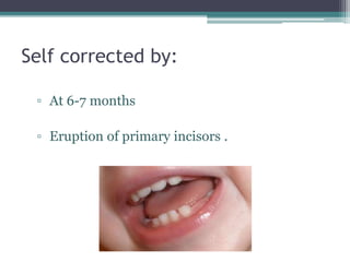
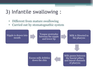
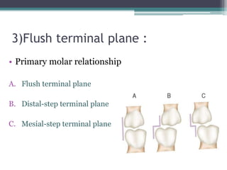
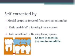
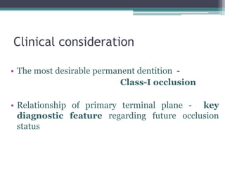
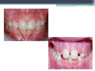
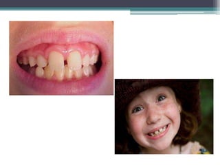
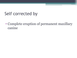
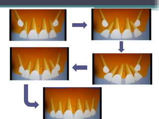
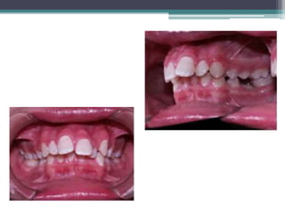
SELF CORRECTING ANOMALIES
MADE BY :
DR. DHARATI PATEL
Content
• Introduction
• Classification
▫ Pre-dental period
▫ Primary dentition
▫ Mixed dentition
▫ Permanent dentition
• Summary
• References
ALSO CALLED TRANSIENT MALOCCLUSION
• Get corrected themselves
• As they pass through the developmental stages
▫ ANOMALY:
Is defined as marked deviation from normal.
▫ SELF CORRECTING ANOMALIES :
Self-correcting anomalies are the anomalies
which arise in the child’s developing dentition during the
period of transition from that of gum pads stage to the onset
of permanent period and get corrected on their own without
any dental treatment.
Classified based on stages of
development of dentition
PREDENTAL PERIOD
PRIMARY DENTITION
MIXED DENTITION
PERMANENT DENTITION
PRE-DENTAL PERIOD
(From birth to the eruption of 1st deciduous teeth)
1) Retrognathic mandible :
• Mandibular lateral sulcus lies posterior to maxillary
lateral sulcus
• Upper gum pad protrudes - about 5 mm
Causes :
• Growth magnitude and duration – greater for anterior
maxilla then anterior mandible
• Upper anterior gum pad > lower anterior gum pad
(intercuspid width ) (intercuspid width )
• Mandible undergoes the largest amount of growth post-
natally rather than prenatally .
Self corrected by :
• within 6-7 months
• By downward and forward growth of the
mandible
Clinical consideration :
▫ Remain till 6 months after birth and helps in
suckling
▫ Can not be used as a reliable diagnostic criteria for
predicting subsequent arch relationship
▫ But if the antero-posterior dimension of gum pads
is greater the possibility of the child developing a
malocclusion is greater.
2) Anterior open bite :
▫ When Upper and lower gum pads are
approximated :-
 Contacts only at the molar region
 Space exist in the anterior region
called anterior open bite
Self corrected by:
▫ At 6-7 months
▫ Eruption of primary incisors .
Clinical consideration
▫ This open bite is considered normal
▫ Helps in suckling
3) Infantile swallowing :
Nipple is drawn into
mouth
Tongue protrudes
between the nipple
and lower lip
Milk is Directed to
the pharynx
Milk passes between
the faucial pillars
and lateral channels
of pharynx
Excess milk dribbles
down the chin
• Different from mature swallowing
• Carried out by stomatognathic system
Features of infantile swallowing
outlined by Moyer(1964) :
• The jaws are apart – tongue placed between upper
and lower gum pads.
• The mandible stabilization – by contraction of
muscles of seventh cranial nerve and interposed
tongue
• Swallow is guided and controlled - Sensory
interchange between lips and tongue
Self corrected by :
▫ Gradually disappears with eruption of teeth
And
▫ As infant begins to eat solid food (approximately
1st year)
▫ Tongue is contained within the dental arch and
mandible is no longer protrude
▫ Indicate the onset of mature swallowing
Clinical consideration
▫ As above two conditions , the anterior region of
the gum pads do not come in contact with each
other
▫ Hence to swallow an infant has to close this space
▫ So tongue is used to close the space by placing it
between the gum pads during swallowing.
▫ During transitional period both swallowing can be
observed
PRIMARY DENTITION
(6 months to 2.5 to 3 years )
1) Anterior deep bite :
▫ Excessive vertical overlapping
▫ The lower incisal edge come in contact with the
cingulum of upper incisor
Causes :
▫ Primary teeth are more upright than their
successors
▫ Have more vertical inclination - Inter-incisal angel
of (about 1500)
▫ Infra-occlusion of partially erupted molars
Self corrected by:
• Complete Eruption of primary molars-Increase
the vertical dimension
• Attrition of incisal edges
• Forward and downward growth of mandible
Clinical consideration
• Should not consider as – Malocclusion
• orthodontic treatment for deep bite – not
required
2) Spacing
• Delabarre (1918) – first described interdental
spacing in primary dentition
Types of spaces in primary dentition
▫ Primate /anthropoid/simian spaces :
▫ Physiological/developmental spaces :
 4 mm in maxilla
 3 mm in mandible
Primate space
Physiologic space
Clinical consideration
• Spaced dentition is supposed to be good -
important for normal development
• Spaces can be utilized for adjustment of
permanent successors which are always larger
• Absence of spaces indicates that crowding may
occur in permanent dentition
Self corrected by :
▫ Eruption of larger permanent successors
▫ Eruption of first permanent molar
(Early mesial shift )
3)Flush terminal plane :
• Primary molar relationship
A. Flush terminal plane
B. Distal-step terminal plane
C. Mesial-step terminal plane
Self corrected by
• Mesial eruptive force of first permanent molar
1. Early mesial shift : By using Primate spaces.
2. Late mesial shift : By using leeway spaces
1.8 mm in maxilla
3.4 mm in mandible
Clinical consideration
• The most desirable permanent dentition -
Class-I occlusion
• Relationship of primary terminal plane - key
diagnostic feature regarding future occlusion
status
Terminal plane prediction
Class-I 56 %
Class-II 44%
Flush
terminal
<2 mm
80% class-I
2 mm
20% class-III
Mesial
step
Class-II
Highly
predictable
Distal
step
Primary terminal
plane (4-5 years )
Initial permanent
first molar
occlusion(6 -7 years )
Final occlusion
(12 years )
1% class-III (< 2mm) 3% class-III
49 % mesial step 27% class- I 59% class- I
17% flush terminal 49% End on
14% distal step 23% class-II 39% class-II
Table: 1 Incidence of Terminal Molar Relationships at Three Stages of
Occlusion Development
4)EDGE TO EDGE BITE :
▫ Due to attrition of primary incisors
▫ By downward growth of mandible over jet
decreased gradually
Self corrected by
• Eruption of permanent incisors
• Having more labial inclination-
inter-incisal inclination 1230
MIXED DENTITION
(6 years to 12-13 years )
1) Anterior deep bite :
▫ Due to large permanent successor incisors
Self corrected by
▫ Complete Eruption of primary molars-
Increase the vertical dimension
2)MANDIBULAR ANTERIOR CROWDING :
• Disproportion between tooth size and arch
length
Cause :
▫ Exchange of larger permanent mandibular
incisors
▫ In narrow lower arch
Self corrected by
• Increased inter-canine width - by jaw expansion
• Tongue pressure – cause forward migration of
lower incisors
Clinical consideration
• Minor crowding - resolves spontaneously by
development
• Moderate crowding – use of leeway space
▫ Preventing mesial movement of permanent molar
3) Ugly-duckling stage:
• Seen at 8-11 years of age
• First described by – H Broadbent (1937)
• Also called Broadbent phenomenon
• Term Indicates Unaesthetic appearance of child
Self corrected by
• Complete eruption of permanent maxillary
canine
Clinical consideration
• During this stage children tends to look ugly
• Parents are often apprehensive and do consult
the dentist
• But this condition is corrected by itself -
pressure is transferred from roots to coronal
area
• So no need of any orthodontic treatment
4) End on relationship:
• Buccal cusp tip of permanent maxillary 1st molar
coincide with buccal cusp of permanent
mandibular 1st molar.
• Obtained in mixed dentition period following
the flush terminal relation in deciduous
dentition.
Class -1 relationship in
1st permanent molars
End on relationship in
1st permanent molars
Self corrected by :
• Eruptive force in mesial direction of
permanent mandibular molars
• Late mesial shift in Non-spaced dentition
▫
Clinical consideration :
Initial
occluding end
on relationship
75% shift
towards class-I
relationship
25% shifts into
class-II
relationship
PERMANENT DENTITION
1) Increased overjet and overbite:
• Overbite : a vertical distance which the incisal edge of
maxillary incisors overlaps the incisal edge of mandibular
incisors.
• Overjet : a horizontal distance between the lingual
aspect of the maxillary incisors and the labial aspect of the
mandibular incisors.
Self corrected by :
▫ By eruption of all permanent molar
▫ Differential growth of mandible
▫ Overbite decreased up to - 0.5 mm by 18 years of age
▫ Overjet decreased up to - o.7 mm between 12 to 20
years of age
Summary :
• Therefore self correcting anomalies
 are part of developing dentition
 not to be considered as any developmental or
pathological abnormality
• If there is any apprehension on part of the parents
 it should be removed promptly
 parents should be explained in brief the physiology
behind these transitional changes
Reference :
▫ Avery D R, McDonald R E.Dentistry for the child and
Adolescent. Mosby, 9th Edition 2012 , 518-520.
▫ Marwah N. Text book of pediatric dentistry scientific
foundation and clinical practice, Mosby, 3rd Edition 2014,
166-178.
▫ Tandan S. Textbook of pediatric dentistry, Paras
publication , 2nd edition ,2009 ,107-117
▫ Singh G. Text book of orthodontics ,Jaypee , 2nd edition
2007,40-45.
▫ Bhalaji S I. Orthodontics - The Art and Science , Medi , 4th
Edition , 2009 , 44-50.
THANK YOU

Self correcting anomalies

  • 1.
    SELF CORRECTING ANOMALIES MADEBY : DR. DHARATI PATEL
  • 2.
    Content • Introduction • Classification ▫Pre-dental period ▫ Primary dentition ▫ Mixed dentition ▫ Permanent dentition • Summary • References
  • 3.
    ALSO CALLED TRANSIENTMALOCCLUSION • Get corrected themselves • As they pass through the developmental stages
  • 4.
    ▫ ANOMALY: Is definedas marked deviation from normal. ▫ SELF CORRECTING ANOMALIES : Self-correcting anomalies are the anomalies which arise in the child’s developing dentition during the period of transition from that of gum pads stage to the onset of permanent period and get corrected on their own without any dental treatment.
  • 5.
    Classified based onstages of development of dentition PREDENTAL PERIOD PRIMARY DENTITION MIXED DENTITION PERMANENT DENTITION
  • 6.
    PRE-DENTAL PERIOD (From birthto the eruption of 1st deciduous teeth) 1) Retrognathic mandible : • Mandibular lateral sulcus lies posterior to maxillary lateral sulcus • Upper gum pad protrudes - about 5 mm
  • 7.
    Causes : • Growthmagnitude and duration – greater for anterior maxilla then anterior mandible • Upper anterior gum pad > lower anterior gum pad (intercuspid width ) (intercuspid width ) • Mandible undergoes the largest amount of growth post- natally rather than prenatally .
  • 9.
    Self corrected by: • within 6-7 months • By downward and forward growth of the mandible
  • 11.
    Clinical consideration : ▫Remain till 6 months after birth and helps in suckling ▫ Can not be used as a reliable diagnostic criteria for predicting subsequent arch relationship ▫ But if the antero-posterior dimension of gum pads is greater the possibility of the child developing a malocclusion is greater.
  • 12.
    2) Anterior openbite : ▫ When Upper and lower gum pads are approximated :-  Contacts only at the molar region  Space exist in the anterior region called anterior open bite
  • 14.
    Self corrected by: ▫At 6-7 months ▫ Eruption of primary incisors .
  • 15.
    Clinical consideration ▫ Thisopen bite is considered normal ▫ Helps in suckling
  • 16.
    3) Infantile swallowing: Nipple is drawn into mouth Tongue protrudes between the nipple and lower lip Milk is Directed to the pharynx Milk passes between the faucial pillars and lateral channels of pharynx Excess milk dribbles down the chin • Different from mature swallowing • Carried out by stomatognathic system
  • 17.
    Features of infantileswallowing outlined by Moyer(1964) : • The jaws are apart – tongue placed between upper and lower gum pads. • The mandible stabilization – by contraction of muscles of seventh cranial nerve and interposed tongue • Swallow is guided and controlled - Sensory interchange between lips and tongue
  • 18.
    Self corrected by: ▫ Gradually disappears with eruption of teeth And ▫ As infant begins to eat solid food (approximately 1st year) ▫ Tongue is contained within the dental arch and mandible is no longer protrude ▫ Indicate the onset of mature swallowing
  • 19.
    Clinical consideration ▫ Asabove two conditions , the anterior region of the gum pads do not come in contact with each other ▫ Hence to swallow an infant has to close this space ▫ So tongue is used to close the space by placing it between the gum pads during swallowing. ▫ During transitional period both swallowing can be observed
  • 20.
    PRIMARY DENTITION (6 monthsto 2.5 to 3 years ) 1) Anterior deep bite : ▫ Excessive vertical overlapping ▫ The lower incisal edge come in contact with the cingulum of upper incisor
  • 21.
    Causes : ▫ Primaryteeth are more upright than their successors ▫ Have more vertical inclination - Inter-incisal angel of (about 1500) ▫ Infra-occlusion of partially erupted molars
  • 23.
    Self corrected by: •Complete Eruption of primary molars-Increase the vertical dimension • Attrition of incisal edges • Forward and downward growth of mandible
  • 24.
    Clinical consideration • Shouldnot consider as – Malocclusion • orthodontic treatment for deep bite – not required
  • 25.
    2) Spacing • Delabarre(1918) – first described interdental spacing in primary dentition
  • 26.
    Types of spacesin primary dentition ▫ Primate /anthropoid/simian spaces : ▫ Physiological/developmental spaces :  4 mm in maxilla  3 mm in mandible
  • 27.
  • 28.
    Clinical consideration • Spaceddentition is supposed to be good - important for normal development • Spaces can be utilized for adjustment of permanent successors which are always larger • Absence of spaces indicates that crowding may occur in permanent dentition
  • 29.
    Self corrected by: ▫ Eruption of larger permanent successors ▫ Eruption of first permanent molar (Early mesial shift )
  • 30.
    3)Flush terminal plane: • Primary molar relationship A. Flush terminal plane B. Distal-step terminal plane C. Mesial-step terminal plane
  • 31.
    Self corrected by •Mesial eruptive force of first permanent molar 1. Early mesial shift : By using Primate spaces. 2. Late mesial shift : By using leeway spaces 1.8 mm in maxilla 3.4 mm in mandible
  • 32.
    Clinical consideration • Themost desirable permanent dentition - Class-I occlusion • Relationship of primary terminal plane - key diagnostic feature regarding future occlusion status
  • 33.
    Terminal plane prediction Class-I56 % Class-II 44% Flush terminal <2 mm 80% class-I 2 mm 20% class-III Mesial step Class-II Highly predictable Distal step
  • 35.
    Primary terminal plane (4-5years ) Initial permanent first molar occlusion(6 -7 years ) Final occlusion (12 years ) 1% class-III (< 2mm) 3% class-III 49 % mesial step 27% class- I 59% class- I 17% flush terminal 49% End on 14% distal step 23% class-II 39% class-II Table: 1 Incidence of Terminal Molar Relationships at Three Stages of Occlusion Development
  • 36.
    4)EDGE TO EDGEBITE : ▫ Due to attrition of primary incisors ▫ By downward growth of mandible over jet decreased gradually
  • 37.
    Self corrected by •Eruption of permanent incisors • Having more labial inclination- inter-incisal inclination 1230
  • 38.
    MIXED DENTITION (6 yearsto 12-13 years ) 1) Anterior deep bite : ▫ Due to large permanent successor incisors
  • 40.
    Self corrected by ▫Complete Eruption of primary molars- Increase the vertical dimension
  • 41.
    2)MANDIBULAR ANTERIOR CROWDING: • Disproportion between tooth size and arch length Cause : ▫ Exchange of larger permanent mandibular incisors ▫ In narrow lower arch
  • 43.
    Self corrected by •Increased inter-canine width - by jaw expansion • Tongue pressure – cause forward migration of lower incisors
  • 44.
    Clinical consideration • Minorcrowding - resolves spontaneously by development • Moderate crowding – use of leeway space ▫ Preventing mesial movement of permanent molar
  • 45.
    3) Ugly-duckling stage: •Seen at 8-11 years of age • First described by – H Broadbent (1937) • Also called Broadbent phenomenon • Term Indicates Unaesthetic appearance of child
  • 47.
    Self corrected by •Complete eruption of permanent maxillary canine
  • 49.
    Clinical consideration • Duringthis stage children tends to look ugly • Parents are often apprehensive and do consult the dentist • But this condition is corrected by itself - pressure is transferred from roots to coronal area • So no need of any orthodontic treatment
  • 50.
    4) End onrelationship: • Buccal cusp tip of permanent maxillary 1st molar coincide with buccal cusp of permanent mandibular 1st molar. • Obtained in mixed dentition period following the flush terminal relation in deciduous dentition.
  • 51.
    Class -1 relationshipin 1st permanent molars End on relationship in 1st permanent molars
  • 52.
    Self corrected by: • Eruptive force in mesial direction of permanent mandibular molars • Late mesial shift in Non-spaced dentition ▫
  • 53.
    Clinical consideration : Initial occludingend on relationship 75% shift towards class-I relationship 25% shifts into class-II relationship
  • 54.
    PERMANENT DENTITION 1) Increasedoverjet and overbite: • Overbite : a vertical distance which the incisal edge of maxillary incisors overlaps the incisal edge of mandibular incisors. • Overjet : a horizontal distance between the lingual aspect of the maxillary incisors and the labial aspect of the mandibular incisors.
  • 56.
    Self corrected by: ▫ By eruption of all permanent molar ▫ Differential growth of mandible ▫ Overbite decreased up to - 0.5 mm by 18 years of age ▫ Overjet decreased up to - o.7 mm between 12 to 20 years of age
  • 57.
    Summary : • Thereforeself correcting anomalies  are part of developing dentition  not to be considered as any developmental or pathological abnormality • If there is any apprehension on part of the parents  it should be removed promptly  parents should be explained in brief the physiology behind these transitional changes
  • 58.
    Reference : ▫ AveryD R, McDonald R E.Dentistry for the child and Adolescent. Mosby, 9th Edition 2012 , 518-520. ▫ Marwah N. Text book of pediatric dentistry scientific foundation and clinical practice, Mosby, 3rd Edition 2014, 166-178. ▫ Tandan S. Textbook of pediatric dentistry, Paras publication , 2nd edition ,2009 ,107-117 ▫ Singh G. Text book of orthodontics ,Jaypee , 2nd edition 2007,40-45. ▫ Bhalaji S I. Orthodontics - The Art and Science , Medi , 4th Edition , 2009 , 44-50.
  • 59.

Editor's Notes

  • #4 These are not true malocclusion but mimic malocclusion during the normal development of dentition and occlusion. They do not need any treatment measures but get corrected themselves as they pass through the developmental stages.
  • #5 Shoba tondon
  • #7 By sillman JH 1938 Range variation – 0-7 mm
  • #8 Growth magnitude and duration is greater in anterior maxilla den anterior mandibular (mc Donald ) So at birth growth of mandible as well as TMJ is not well develop as maxilla so mandible is smaller in size and retruded
  • #10 Bhalaji ramus – anterior surface resorption (-) posterior surface (+) so facilities lengthening of mandible body body – lengthens by growth of ramus angle – lingual side posterio-inferior (-) anterio-superior (+) buccal side anterio-superior (-) posterio-superior (+) alveolar – as teeth erupts increase in height by deposition at margins condyle – condylar cartilage bone deposition at condylar cartilage so condyle grows towards cranial base as the condyle pushes against cranial base the entire mandible gets displaced forward and downward
  • #12 M.C. Donald
  • #13 Bhalaji
  • #17 The ability to feed from breast is present , automatic perastaltic movement of toungue , regular breathing continues. Stamatognathic system structure – jaw tongue teeth musculature Functions – mastication deglutination respiration speech
  • #18 Bhalaji
  • #19 There is distinct changes in swallowing pattern
  • #20 Cessation of infantile swallow and the appearance of mature swallow occurs gradually Bhalaji
  • #21 Primary incisors erupts into anterior space of the gum pads thus acconting for the apparent overbite
  • #24 Molars came into occlusion
  • #25 Skeletal deep bite – upward forward rotation of the mandible and worsen by downward forward growth of maxilla horizontal growth pattern Dental deep bite – over eruption of incisors and infra occlusion
  • #27 Primate – present in primate species ,very prominent between upper lateral and canine and lower canine and 1st molar Physiologic – between all primary teeth and important role in development of normal permanent occlusion Leeway difference between mesial-distal width of the primary canine molars and their permanent successors
  • #32 Sum of Mesiodistal width of primary canine and first and second molar and sum of mesiodistal width of first and second pre molar
  • #38 So there is 2 mm of overjet and overbite is present
  • #44 Cause tipping movement of incisor in to labial direction
  • #45 By lingual arch and lip bumper - cause control position of molar , distal tipping of molars and labial tipping of incisors There by utilizing this space to decrowd the anteriors Bhalaji – pg no 411
  • #46 Personal transformeation for better – Hans Anderson 1844-45 fairy tail
  • #50 Bhalaji pg no – 51 When canine erupts in oral cavity and the pressure is transferred from the roots to the coronal area of the incisors