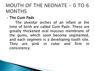
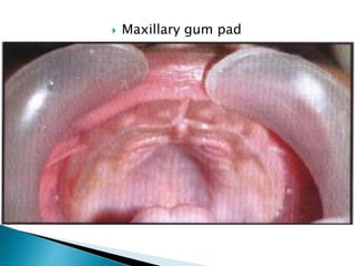
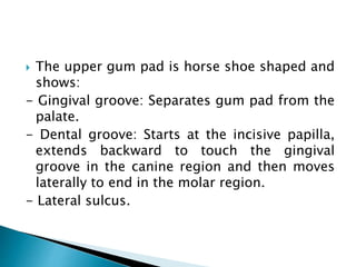
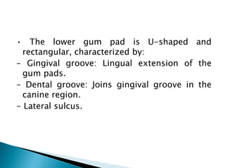
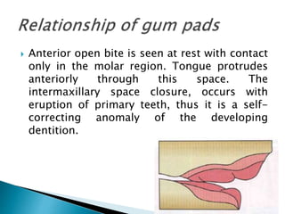
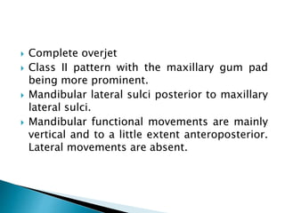
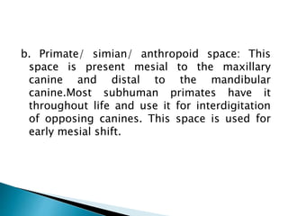
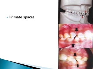
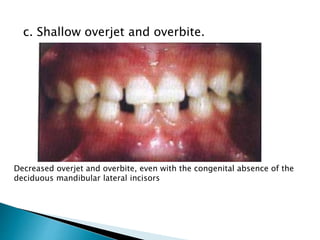
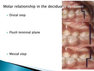
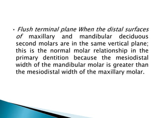
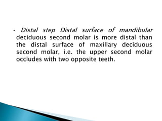
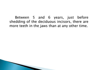
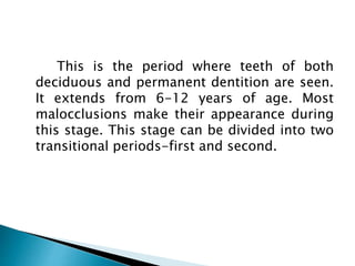
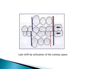
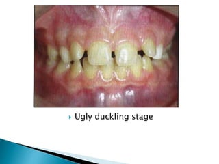
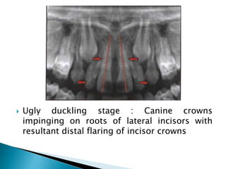
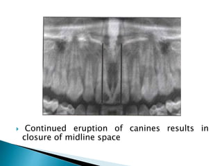
5. The Gum Pads
The alveolar arches of an infant at the
time of birth are called Gum Pads. These are
greatly thickened oral mucous membrane of
the gums, which soon become segmented,
and each segment is a developing tooth site.
They are pink in color and firm in
consistency.
10. The pads get divided into a labio/buccal
and a lingual portion which differentiates
later. Transverse grooves separate the gum
pads into 10 segments. The groove between
the canine and the first molar region is called
the lateral sulcus, which helps to judge the
inter-arch relationship.
11. The upper gum pad is horse shoe shaped and
shows:
- Gingival groove: Separates gum pad from the
palate.
- Dental groove: Starts at the incisive papilla,
extends backward to touch the gingival
groove in the canine region and then moves
laterally to end in the molar region.
- Lateral sulcus.
12. • The lower gum pad is U-shaped and
rectangular, characterized by:
- Gingival groove: Lingual extension of the
gum pads.
- Dental groove: Joins gingival groove in the
canine region.
- Lateral sulcus.
13. Anterior open bite is seen at rest with contact
only in the molar region. Tongue protrudes
anteriorly through this space. The
intermaxillary space closure, occurs with
eruption of primary teeth, thus it is a self-
correcting anomaly of the developing
dentition.
14. Complete overjet
Class II pattern with the maxillary gum pad
being more prominent.
Mandibular lateral sulci posterior to maxillary
lateral sulci.
Mandibular functional movements are mainly
vertical and to a little extent anteroposterior.
Lateral movements are absent.
15. A precise "bite" or jaw relationship is not
yet seen. Therefore, neonatal jaw relationship
cannot be used as a diagnostic criterion for
reliable prediction of subsequent occlusion in
the primary dentition.
16. Occasionally a child is born with teeth
already present in the mouth. Natal teeth are
present at birth whereas neonatal teeth erupt
during the first month. Pre-erupted teeth
erupt during the second or third month.
18. The incidence of natal and neonatal teeth
is estimated to be 1:1000 and 1:30000
respectively. These teeth are almost always
mandibular incisors, which frequently display
enamel hypoplasia. There are familial
tendencies for such teeth. They should not be
removed if normal but removed if
supernumerary or mobile.
19. At birth, both maxilla and mandible are
small compared to the rest of the face.
Extensive early transverse and ventral
development of both jaws occurs leading to
an anteroposterior relation between the jaws.
20. Overjet diminishes markedly during the
first 6 months. Increase in jaw size provides
enough space for harmonious arrangement of
deciduous teeth. Thus crowding seen in the
pads disappears when the teeth erupt.
21. Eruption of deciduous teeth commences at
about 6 months of age. Occlusion starts
developing posteriorly when deciduous first
molars attain contact. By the time the first
molars have settled, occlusion in the
posterior region is established.
22. The deciduous dentition stage starts from
the eruption of the first deciduous tooth,
usually the deciduous mandibular central
incisors and ends with the eruption of the
first permanent molar, i.e. from 6 months to
6 years of postnatal life. By 2 years of age,
deciduous dentition is usually complete and
in full function. Root formation of all
deciduous teeth is complete by 3 years of
age.
23. Normal Signs of Primary Dentition
a. Spaced anteriors: Spacing is usually seen in
the deciduous dentition to accommodate
larger permanent teeth in the jaws.
Spacing in deciduous dentition
24. b. Primate/ simian/ anthropoid space: This
space is present mesial to the maxillary
canine and distal to the mandibular
canine.Most subhuman primates have it
throughout life and use it for interdigitation
of opposing canines. This space is used for
early mesial shift.
26. c. Shallow overjet and overbite.
Decreased overjet and overbite, even with the congenital absence of the
deciduous mandibular lateral incisors
27. d. Almost vertical inclination of the anteriors.
e. Ovoid arch form.
f. Straight/flush terminal plane relation: The
molar relationship in the primary dentition
can be classified into 3 types:
28. Distal step
Flush tenninal plane
Mesial step
Molar relationship in the deciduous dentition
29. • Flush terminal plane When the distal surfaces
of maxillary and mandibular deciduous
second molars are in the same vertical plane;
this is the normal molar relationship in the
primary dentition because the mesiodistal
width of the mandibular molar is greater than
the mesiodistal width of the maxillary molar.
30. • Mesial step Distal surface of mandibular
deciduous second molar is mesial to the
distal surface of maxillary deciduous second
molar.
31. • Distal step Distal surface of mandibular
deciduous second molar is more distal than
the distal surface of maxillary deciduous
second molar, i.e. the upper second molar
occludes with two opposite teeth.
32. Between 5 and 6 years, just before
shedding of the deciduous incisors, there are
more teeth in the jaws than at any other time.
33.
34. This is the period where teeth of both
deciduous and permanent dentition are seen.
It extends from 6-12 years of age. Most
malocclusions make their appearance during
this stage. This stage can be divided into two
transitional periods-first and second.
35. Emergence of the first permanent molars
and transition of incisors .The following
events take place during this period :
36. Eruption of Permanent First Molars
The first permanent molars erupt at 6
years. They play an important role in the
establishing and in the functioning of
occlusion, in the permanent dentition.
37. Anteroposterior positioning of the permanent
molars is influenced by:
1. Terminal plane relationship
Occlusal relationships of
primary and permanent
molars
38. When the deciduous second molars are in a
fIush terminal plane, the permanent first
molar erupts initially into a cusp-to-cusp
relationship, which later transforms into a
Class I molar relation using the primate
spaces.
Early shift of the erupting first
permanent molars moving utilizing the
primate spaces
39. Later, cusp-to-cusp relationship of the
permanent first molar can be converted to a
Class I relationship by the mesial shift of the
permanent first molar following exfoliation of
the primary molar and thus making use of the
Leeway space (late mesial shift).
Late shift by utilization of the Leeway
space
40. When the deciduous second molars are in a
distal step, the permanent first molar will
erupt into a Class II relation. This molar
configuration is not self-correcting and will
cause a Class II malocclusion despite Leeway
space and differential growth.
41. Primary second molars in mesial step
relationship lead to a Class I molar relation in
mixed dentition. This may remain or progress
to a half or full cusp Class III with continued
mandibular growth.
42. 2. Early mesial shift in arch with physiologic
spacing: In a spaced arch, eruptive force of
the permanent molars causes closing of any
spaces between the primary molars or
primate spaces, thus allowing molars to shift
mesially.
44. Leeway space of Nance The combined
mesiodistal widths of deciduous canine, first and
second molars is more than that of the combined
mesiodistal width of permanent canine, first and
second premolar. The difference between the two
is called the Leeway space.
Maxilla 0.9 mm/segment = 1.8 mm total
Mandible 1.7 mm/ segment = 3.4 mm total.
47. Incisor Eruption
Permanent incisors develop lingual to the
primary incisors. For incisors to erupt in
normal alignment, there is an obligate space
requirement in the anterior part of both the
arches which is termed as incisor liability, i.e.
the total sum of the mesiodistal width of four
permanent incisors is larger than that of
primary incisors by 7.6 mm in maxilla and 6
mm in mandible (Wayne). This obligate space
is provided by:
48. Interdental physiologic spacing in the primary
incisor region: 4 mm in maxillary arch; 3 mm
in mandibular arch.
49. Increase in the inter-canine arch width:
Significant amount of growth occurs with the
eruption of incisors and canines.
50. Increase in the anterior length of the dental
arches: Permanent incisors erupt labial to the
primary incisors to obtain an added space of
2-3 mm.
51. Change in inclination of permanent incisors:
Primary teeth are upright but permanent
teeth incline to the labial surface thus
decreasing the inter-incisal angle from about
150 degrees in the deciduous dentition to
123 degrees in the permanent dentition. This
increases the arch perimeter.
52. Comparison of the angulation of the permanent and primary teeth
53. The Broadbent's Phenomenon- Ugly Duckling
Stage (7-14 years)
Around the age of 8 years, a midline diastema
is commonly seen in the upper arch, which is
usually misinterpreted by the parents as a
malocclusion. Crowns of canines in young jaws
impinge on developing lateral incisor roots, thus
driving the roots medially and causing the
crowns to flare laterally. The roots of the central
incisors are also forced together thus causing a
maxillary midline diastema. The period from the
eruption of lateral incisor to canine is termed as
the Ugly Duckling stage. It is an unaesthetic
metamorphosis, which eventually leads to an
aesthetic result.
55. With eruption of canines, the impingement
from the roots shifts incisally thus driving the
incisor crowns medially, resulting in closure
of the diastema.
56. Ugly duckling stage : Canine crowns
impinging on roots of lateral incisors with
resultant distal flaring of incisor crowns
58. This is a stable phase where little changes
take place in the dentition. The teeth present
are the permanent incisors and first molar
along with the deciduous canines and molars.
Some of the features of this stage are:
59. 1. Any asymmetry in emergence and
corresponding differences in height levels or
crown lengths between the right and left side
teeth are made up(marked).
60. 2. Occlusal and interproximal wear of
deciduous teeth causes occlusal morphology
to approach that of a plane.
62. 4. Root formation of emerged incisors, canines
and molars continues, along with
concomitant increase in alveolar process
height.
63. 5. Resorption of roots of deciduous molars.
This phase prepares for the second
transitional phase.
64. The following events take place:
1. Exfoliation of primary molars and canines.
At around 10 years of age, the first deciduous
tooth in the posterior region, usually the
mandibular canine sheds and marks the
beginning of the second transitional period.
Usually no crowding is seen before
emergence except maybe between the
maxillary first premolar and canine.
65. 2. Eruption of permanent canines and
premolars. These teeth erupt after a pause of
1-2 years following incisor eruption. The first
posterior teeth to erupt are the mandibular
canine and first premolar (9-10 years)
followed by maxillary premolars and canine
around 11-12 years.
67. Most common eruption sequence is 4-5-3
in the maxilla and 3-4-5 in the mandible.
Favorable occlusion in this region is largely
dependent on:
Favorable eruption sequence.
Satisfactory tooth size- available space ratio.
Attainment of normal molar relation with
minimum diminution of space available for
bicuspids.
68. 3. Eruption of permanent second molars.
Before emergence second molars are oriented
in a mesial and lingual direction. These teeth
are formed palatally and are guided into
occlusion by the Cone Funnel mechanism (the
upper palatal cusp/cone slides into the lower
occlusal fossa/funnel). The arch length is
reduced prior to second molar eruption by
the mesial eruptive forces. Therefore,
crowding if present is accentuated.
71. The permanent dentition forms within the
jaws soon after birth. Calcification begins at
birth with the calcification of the cusps of the
first permanent molar and extends as late as
the 25th year of life. Complete calcification of
incisor crowns takes place by 4 to 5 years and
of the other permanent teeth by 6 to 8 years
except for the third molars. Therefore the
total calcification period is about 10 years.
The permanent incisors develop lingual to the
deciduous incisors and move labially as they
erupt. The premolars develop below the
diverging roots of the deciduous molars.
72. Nolla (1960) gave stages
of tooth development to
make a meaningful
assessment of eruption from
panoramic/ posteroanterior
radiographs which is given in
Figure (right) and elaborated
in Table (next).
74. At approximately 13 years of age all
permanent teeth except third molars are fully
erupted. Before the deciduous incisors are
shed, there are 48 teeth / parts of teeth
present in the jaws.
75. Features of the permanent dentition:
Coinciding midline.
Class 1molar relationship of the permanent
first molar.
Vertical overbite of about one-third the
clinical crown height of the mandibular
central incisors.
76. Overjet: Overjet and over bi te decreases
throughout the second decade of life due to
greater forward growth of the mandible.
Curve of Spee: Develops during transition and
stabilizes in adulthood.
77.
78. Eruption is the developmental process that
moves a tooth from its crypt position through
the alveolar process into the oral cavity and
to occlusion with its antagonist. During
eruption of succedaneous teeth:
Primary tooth resorbs
Roots of the permanent teeth lengthen
Increase in the alveolar process height
Permanent teeth move through the bone.
79. Teeth do not begin to move occlusally
until crown formation is complete. It takes 2-
5 years for posterior teeth to reach the
alveolar crest following crown completion and
12-20 months to reach occlusion after
reaching alveolar margin.
80. Tencate divided tooth eruption into the
following 3 stages:
1. Pre-eruptive tooth movement: Movement of
tooth germs within the jaws before they
begin to erupt.
2. Eruptive tooth movement: Tooth movement
from its position within the jaws to its
functional position.
3. Post-eruptive tooth movement: Maintaining
the position of erupted tooth in occlusion
while the jaws continue to grow.
81. Tooth eruption can also be divided into:
1. Pre-emergent eruption During the stage of
crown formation, there is very slow labial or
buccal drift of the tooth follicle within the
bone. However, there is no eruptive
movement.
82. Two processes are necessary for a tooth
to erupt intra-osseously:
Resorption of bone and primary tooth roots
overlying the crown of the erupting tooth.
Eruption mechanism itself must move the
tooth in the direction where the path has
been cleared.
Resorption is the rate-limiting factor in
preemergent eruption.
83. 2. Post-emergent eruption Once a tooth
emerges into the mouth, it erupts rapidly
(about 4 mm in 14 weeks) until it
approaches the occlusal level and is
subjected to the forces of mastication. This
stage of relatively rapid eruption is called as
the postemergent spurt. This is followed by
a phase of Juvenile Occlusal Equilibrium, i.e.
after teeth reach the occlusal level; eruption
becomes almost imperceptibly slow
although it definitely continues.
84. After the teeth are in function, they
erupt at the same rate as the vertical
growth of the mandibular ramus unless
there is occlusal wear or the antagonist
tooth is lost at any age, in which case
additional eruption occurs. When the
pubertal growth ends, a final phase of tooth
eruption takes place known as Adult
Occlusal Equilibrium.
87. 2. Intra-alveolar Tooth position is affected by-
Presence or absence of adjacent teeth
Rate of resorption of primary teeth
Early loss of primary teeth
Localized pathologic conditions.
88. 3. Tntraoral stage Tooth can be moved by lip,
cheek, tongue muscles or external objects
and drift into spaces.
89. 4. Occlusal stage Muscles of mastication exert
influence through interdigitation of cusps.
The periodontal ligament disseminates the
strong forces of chewing to the alveolar bone.
90. There is wide variability in the sequence of
arrival of teeth in the mouth.
Maxilla 6-1-2-4-3-5-7 or 6-1-2-4-5-3-7
(most common)
Mandible 6-1-2-4-5-3-7or 6-1-2-3-4-5-7
(most common)
91. Dental age 6: First stage of eruption
Eruption of mandibular central incisor and
permanent first molar.
Mandibular molar eruption precedes
maxillary molar.
92. Dental age 7
Eruption of maxillary central and mandibular
lateral incisor.
Root formation of maxillary lateral incisor
well advanced.
Crown completion of canines and premolars.
93. Dental age 8
Eruption of maxillary lateral incisor.
Delay of 2-3 years before any further teeth
erupt.
94. Dental age 9
One-third root formation of mandibular
canine and first premolar is complete.
Root development of mandibular second
premolar begins.
95. Dental age 10
One-half root formation of mandibular canine
and first premolar is complete.
Significant root development of maxillary and
mandibular second premolar as well as
maxillary canine.
Root completion of mandibular incisors and
near completion of maxillary laterals.
According to Movers, mandibular canine
erupts between 9 and 10 years.
96. Dental age 11
Eruption of mandibular canine (according to
Proffit), mandibular first premolar and
maxillary first premolar.
Maxillary first premolar erupts ahead of
canine and second premolar.
97. Dental age 12
Remaining succedaneous teeth erupt.
Second permanent molars nearing eruption
Early beginnings of third molar
98. Dental age 13,14,15
Completion of roots of permanent teeth
Third molars apparent on the radiograph
99. Change in eruption sequence is a reliable
sign of disturbance in normal development of
the dentition.
Certain normal variations with important
clinical significance:
100. Eruption of second molars ahead of
premolars in the mandibular arch. This
decreases the space for second premolars,
which get partially blocked out.
Eruption of maxillary canines ahead of
premolars will cause the canines to be forced
out labially.
Asymmetries in eruption between the right
and left sides occurs when there is lack of
space to accommodate erupting teeth due to
different pattern of mechanical obstruction,
decreased space on one side compared to the
other.
101.
102. The usual arch dimensions measured are:
1. Widths of the canines, primary molars
(premolars) and first permanent molars:
a. Dimensional increase in width involves
alveolar process growth almost totally, since
there is little skeletal width increase at this
time.
103. b. Clinically significant differences exist in the
manner and magnitude of width changes in
the maxilla and mandible. Width increase
correlates highly with vertical alveolar process
growth. Maxillary alveolar processes diverge
while mandibular alveolar processes are more
parallel. Thus, maxillary width increases more
and can be easily altered in treatment.
104. c. The only significant increase in mandibular
inter-canine width occurs during eruption of
incisors when primary cuspids are moved
distally into primate spaces and does not
increase significantly thereafter.
105. d. Maxillary arch width increase is timed with
periods of active eruption of teeth. Eruption
of maxillary permanent canines is an
important factor in widening of the arch.
106. e. Maxillary premolar width increase is
coincidental with vertical growth whereas
mandibular premolar width increase occurs
beca use of further buccal placement of
premolar crowns.
107. 2. Length or depth: Arch length or depth is
measured at the midline from a point midway
between central incisors to a tangent
touching distal surfaces of second primary
molars or premolars. Any changes in arch
length are coarse reflections of changes in
perimeter.
108. 3. Arch circumference or perimeter: Measured
from distal surface of second primary molar
or mesial surface of first permanent molar
around the arch over contact points and
incisal edges in a smoothened curve to the
distal of second primary molar or mesial
surface of first permanent molar of the
opposite side.
109. The reduction in mandibular arch
circumference during transitional and early
adolescent dentition is a result of:
a) Late mesial shift of first permanent molar as
"Leeway space" is pre-empted.
b) Mesial drifting tendency of posterior teeth
throughout life.
c) Slight interproximal wear of teeth.
d) Lingual positioning of incisors.
e) Original tipped position of incisors and
molars.
112. 1. Ectodermal dysplasia Complete or partial
anodontia of both the dentitions along with
the presence of malformed teeth.
Orthopantomogram of a case with ectodermal dysplasia
113. 2. Anodontia Absence of 1 or more teeth due
to failure of tooth bud initiation. Most
commonly missing teeth are third molars
(16%) followed by mandibular second
premolars (4.4%), maxillary lateral incisor
(1.7%) and maxillary second premolars (1.6%).
114. 3. Supemumerarv and supplemental teeth
Teeth in excess of the normal complement of
teeth. The difference between the two is that
supplemental teeth resemble normal teeth
whereas supernumerary teeth do not, e.g. of
supernumerary teeth:
Mesiadens between maxillary central incisors.
Pendens located buccal to the arch
Distomalar distal to the third molar.
Paramolar located buccal or lingual to molars.
115. 4. Natal and neanatal teeth These may be
either supernumerary or deciduous teeth.
119. 2. Mulberry molars Occlusal surface is
narrower than the cervical margin and is
made up of agglomerate mass of globules;
seen in congenital syphilis.
120. 3. Peg shaped laterals Proximal surfaces of the
crown converge giving the tooth a conical
shape.
121. 4. Macrodontia Teeth larger than normal. It
may be true or relative generalized.
122. 5. Microdontia Teeth smaller than normal. It
may be true or relative generalized; most
commonly the lateral incisor and third
molars.
123. 6. Dens in dente Tooth invaginates before
calcification, e.g. permanent maxillary lateral
incisor.
124. 7. Dens evaginatus A tubercle or protruberance
from the involved surface of the affected
tooth; occurs due to proliferation or
evagination of part of the inner enamel
epithelium into the stellate reticulum. Seen in
premolars.
125. 8. Gemination Single tooth germ splits into
partially or fully separated crowns but with a
common root and root canal.
126. 9. Fusion Two tooth germs mute to form a
single large crown with two root canals; seen
in incisors.
128. 11. Taurodontism Enlargement of the body and
pulp chamber of a multi-rooted tooth with
apical displacement of the pulpal floor and
bifurcation of the roots.
129. 1. Enamel hypoplasia Reduction in the amount
of enamel formed.
Local enamel hypoplasia Periapical infection
or trauma (Turner's tooth)
Systemic enamel hypoplasia Rickets, German
measles, fluoride ingestion.
Hereditary enamel hypoplasia Tooth appears
yellow due to reduced enamel thickness.
130. 2. Amelogenesis imperfecta Hereditary
disorder wherein the quality and quantity of
enamel formed is altered. Three types:
Hypoplastic Defective matrix formation
Hypocalclfication Defective mineralization of
matrix.
Hypomaturation Immature enamel crystals.
131. 3. Dentinogenesis imperfecta Hereditary
developmental disorder of the dentine. The
dentine appears grey to brownish violet,
enamel frequently separates from the
defective dentine, roots become short, canals
get obliterated, rapid attrition is seen.
137. 9. Enamel pearls Attached to the furcation area
of maxillary molars.
138. 1. Enamel hypocalcijication Calcification is
subnormal. It may be local, systemic or
hereditary.
2. Interglobular dentine Areas of partially
calcified dentine.
139. 1. Concrescence Cemental union of two teeth.
2. Retarded eruption Due to endocrine
disturbances, vitamin deficiencies, local
causes.
3. Ankylosed teeth: Teeth fail to erupt to the
occlusal level as they are fused to the bone.
140.
141. Development of dentition in humans is
complex and depends on many variables.
Development of dentition deviates markedly
from that of other parts and structures of the
body. Crowns of teeth are formed directly to
adult size and housed within the jaws years
before they emerge.
142. To determine an abnormal course of
development, it is the responsibility of an
orthodontist to have adequate knowledge on
the subject to differentiate abnormal from
normal before initiating therapy.
143.
144. I. Bishara SE, Hoppens BL Jakobsen JR, Kohout FJ. Changes
in molar relationships between the deciduous and
permanent dentitions: a longitudinal study, Am J Orthod
Dentofac Orthoped 1988;93:19.
2. Bjork A, Skeiller V. Facial development and tooth
eruption. An implant study at the age of puberty, Am J
Orthod 1972;62:339-83.
3. British Standards Tnstitute.Glossary of Dental terms (SS
4492). London: BSI, 1983.
4. Broadbent BH. Ontogenetic development of occlusion,
Angle Orthod, 1941;11:223-41.
5. elements EMB, et al. Age at which deciduous teeth arc
shed, Brit Med J 1957;1:1508.
6. Friel S. The development of ideal occlusion of the gum
pads and teeth, Am J Orthodint 1954;40:1963.
145. 7. HellmanM. Factors influencing occlusion, In
Gregory WK, Broadbent BH, Hellman M, editors:
Development of Occlusion, Philadelphia, 1941,
University of Pennsylvania Press.
8. Inuzuka K. Changes in molar relationships
between the deciduous and permanent
dentitions: a longitudinal study, Am J Orthod
Dentofac Orthoped 1990;93:19.
9. Marks se [r, Schroeder HE. Tooth eruption:
theories and facts, Anat Rec 1996;245:374-93.
10. Marks SC Jr. The basic and applied biology of
tooth eruption, Connective Tissue Res
1995;32:149-57.
146. 11. Moorrees C. The dentition of the growing
child: a longitudinal study of dental development
between 3 and 18 years of age, Cambridge,
Mass, 1959, Harvard University Press.
12. Moorrees CF, Chadha JM. Available space for
the incisors during dental development, Angle
Orthod, 1965;35:12-22.
13. Sillman JH. Dimensional changes of dental
arches: longitudinal studies from bi rth to 25
years, Am J Orthod 1964;50:824-42.
14. Van der Linden, Dutterloo HS. Development
of Human Dentition: an atlas: Harper and Row,
1976.