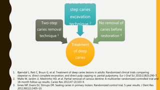
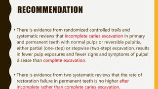
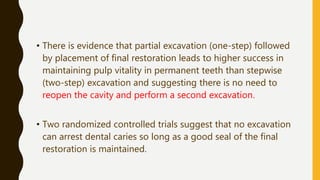
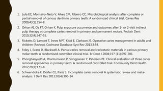
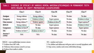
AAPD GUIDE LINES
FOR RESTORATIVE
DENTISTRY
ADOPTED 1991
REVISED 2014
BY DR.DHARATI
PATEL
PURPOSE
W H E N I T I S N E C E S S A R Y TO T R E AT ? ? ?
W H AT T H E A P P R O P R I AT E M AT E R I A L S A N D T E C H N I Q U E S
? ? ?
METHOD
• Electronic database and hand searches, for
the most part between the years 1995-2013,
were conducted
• Those papers that were used to evaluate
clinical efficacy on specific restorative
dentistry topics initially were evaluated by
abstract by two individuals.
• Full evaluation and abstraction included
examination of the research methods and
potential for study bias
• Research that was considered deficient or
had high bias was eliminated.
• This strategy yielded 35 meta-
analyses/systemic reviews and 62
randomized controlled clinical trials that
primarily made up the evidence for this
guideline.
Assessment
based on a modification of the American Dental
Association’s grading of recommendations
STRONG
EVIDENCE
(based on well-
executed randomized
control trials, meta-
analyses, systematic
EVIDENCE IN
FAVOR
(based on weaker
evidence from
clinical trials)
EXPERT OPINION
(based on retrospective
trials, case reports, in vitro
studies, and opinions from
clinical researchers)
WHEN TO
RESTORE
• The management of dental caries was based on the belief
that caries was a progressive disease that eventually
destroyed the tooth unless there was surgical and restorative
intervention.1
• Primary teeth may require different criteria due to thinner
enamel and dentin and broader proximal 2 contacts and
primary teeth may be more susceptible to restoration failures
than permanent teeth.3
1. Tinanoff, N, Douglass J.M. Clinical decision-making for caries management of primary teeth. J Dent Ed
2001;65(10):1133-42
2. Nelson SJ. Wheeler’s Dental Anatomy, Physiology, and Occlusion. 9th ed. Philadelphia. WB Saunders; 2010.
3. Hickel R, Kaaden C, Paschos E, Buerkle V, GarcĂ­a-Godoy F, Manhart J. Longevity of occlusally-stressed
restorations in posterior primary teeth. Am J Dent 2005;18: 198-211.
therapy include
• Removing cavitation or defects
to eliminate areas that are
susceptible to caries
• Stopping the progression of
tooth demineralization and
infection into the dental pulp
• Restoring the integrity of tooth
structure
• Preventing the shifting of teeth
due to loss of tooth structure.
therapy include
• More susceptible to fracture,
recurrent lesions, restoration
failure
• Pulp exposure during caries
excavation
• Future pulpal complications
• Iatrogenic damage to adjacent
teeth
1. Downer MC, Azli NA, Bedi R, Moles DR, Setchell DJ. How long do routine dental restorations last? A systematic review. Brit Dent J
1999;187(8):432-9.
2. Lenters M, van Amerongen WE, Mandari GJ. Iatrogenic damage to the adjacent surface of primary molars in three different ways of
cavity preparation. Eur Archives Paed Dent 2006;1(1):6 10.
3. Ricketts D, Lamont T, Innes NPT, Kidd E, Clarkson JE. Techniques for managing decay in teeth. The Cochrane Summaries, March 28,
RECOMMENDATIONS
• Management of dental caries includes
– Identification of an individual’s risk for caries progression
– Understanding of the disease process
– And manage with appropriate preventive services when indicated.1
• Decisions for when to restore carious lesions should include at
least
– clinical criteria of visual detection of enamel cavitation
– visual identification of shadowing of the enamel
– radiographic recognition of enlargement of lesions over time.2,3
1. American Academy of Pediatric Dentistry. Guideline on caries risk assessment and management for infants, children, and adolescents.
Pediatr Dent 2014;36(special issue):127-34.
2. Ismail AI, Sohn W, Tellez M, et al. The international caries detection and assessment system (ICDAS): An integrated system for
measuring dental caries. Community Dent Oral Epidemiol 2007;35(3):170-8.
3. Beauchamp J, Caufield PW, Crall JJ, et al. Evidence-based clinical recommendations for the use of pit-and-fissure sealants: A report of
the American Dental Association Council on Scientific Affairs. J Am Dent Assoc 2008;139 (3):257-68.
DEEP CARIES
EXCAVATION AND
RESTORATION
• The AAPD Guideline on Pulp Therapy for Primary and Immature
Permanent Teeth stated that
……..the treatment objective for a tooth affected by caries is to
maintain pulpal vitality, especially in immature permanent teeth for
continued apexogenesis. 1
1. American Academy of Pediatric Dentistry. Guideline on pulp therapy for primary and immature
permanent teeth. Pediatr Dent 2013;35(special issue):235-42.
Treatment
of deep
caries
Two-step
caries removal
technique 1
Partial or one-
step caries
excavation
technique 2 No removal of
caries before
restoration 3
1. Bjørndal L, Reit C, Bruun G, et al. Treatment of deep caries lesions in adults: Randomized clinical trials comparing
stepwise vs. direct complete excavation, and direct pulp capping vs. partial pulpotomy. Eur J Oral Sci 2010;118(3):290-7.
2. Maltz M, Jardim JJ, Mestrinho HD, et al. Partial removal of carious dentine: A multicenter randomized controlled trial and
18-month follow-up results. Caries Res 2013;47 (2):103-9.
3. Innes NP, Evans DJ, Stirrups DR. Sealing caries in primary molars: Randomized control trial, 5-year results. J Dent Res
2011;90(12):1405-10.
RECOMMENDATION
• There is evidence from randomized controlled trails and
systematic reviews that incomplete caries excavation in primary
and permanent teeth with normal pulps or reversible pulpitis,
either partial (one-step) or stepwise (two-step) excavation, results
in fewer pulp exposures and fewer signs and symptoms of pulpal
disease than complete excavation.
• There is evidence from two systematic reviews that the rate of
restoration failure in permanent teeth is no higher after
incomplete rather than complete caries excavation.
• There is evidence that partial excavation (one-step) followed
by placement of final restoration leads to higher success in
maintaining pulp vitality in permanent teeth than stepwise
(two-step) excavation and suggesting there is no need to
reopen the cavity and perform a second excavation.
• Two randomized controlled trials suggest that no excavation
can arrest dental caries so long as a good seal of the final
restoration is maintained.
1. Lula EC, Monteiro-Neto V, Alves CM, Ribeiro CC. Microbiological analysis after complete or
partial removal of carious dentin in primary teeth: A randomized clinical trial. Caries Res
2009;43(5):354-8.
2. Orhan AI, Oz FT, Orhan K. Pulp exposure occurrence and outcomes after 1- or 2-visit indirect
pulp therapy vs complete caries removal in primary and permanent molars. Pediatr Dent
2010;32(4):347-55.
3. Ricketts D, Lamont T, Innes NPT, Kidd E, Clarkson JE. Operative caries management in adults and
children (Review). Cochrane Database Syst Rev 2013;3:54.
4. Foley J, Evans D, Blackwell A. Partial caries removal and cariostatic materials in carious primary
molar teeth: A randomised controlled clinical trial. Br Dent J 2004;197 (11):697-701.
5. Phonghanyudh A, Phantumvanit P, Songpaisan Y, Petersen PE. Clinical evaluation of three caries
removal approaches in primary teeth: A randomised controlled trial. Community Dent Health
2012;29(2):173-8.
6. Schwendicke F, Dorfer CE, Paris S. Incomplete caries removal A systematic review and meta-
analysis. J Dent Res 2013;92(4):306-14.
PIT AND FISSURE
SEALANT
• Pit and fissure caries account 80 to 90 % of all caries in permanent
posterior teeth and 44 % in primary teeth.1
• According to a meta-analysis of 24 studies, the overall effectiveness of
auto- polymerized fissure sealants in preventing dental decay was
71%.2
• Another Cochrane review calculated that placement of resin-based
sealant in children and adolescent reduces caries incidence of 86 %
after one year and 57 % at 48 to 54 months. 3
1. Beauchamp J, Caufield PW, Crall JJ, et al. Evidence-based clinical recommendations for the use of pit-and-fissure
sealants: A report of the American Dental Association Council on Scientific Affairs. J Am Dent Assoc 2008;139
(3):257-68.
2. Llodra JC, Bravo M, Delgado-Rodriguez M, Baca P, Galvez R. Factors influencing the effectiveness of sealants: A
meta-analysis. Community Dent Oral Epidemiol 1993; 21(5):261-8.
3. Ahovuo-Saloranta A, Hijri A, Nordblad A, Worthington H, Makela M. Pit and fissure sealants for preventing dental
• Sealants must be retained on the tooth and should be
monitored to be most effective. Studies incorporating recall
and maintenance have reported sealant success levels of 80 to
90 % after 10 or more years.
• Pit and fissure sealants lower the number of viable bacteria,
including Streptococcus mutans and lactobacilli, by at least
100-fold and reduced the number of lesions with any viable
bacteria by about 50% .
Griffin SO, Gray SK, Malvitz DM, Gooch BF. Caries risk in formerly sealed teeth. J Am Dent Assoc 2009;140(4): 415-
23.
RECOMMENDATION
1. Based on a meta-analysis sealants should be placed on pit and
fissure surfaces judged to be at risk for dental caries or surfaces
that already exhibit incipient, non-cavitated carious lesions to
inhibit lesion progression.
ď‚§ The best evaluation of high caries risk is done by an experienced
clinician using indicators of low socio-economic status, high
frequency of sugar consumption, prior caries, active white spot
lesions and enamel defects, and low salivary flow.
American Academy of Pediatric Dentistry. Guideline on caries risk assessment and management
for infants, children, and adolescents. Pediatr Dent 2014;36(special issue):127-34.
RECOMMENDATION
2. Sealant placement methods should include careful cleaning
of the pits and fissures without mechanical tooth
preparation.
ď‚§ Two systematic reviews have shown that teeth cleaned prior to
sealant application with a tooth brush prophylaxis exhibited
similar or higher success rate compared to those sealed after
hand piece prophylaxis.
ď‚§ There is evidence that mechanical preparation may make a
tooth more prone to caries in case of resin-based sealant loss.
• Gray SK, Griffin SO, Malvitz DM, Gooch BF. A comparison of the effects of toothbrushing and handpiece prophylaxis
on retention of sealants. J Am Dent Assoc 2009; 140(1):38-46.
• Dhar V, Chen H. Evaluation of resin based and glass ionomer based sealants placed with or without tooth
preparation- A two year clinical trial. Pediatr Dent 2012;34(1):46-50.
RECOMMENDATION
3. Based on a systematic review, resin-based sealants require
placement in a moisture controlled environment, often facilitated
by four-handed technique.
ď‚§ According to a systematic review, isolation of the tooth is an
important aspect of sealant placement and use of rubber dam
improves the retention rates of light-cured resin based sealants.1
ď‚§ Another systematic review has shown that four-handed technique
has been associated with higher retention of resin based sealants.2
1. Muller-Bolla M, Lupi-Pégurier L, Tardieu C, Velly AM, Antomarchi C. Retention of resin-based pit and fissure sealants:
A systematic review. Community Dent Oral Epidemiol 2006;34(5):321-36.
2. Griffin SO, Jones K, Gray SK, Malvitz DM, Gooch BF. Exploring four-handed delivery and retention of resin-based
sealants. J Am Dent Assoc 2008;139(3):281-89.
RECOMMENDATION
4. With regard to primer placement before sealant application,
there is one randomized clinical trial that suggests that acetone
or ethanol solvent based primers enhanced the retention of
sealants whereas water-based primers were found to drastically
reduce the retention of sealants.1
5. A systematic review found that self-etch bonding agents may not
provide as good retention as acid etch technique.2
1. Feigal RJ, Musherure P, Gillespie B, Levy-Polack M, Quelhas Hebling J. Improved sealant retention with bonding
agents: A clinical study of two-bottle and single-bottle systems. J Dent Res 2000;79(11):1850-6.
2. Muller-Bolla M, Lupi-Pégurier L, Tardieu C, Velly AM, Antomarchi C. Retention of resin-based pit and fissure sealants:
A systematic review. Community Dent Oral Epidemiol 2006;34(5):321-36.
3. Maher MM, Elkashlan HI, El-Housseiny AA. Effectiveness of a self-etching adhesive on sealant retention in primary
teeth. Pediatr Dent 2013;35(4):351-4.
RECOMMENDATION
6. There is evidence that resin-based materials achieve better
retention and therefore, may be preferred as dental sealants, but
glass ionomer sealants could be used as transitional sealants when
moisture control is not possible.
ď‚§ One trial reported retention rate of 76.5 % for light polymerized fissure
sealants in the follow-up time of 2.8 years. Another randomized clinical
trial studied effectiveness of glass ionomer sealants in primary molars
and found retention rate as low as 18.7 % in 1.38 years
1. Hotuman E, Rølling I, Poulsen S. Fissure sealants in a group of 3-4-year-old children. Int J Paediatr Dent 1998;
8(2):159-60.
2. Chadwick BL, Treasure ET, Playle RA. A randomized controlled trial to determine the effectiveness of glass
ionomer sealants in pre-school children. Caries Res 2005; 39(1):34-40.
7. There is evidence from a randomized clinical trial that a low-
viscosity hydrophilic material bonding layer, as part of or under
the actual sealant, is better for long-term retention and
effectiveness.
7. There is insufficient data to support use of fissure sealants in
primary teeth.
RESIN
INFILTRATION
• Resin infiltration is an innovative approach primarily to arrest the
progression of non-cavitated interproximal caries lesions.
• The aim of the resin infiltration technique is to allow penetration
of a low viscosity resin into the porous lesion body of enamel
caries.
• The review concluded that resin infiltration has a potential
consistent benefit in slowing the progression or reversing non-
cavitated carious lesions.1
• An additional use of resin infiltration has been suggested to
restore white spot lesions formed during orthodontic
treatment.
• Based on a randomized clinical trial, resin infiltration
significantly improved the clinical appearance of such white
spot lesions and visually reduced their size.2
1. Tellez M, Gomez J, Kaur S; Pretty IA, Ellwood R, Ismail AI. Non-surgical management methods of noncavitated carious
lesions. Community Dent Oral Epidemiol 2013; 41(1):79-96.
2. Senestraro SV, Crowe JJ, Wang M, et al. Minimally invasive resin infiltration of arrested white-spot lesions. J Am Dent
Assoc 2013;144(9):997-1005.
RECOMMENDATION
1. From randomized controlled trials, there is evidence in favor of
resin infiltration as a treatment option for small, non-cavitated
interproximal carious lesions in permanent teeth.
DENTAL
AMALGAM
• Dental amalgam has been the most commonly used restorative material in
posterior teeth for over 150 years and is still widely used throughout the
world today.
• Amalgam contains a mixture of metals such as silver, copper, and tin, in
addition to approximately 50 % mercury.
• With regard to safety of dental amalgam, a comprehensive literature review
of dental studies published between 2004 and 2008 found insufficient
evidence of associations between mercury release from dental amalgam and
the various medical complaints.
• Two independent randomized controlled trials in children have examined the
effects of mercury release from amalgam restorations and found no effect on
the central and peripheral nervous systems and kidney function.
On July 28, 2009, The food and drug
administration (FDA) issued a final rule
Reclassified dental amalgam to a class II device (having some
risk) and designated guidance that included warning labels
Possible harm of
mercury vapor
Disclosure of mercury
content
Contraindications for
persons with known
mercury sensitivity.
Class I amalgam
restorations
In primary teeth
success rate of 85 to
96 % for up to 7
years, with an
average annual
failure rate of 3.2 %
In permanent teeth
of children success
rate is 89.8 to 98.8 %
for up to 7 yrs.
Class II restorations
In primary teeth
survive a minimum
of 3.5 years and
potentially in
excess of 7 years.
In permanent teeth
The mean annual
failure rates of
amalgam and
composite are equal
at 2.3 %
• The meta-analysis comparing amalgam and composite Class II
restorations in permanent teeth suggests that higher
replacement rates of composite
• This meta-analysis also concludes that the median success
rate of composite and amalgam are statistically equivalent
after ten years, at 92 percent and 94 percent respectively.
Heintze SD, Rousson V. Clinical effectiveness of direct Class II restorations – A meta-analysis. J Adhes Dent 2012; 14:407-
31
RECOMMENDATION
1. There is strong evidence that dental amalgam is efficacious in the
restoration of Class I and Class II cavity restorations in primary
and permanent teeth.
COMPOSITE
• Resin-based composite restorations were introduced in dentistry about a
half century ago as an esthetic restorative material
• Several factors contribute to the longevity of resin composites, including
operator experience, restoration size, and tooth position.
• Resins are more technique sensitive than amalgams and require longer
placement time.
• In cases where isolation or patient cooperation is in question, resin-based
composite may not be the restorative material of choice
1. Bernardo M, Luis H, Martin MD, et al. Survival and reasons for failure of amalgam versus composite posterior
restorations placed in a randomized clinical trial. J Am Dent Assoc 2007;138(6):775-83.
2. Antony K, Genser D, Hiebinger C, Windisch F. Longevity of dental amalgam in comparison to composite
materials. GMS Health Technol Assess 2008;13(4)
BPA TOXICITY
• Bisphenol A (BPA) and its derivatives are components of resin-
based dental sealants and composites.
• Trace amounts of BPA derivatives are released from dental
resins through salivary enzymatic hydrolysis and may be
detectable in saliva up to three hours after resin placement.
• BPA exposure reduction is achieved by cleaning filling
surfaces with pumice, cotton roll, rubber dam and rinsing.
Fleisch AF, Sheffield PE, Chinn C, Edelstein BL, Landrigan PJ. Bisphenol A and related compounds in dental materials.
Pediatrics 2010;126(4):760-8.
• There is strong evidence from a meta-analysis of 59 randomized clinical
trials of Class I and II composite and amalgam restorations showing an
overall success rate about 90 percent after 10 years for both materials, with
rubber dam use significantly increasing restoration longevity.
• In permanent molars, composite replacement after 3.4 years was no
different than amalgam but after 7 to 10 years the replacement rate was
higher for composite. Secondary caries rate was reported as 3.5 times
greater for composite versus amalgam.
• Regarding different types of composites (packable, hybrid, Nano, macro,
and micro filled) there is strong evidence showing similar overall clinical
performance for these materials.
1. Heintze SD, Rousson V. Clinical effectiveness of direct Class II restorations - A meta-analysis. J Adhes Dent 2012;
14(5):407-31.
2. Dijken JW, Pallesen U. A six-year prospective randomized study of a nano-hybrid and a conventional hybrid resin
RECOMMENDATION
• In primary molars, there is strong evidence from randomized controlled
trials that composite resins are successful when used in Class I restorations.
• In permanent molars, there is strong evidence from meta-analyses that
composite resins can be used successfully for Class I and II restorations.
• Evidence from a meta-analysis shows enamel and dentin bonding agents
decrease marginal staining and detectable margins for the different types
of composites.
COMPOMER
• Polyacid-modified resin-based composites or Compomers were
introduced into dentistry in the mid-1990s.
• They contain 72 percent (by weight) strontium fluorosilicate glass and
the average particle size is 2.5 micrometers
• Considering the ability to release fluoride, esthetic value, and simple
handling properties of Compomer , it can be useful in pediatric
dentistry.
• Based on a recent randomized clinical trial, the longevity of Class I Compomer
restorations in primary teeth was not statistically different compared to
amalgam, but Compomers were found to need replacement more frequently
due to recurrent caries.1
• In Class II Compomer restorations in primary teeth, the risk of developing
secondary caries and failure did not increase over a two-year period in primary
molars.2
• Most randomized clinical trials showed that Compomer tends to have better
physical properties compared to glass ionomer and resin modified glass
ionomer cements and in primary teeth.3
1. Soncini JA, Meserejian NN, Trachtenberg F, Tavares M, Hayes C. The longevity of amalgam versus compomer/ composite restorations
in posterior primary and permanent teeth: Findings from the New England Children’s Amalgam Trial. J Am Dent Assoc 2007;138(6):763-
72.
2. Daou MH, Attin T, Göhring TN. Clinical success of compomer and amalgam restorations in primary molars: Follow up in 36 months.
Schweiz Monatsschr Zahnmed 2009;119(11):1082-8
3. Welbury RR, Shaw AJ, Murray JJ, Gordon PH, McCabe JF. Clinical evaluation of paired compomer and glass ion- omer restorations in
RECOMMENDATIONS
• Compomers can be an alternative to other restorative materials in the
primary dentition in Class I and Class II restorations.
• There is not enough data comparing Compomers to other restorative
materials in permanent teeth of children.
GIC
G L A S S I O N O M E R S C E M E N T S H AV E B E E N U S E D I N
D E N T I S T R Y A S R E S TO R AT I V E C E M E N T S , C AV I T Y
L I N E R / B A S E , A N D L U T I N G C E M E N T S I N C E T H E E A R LY
1 9 7 0 S
All glass ionomers
have several
properties that
make them
favorable for use
in children
including
chemical
bonding
to both
enamel
and
dentin
Thermal
expansion
similar to
that of
tooth
structure
Biocompa
tibility
Uptake
and
release of
decreased
moisture
sensitivity
when
compared to
resins
FLUORIDE RELEASE
• Fluoride is released from glass ionomer and taken up by the
surrounding enamel and dentin, resulting in teeth that are less
susceptible to acid challenge.1
• One study has shown that fluoride release can occur for at least one
year.2
• Glass ionomer can act as a reservoir of fluoride, as uptake can occur
from dentifrices, mouth rinses, and topical fluoride applications.3
1. Tyas MJ. Cariostatic effect of glass ionomer cements: A 5-year clinical study. Aust Dent J 1991;36(3):236-9.
2. Swartz ML, Phillips RW, Clark HE. Long-term fluoride release from glass ionomer cements. J Dent Res 1984;63
(2):158-60. Forsten L.
3. Fluoride release and uptake by glass ionomers and related materials and its clinical effect. Biomaterials
IN PRIMARY TEETH……….
• One randomized clinical trial showed the overall median time from
treatment to failure of glass ionomer restored teeth was 1.2 years.
• Based on findings of a systematic review conventional glass ionomers
are not recommended for Class II restorations in primary molars.1
• Conventional glass ionomer restorations have other drawbacks such as
poor anatomical form and marginal integrity.2
• Composite restorations were more successful than glass ionomer
cements where moisture control was not a problem.1
1. Toh SL, Messer LB. Evidence-based assessment of tooth-colored restorations in proximal lesions of primary
molars. Pediatr Dent 2007;29(1):8-15.
2. Daou MH, Tavernier B, Meyer JM. Two-year clinical evaluation of three restorative materials in primary molars. J
Clin Pediatr Dent 2009;34(1):53-8.
RMGIC…!!!!!
• Based on a meta-analysis, RMGIC is more successful than conventional
glass ionomer as a restorative material.1
• A systematic review supports the use of RMGIC in small to moderate
sized Class II cavities.2
• Class II RMGIC restorations are able to withstand occlusal forces on
primary molars for at least 1 year.
• Because of fluoride release, RMGIC may be considered for Class I and
Class II restorations of primary molars in a high caries risk population.3
1. Chadwick BL, Evans DJ. Restoration of Class II cavities in primary molar teeth with conventional and resin modified
glass ionomer cements: A systematic review of the literature. Eur Arch Paediatr Dent 2007;8(1):14-21.
2. Toh SL, Messer LB. Evidence-based assessment of tooth-colored restorations in proximal lesions of primary molars.
Pediatr Dent 2007;29(1):8-15.
3. Mickenautsch S, Yengopal V, Leal SC, Oliveira LB, Bezerra AC, Bonecker M. Absence of carious lesions at margins of
glass-ionomer and amalgam restorations: A meta-analysis. Eur J Paediatr Dent 2009;10(1):41-6.
IN PERMANENT TEETH..
• Data from a meta- analysis shows that RMGIC is more caries preventive
than composite resin with or without fluoride.
• Another meta- analysis showed that cervical restorations (Class V) with
glass ionomers may have a good retention rate, but poor esthetics.
• For Class II restorations in permanent teeth, one randomized clinical trial
showed unacceptable high failure rates of conventional glass ionomers,
irrespective of cavity size.
1. Yengopal V, Mickenautsch S, Caries-preventive effect of resin modified glass-ionomer cement (RM-GIC) versus
composite resin: A quantitative systematic review. Eur Arch Paediatr Dent 2011;12(1):5-14.
2. Heintze SD, Ruffieux C, Rousson V. Clinical performance of cervical restorations- A meta-analysis. Dent Mater
2010;26(10):993-1000.
RECOMMENDATION
• There is evidence in favor of glass ionomer cements for Class I
restorations in primary teeth.
• There is strong evidence that resin-modified glass ionomer cements for
Class I restorations are efficacious, and expert opinion supports Class II
restorations in primary teeth.
• There is strong evidence that interim therapeutic restoration/atraumatic
restorative technique (ITR/ART) using high viscosity glass ionomer
cements has value as single surface temporary restoration for both
primary and permanent teeth.
PRE-FORMED
METAL CROWNS
S TA I N L E S S S T E E L C R O W N S
• Preformed metal crowns (also known as stainless steel crowns) are
prefabricated metal crown forms that are adapted to individual teeth
and cemented with a biocompatible luting agent.
• Evidence suggests that preformed metal crowns showed greater
longevity than amalgam restorations.
• Five studies which retrospectively compared Class II amalgam to
preformed metal crowns showed an average five year failure rate of 26
% for amalgam and 7 % for preformed metal crowns.
Randall RC. Preformed metal crowns for primary and permanent molar teeth: Review of the literature.
Pediatr Dent 2002;24(5):489-500.
GINGIVAL HEALTH…
• With regards to gingival health adjacent to preformed metal crowns, a
one year randomized controlled trial showed no difference in gingival
inflammation between preformed metal crowns and composite
restorations after pulpotomy.
• Yet, a two year randomized clinical study showed more gingival bleeding
for preformed metal crowns vs. composite/glass ionomer restorations.
• Inadequately contoured crown and residues of set cement remaining in
contact with the gingival sulcus are suggested as reasons for gingivitis
associated with preformed metal crowns, and a preventive regime
including oral hygiene instruction is recommended to be incorporated
• There is one randomized control trial on preformed metal crowns versus
cast crowns placed on permanent teeth and this report found no
difference between the two restoration types for quality and longevity
after 24 months. 3
• The indications include teeth with severe genetic/ developmental
defects, grossly carious teeth, traumatized teeth, along with tooth
developmental stage or financial considerations that require semi-
permanent restoration instead of a permanent cast restoration.1,2,3
1. Attari N, Roberts JF. Restoration of primary teeth with crowns: A systematic review of the literature. Eur Arch
Paediatr Dent 2006;7(2):58-62.
2. Randall RC, Vrijhoef MM, Wilson NH. Efficacy of preformed metal crowns vs. amalgam restorations in primary
molars: A systematic review. J Am Dent Assoc 2000;131 (3):337-43.
3. Zagdwon AM, Fayle SA, Pollard MA. A prospective clinical trial comparing preformed metal crowns and cast
restorations for defective first permanent molars. Eur J Paediatr Dent 2003;4(3):138-42
RECOMMENDATIONS
• There is evidence from retrospective studies showing greater longevity
of preformed metal crown restorations compared to amalgam
restorations for the treatment of carious lesions in primary teeth.
• There is evidence from case reports and one randomized controlled trial
supporting the use of preformed metal crowns in permanent teeth as a
semi-permanent restoration for the treatment of severe enamel defects
or grossly carious teeth.
ANTERIOR
AESTHETIC
RESTORATION IN
PRIMARY TEETH
Esthetic restoration
of primary anterior
teeth can be
especially
challenging due to
The small size of the teeth
Close proximity of the pulp to
the tooth surface
Relatively thin enamel; lack of
surface area for bonding
Issues related to child behavior
CLASS-III RESTORATION
• Class III (interproximal) restorations of primary incisors are often
prepared with labial or lingual dovetails to incorporate a large surface
area for bonding to enhance retention.1
• Resin-based restorations are appropriate for anterior teeth that can be
adequately isolated from saliva and blood. 2
• Resin-modified glass ionomer cements have been suggested for this
category, especially when adequate isolation is not possible.3
1. Waggoner WF. Restoring primary anterior teeth. Pediatr Dent 2002;24(5):511-6.
2. Croll TP, Bar-Zion Y, Segura A, Donly KJ. Clinical per-formance of resin-modified glass ionomer cement restorations in
primary teeth. A retrospective evaluation. J Am Dent Assoc 2001;132(8):1110-6.
3. Donly KJ. Restorative dentistry for children. Dent Clin North Am 2013;57(1):75-82.
CLASS-V RESTORATION
• In Class V restorations it is sometimes impossible to isolate teeth for the
placement of composite restorations. In these cases, glass ionomer
cement or resin-modified glass ionomer cement is suggested. 1,2
1. Croll TP, Bar-Zion Y, Segura A, Donly KJ. Clinical per-formance of resin-modified glass ionomer cement
restorations in primary teeth. A retrospective evaluation. J Am Dent Assoc 2001;132(8):1110-6.
2. Donly KJ. Restorative dentistry for children. Dent Clin North Am 2013;57(1):75-82.
FULL CORONAL RESTORATION
(1) caries is present on multiple surfaces,
(2)incisal edge is involved
(3) there is extensive cervical decalcification
(4) pulpal therapy is indicated
(5) caries may be minor, but oral hygiene is very poor
(6) the child’s behavior makes moisture control very difficult
• A retrospective study showed that 80 percent of strip crowns were
completely retained after three years, and 20 percent were partially
retained, with none being completely lost.1
• Another retrospective study, with 24-74 months follow-up, reported 80
percent retention of strip crowns.2
• Pre-formed stainless steel crowns and open-faced stainless steel crowns
are other options; however, there appears to be no published data on
the use of either crown on primary anterior teeth.3
1. Kupietzky A, Waggoner WE, Galea J. Long-term photographic and radiographic assessment of bonded resin
composite strip crowns for primary incisors: Results after 3 years. Pediatr Dent 2005;27(3):221-5.
2. Ram D, Fuks AB. Clinical performance of resin-bonded composite strip crowns in primary incisors: A retrospective
study. Int J Paediatr Dent 2006;16(1):49-54
3. Waggoner WF. Anterior crowns for primary anterior teeth: An evidence based assessment of the literature. Eur
Arch Paediatr Dent 2006;7(2):53-7.
RECOMMENDATION
• There is expert opinion that suggests the use of resin based composites
as a treatment option for Class III and Class V restorations in the primary
and permanent dentition.
• There is expert opinion that suggests the use of resin modified glass
ionomer cement as a treatment option for Class III and Class V
restorations for primary teeth, particularly in circumstances where
adequate isolation of the tooth to be restored is difficult.
• There is expert opinion that suggests that strip crowns, pre-veneered
stainless steel crowns, preformed stainless steel crowns, and open-faced
stainless steel crowns are a treatment option for full coronal coverage
restorations in primary anterior teeth
@ .....CLIMAX
REFERENCES
1. Weyant RJ, Tracy SL, Anselmo T, et al. Topical fluoride for caries prevention: Executive
summary of the updated recommendations and supporting systematic review. J Am Dent
Assoc 2013;144(11):1279-91.
2. Tinanoff, N, Douglass J.M. Clinical decision-making for caries management of primary
teeth. J Dent Ed 2001;65(10):1133-42.
3. Sheiham A. Impact of dental treatment on the incidence of dental caries in children and
adults. Community Dent Oral Epidemiol 1997;25(1):104-12.
18. Innes NP, Evans DJ, Stirrups DR. Sealing caries in pri-mary molars: Randomized control
trial, 5-year results. J Dent Res 2011;90(12):1405-10.
19. Lula EC, Monteiro-Neto V, Alves CM, Ribeiro CC. Microbiological analysis after
complete or partial removal of carious dentin in primary teeth: A randomized clinical trial.
Caries Res 2009;43(5):354-8.
20. Orhan AI, Oz FT, Orhan K. Pulp exposure occurrence and outcomes after 1- or 2-visit
indirect pulp therapy vs complete caries removal in primary and permanent molars. Pediatr
Dent 2010;32(4):347-55.
Croll TP, Bar-Zion Y, Segura A, Donly KJ. Clinical per-formance of resin-modified glass
ionomer cement restora-tions in primary teeth. A retrospective evaluation. J Am Dent Assoc
2001;132(8):1110-6.
130. Donly KJ. Restorative dentistry for children. Dent Clin North Am 2013;57(1):75-82.
131. Lee JK. Restoration of primary anterior teeth: Review of the literature. Pediatr Dent
2002;24(5):506-10.
132. Kupietzky A, Waggoner WE, Galea J. Long-term photographic and radiographic
assessment of bonded resin composite strip crowns for primary incisors: Results after 3 years.
Pediatr Dent 2005;27(3):221-5.
133. Ram D, Fuks AB. Clinical performance of resin-bonded composite strip crowns in primary
incisors: A retro-spective study. Int J Paediatr Dent 2006;16(1):49-54.
134. Roberts C, Lee JY, Wright JT. Clinical evaluation of and parental satisfaction with resin-
faced stainless steel crowns. Pediatr Dent 2001;23(1):28-31.
135. MacLean J, Champagne C, Waggoner W, Ditmyer M, Casamassimo P. Clinical outcomes
for primary anterior teeth treated with preveneered stainless steel crowns. Pediatr Dent
2007;29(5):377-82.
THANK YOU
PULPOTOMIZED TEETH …
• A two-year randomized control trial regarding restoration of primary
teeth that had undergone a pulpotomy procedure found a non-
significant difference in survival rate for teeth restored with preformed
metal crowns (95 percent) versus resin modified glass
ionomer/composite restoration (92.5 percent).
• However, a systematic review did not show strong evidence that
preformed metal crowns were superior over other restorations for
pulpotomized teeth.
1. Bazargan H, Chopra S, Gatonye L, Jones H, Kaur T. Permanent restorations on pulpotomized primary molars: An
evidence based review of the literature. 2007.
2. Atieh M. Stainless steel crown versus modified open- sandwich restorations for primary molars: A 2-year
randomized clinical trial. Int J Paediatr Dent 2008;18(5):325-32.

AAPD guidelines for restorative dentistry

  • 1.
    AAPD GUIDE LINES FORRESTORATIVE DENTISTRY ADOPTED 1991 REVISED 2014 BY DR.DHARATI PATEL
  • 2.
    PURPOSE W H EN I T I S N E C E S S A R Y TO T R E AT ? ? ? W H AT T H E A P P R O P R I AT E M AT E R I A L S A N D T E C H N I Q U E S ? ? ?
  • 3.
  • 4.
    • Electronic databaseand hand searches, for the most part between the years 1995-2013, were conducted • Those papers that were used to evaluate clinical efficacy on specific restorative dentistry topics initially were evaluated by abstract by two individuals.
  • 5.
    • Full evaluationand abstraction included examination of the research methods and potential for study bias • Research that was considered deficient or had high bias was eliminated. • This strategy yielded 35 meta- analyses/systemic reviews and 62 randomized controlled clinical trials that primarily made up the evidence for this guideline.
  • 6.
    Assessment based on amodification of the American Dental Association’s grading of recommendations STRONG EVIDENCE (based on well- executed randomized control trials, meta- analyses, systematic EVIDENCE IN FAVOR (based on weaker evidence from clinical trials) EXPERT OPINION (based on retrospective trials, case reports, in vitro studies, and opinions from clinical researchers)
  • 7.
  • 8.
    • The managementof dental caries was based on the belief that caries was a progressive disease that eventually destroyed the tooth unless there was surgical and restorative intervention.1 • Primary teeth may require different criteria due to thinner enamel and dentin and broader proximal 2 contacts and primary teeth may be more susceptible to restoration failures than permanent teeth.3 1. Tinanoff, N, Douglass J.M. Clinical decision-making for caries management of primary teeth. J Dent Ed 2001;65(10):1133-42 2. Nelson SJ. Wheeler’s Dental Anatomy, Physiology, and Occlusion. 9th ed. Philadelphia. WB Saunders; 2010. 3. Hickel R, Kaaden C, Paschos E, Buerkle V, García-Godoy F, Manhart J. Longevity of occlusally-stressed restorations in posterior primary teeth. Am J Dent 2005;18: 198-211.
  • 9.
    therapy include • Removingcavitation or defects to eliminate areas that are susceptible to caries • Stopping the progression of tooth demineralization and infection into the dental pulp • Restoring the integrity of tooth structure • Preventing the shifting of teeth due to loss of tooth structure. therapy include • More susceptible to fracture, recurrent lesions, restoration failure • Pulp exposure during caries excavation • Future pulpal complications • Iatrogenic damage to adjacent teeth 1. Downer MC, Azli NA, Bedi R, Moles DR, Setchell DJ. How long do routine dental restorations last? A systematic review. Brit Dent J 1999;187(8):432-9. 2. Lenters M, van Amerongen WE, Mandari GJ. Iatrogenic damage to the adjacent surface of primary molars in three different ways of cavity preparation. Eur Archives Paed Dent 2006;1(1):6 10. 3. Ricketts D, Lamont T, Innes NPT, Kidd E, Clarkson JE. Techniques for managing decay in teeth. The Cochrane Summaries, March 28,
  • 10.
    RECOMMENDATIONS • Management ofdental caries includes – Identification of an individual’s risk for caries progression – Understanding of the disease process – And manage with appropriate preventive services when indicated.1 • Decisions for when to restore carious lesions should include at least – clinical criteria of visual detection of enamel cavitation – visual identification of shadowing of the enamel – radiographic recognition of enlargement of lesions over time.2,3 1. American Academy of Pediatric Dentistry. Guideline on caries risk assessment and management for infants, children, and adolescents. Pediatr Dent 2014;36(special issue):127-34. 2. Ismail AI, Sohn W, Tellez M, et al. The international caries detection and assessment system (ICDAS): An integrated system for measuring dental caries. Community Dent Oral Epidemiol 2007;35(3):170-8. 3. Beauchamp J, Caufield PW, Crall JJ, et al. Evidence-based clinical recommendations for the use of pit-and-fissure sealants: A report of the American Dental Association Council on Scientific Affairs. J Am Dent Assoc 2008;139 (3):257-68.
  • 11.
  • 12.
    • The AAPDGuideline on Pulp Therapy for Primary and Immature Permanent Teeth stated that ……..the treatment objective for a tooth affected by caries is to maintain pulpal vitality, especially in immature permanent teeth for continued apexogenesis. 1 1. American Academy of Pediatric Dentistry. Guideline on pulp therapy for primary and immature permanent teeth. Pediatr Dent 2013;35(special issue):235-42.
  • 13.
    Treatment of deep caries Two-step caries removal technique1 Partial or one- step caries excavation technique 2 No removal of caries before restoration 3 1. Bjørndal L, Reit C, Bruun G, et al. Treatment of deep caries lesions in adults: Randomized clinical trials comparing stepwise vs. direct complete excavation, and direct pulp capping vs. partial pulpotomy. Eur J Oral Sci 2010;118(3):290-7. 2. Maltz M, Jardim JJ, Mestrinho HD, et al. Partial removal of carious dentine: A multicenter randomized controlled trial and 18-month follow-up results. Caries Res 2013;47 (2):103-9. 3. Innes NP, Evans DJ, Stirrups DR. Sealing caries in primary molars: Randomized control trial, 5-year results. J Dent Res 2011;90(12):1405-10.
  • 14.
    RECOMMENDATION • There isevidence from randomized controlled trails and systematic reviews that incomplete caries excavation in primary and permanent teeth with normal pulps or reversible pulpitis, either partial (one-step) or stepwise (two-step) excavation, results in fewer pulp exposures and fewer signs and symptoms of pulpal disease than complete excavation. • There is evidence from two systematic reviews that the rate of restoration failure in permanent teeth is no higher after incomplete rather than complete caries excavation.
  • 15.
    • There isevidence that partial excavation (one-step) followed by placement of final restoration leads to higher success in maintaining pulp vitality in permanent teeth than stepwise (two-step) excavation and suggesting there is no need to reopen the cavity and perform a second excavation. • Two randomized controlled trials suggest that no excavation can arrest dental caries so long as a good seal of the final restoration is maintained.
  • 16.
    1. Lula EC,Monteiro-Neto V, Alves CM, Ribeiro CC. Microbiological analysis after complete or partial removal of carious dentin in primary teeth: A randomized clinical trial. Caries Res 2009;43(5):354-8. 2. Orhan AI, Oz FT, Orhan K. Pulp exposure occurrence and outcomes after 1- or 2-visit indirect pulp therapy vs complete caries removal in primary and permanent molars. Pediatr Dent 2010;32(4):347-55. 3. Ricketts D, Lamont T, Innes NPT, Kidd E, Clarkson JE. Operative caries management in adults and children (Review). Cochrane Database Syst Rev 2013;3:54. 4. Foley J, Evans D, Blackwell A. Partial caries removal and cariostatic materials in carious primary molar teeth: A randomised controlled clinical trial. Br Dent J 2004;197 (11):697-701. 5. Phonghanyudh A, Phantumvanit P, Songpaisan Y, Petersen PE. Clinical evaluation of three caries removal approaches in primary teeth: A randomised controlled trial. Community Dent Health 2012;29(2):173-8. 6. Schwendicke F, Dorfer CE, Paris S. Incomplete caries removal A systematic review and meta- analysis. J Dent Res 2013;92(4):306-14.
  • 17.
  • 18.
    • Pit andfissure caries account 80 to 90 % of all caries in permanent posterior teeth and 44 % in primary teeth.1 • According to a meta-analysis of 24 studies, the overall effectiveness of auto- polymerized fissure sealants in preventing dental decay was 71%.2 • Another Cochrane review calculated that placement of resin-based sealant in children and adolescent reduces caries incidence of 86 % after one year and 57 % at 48 to 54 months. 3 1. Beauchamp J, Caufield PW, Crall JJ, et al. Evidence-based clinical recommendations for the use of pit-and-fissure sealants: A report of the American Dental Association Council on Scientific Affairs. J Am Dent Assoc 2008;139 (3):257-68. 2. Llodra JC, Bravo M, Delgado-Rodriguez M, Baca P, Galvez R. Factors influencing the effectiveness of sealants: A meta-analysis. Community Dent Oral Epidemiol 1993; 21(5):261-8. 3. Ahovuo-Saloranta A, Hijri A, Nordblad A, Worthington H, Makela M. Pit and fissure sealants for preventing dental
  • 19.
    • Sealants mustbe retained on the tooth and should be monitored to be most effective. Studies incorporating recall and maintenance have reported sealant success levels of 80 to 90 % after 10 or more years. • Pit and fissure sealants lower the number of viable bacteria, including Streptococcus mutans and lactobacilli, by at least 100-fold and reduced the number of lesions with any viable bacteria by about 50% . Griffin SO, Gray SK, Malvitz DM, Gooch BF. Caries risk in formerly sealed teeth. J Am Dent Assoc 2009;140(4): 415- 23.
  • 20.
    RECOMMENDATION 1. Based ona meta-analysis sealants should be placed on pit and fissure surfaces judged to be at risk for dental caries or surfaces that already exhibit incipient, non-cavitated carious lesions to inhibit lesion progression. ď‚§ The best evaluation of high caries risk is done by an experienced clinician using indicators of low socio-economic status, high frequency of sugar consumption, prior caries, active white spot lesions and enamel defects, and low salivary flow. American Academy of Pediatric Dentistry. Guideline on caries risk assessment and management for infants, children, and adolescents. Pediatr Dent 2014;36(special issue):127-34.
  • 21.
    RECOMMENDATION 2. Sealant placementmethods should include careful cleaning of the pits and fissures without mechanical tooth preparation.  Two systematic reviews have shown that teeth cleaned prior to sealant application with a tooth brush prophylaxis exhibited similar or higher success rate compared to those sealed after hand piece prophylaxis.  There is evidence that mechanical preparation may make a tooth more prone to caries in case of resin-based sealant loss. • Gray SK, Griffin SO, Malvitz DM, Gooch BF. A comparison of the effects of toothbrushing and handpiece prophylaxis on retention of sealants. J Am Dent Assoc 2009; 140(1):38-46. • Dhar V, Chen H. Evaluation of resin based and glass ionomer based sealants placed with or without tooth preparation- A two year clinical trial. Pediatr Dent 2012;34(1):46-50.
  • 22.
    RECOMMENDATION 3. Based ona systematic review, resin-based sealants require placement in a moisture controlled environment, often facilitated by four-handed technique.  According to a systematic review, isolation of the tooth is an important aspect of sealant placement and use of rubber dam improves the retention rates of light-cured resin based sealants.1  Another systematic review has shown that four-handed technique has been associated with higher retention of resin based sealants.2 1. Muller-Bolla M, Lupi-Pégurier L, Tardieu C, Velly AM, Antomarchi C. Retention of resin-based pit and fissure sealants: A systematic review. Community Dent Oral Epidemiol 2006;34(5):321-36. 2. Griffin SO, Jones K, Gray SK, Malvitz DM, Gooch BF. Exploring four-handed delivery and retention of resin-based sealants. J Am Dent Assoc 2008;139(3):281-89.
  • 23.
    RECOMMENDATION 4. With regardto primer placement before sealant application, there is one randomized clinical trial that suggests that acetone or ethanol solvent based primers enhanced the retention of sealants whereas water-based primers were found to drastically reduce the retention of sealants.1 5. A systematic review found that self-etch bonding agents may not provide as good retention as acid etch technique.2 1. Feigal RJ, Musherure P, Gillespie B, Levy-Polack M, Quelhas Hebling J. Improved sealant retention with bonding agents: A clinical study of two-bottle and single-bottle systems. J Dent Res 2000;79(11):1850-6. 2. Muller-Bolla M, Lupi-Pégurier L, Tardieu C, Velly AM, Antomarchi C. Retention of resin-based pit and fissure sealants: A systematic review. Community Dent Oral Epidemiol 2006;34(5):321-36. 3. Maher MM, Elkashlan HI, El-Housseiny AA. Effectiveness of a self-etching adhesive on sealant retention in primary teeth. Pediatr Dent 2013;35(4):351-4.
  • 24.
    RECOMMENDATION 6. There isevidence that resin-based materials achieve better retention and therefore, may be preferred as dental sealants, but glass ionomer sealants could be used as transitional sealants when moisture control is not possible.  One trial reported retention rate of 76.5 % for light polymerized fissure sealants in the follow-up time of 2.8 years. Another randomized clinical trial studied effectiveness of glass ionomer sealants in primary molars and found retention rate as low as 18.7 % in 1.38 years 1. Hotuman E, Rølling I, Poulsen S. Fissure sealants in a group of 3-4-year-old children. Int J Paediatr Dent 1998; 8(2):159-60. 2. Chadwick BL, Treasure ET, Playle RA. A randomized controlled trial to determine the effectiveness of glass ionomer sealants in pre-school children. Caries Res 2005; 39(1):34-40.
  • 25.
    7. There isevidence from a randomized clinical trial that a low- viscosity hydrophilic material bonding layer, as part of or under the actual sealant, is better for long-term retention and effectiveness. 7. There is insufficient data to support use of fissure sealants in primary teeth.
  • 26.
  • 27.
    • Resin infiltrationis an innovative approach primarily to arrest the progression of non-cavitated interproximal caries lesions. • The aim of the resin infiltration technique is to allow penetration of a low viscosity resin into the porous lesion body of enamel caries.
  • 28.
    • The reviewconcluded that resin infiltration has a potential consistent benefit in slowing the progression or reversing non- cavitated carious lesions.1 • An additional use of resin infiltration has been suggested to restore white spot lesions formed during orthodontic treatment. • Based on a randomized clinical trial, resin infiltration significantly improved the clinical appearance of such white spot lesions and visually reduced their size.2 1. Tellez M, Gomez J, Kaur S; Pretty IA, Ellwood R, Ismail AI. Non-surgical management methods of noncavitated carious lesions. Community Dent Oral Epidemiol 2013; 41(1):79-96. 2. Senestraro SV, Crowe JJ, Wang M, et al. Minimally invasive resin infiltration of arrested white-spot lesions. J Am Dent Assoc 2013;144(9):997-1005.
  • 29.
    RECOMMENDATION 1. From randomizedcontrolled trials, there is evidence in favor of resin infiltration as a treatment option for small, non-cavitated interproximal carious lesions in permanent teeth.
  • 30.
  • 31.
    • Dental amalgamhas been the most commonly used restorative material in posterior teeth for over 150 years and is still widely used throughout the world today. • Amalgam contains a mixture of metals such as silver, copper, and tin, in addition to approximately 50 % mercury. • With regard to safety of dental amalgam, a comprehensive literature review of dental studies published between 2004 and 2008 found insufficient evidence of associations between mercury release from dental amalgam and the various medical complaints. • Two independent randomized controlled trials in children have examined the effects of mercury release from amalgam restorations and found no effect on the central and peripheral nervous systems and kidney function.
  • 32.
    On July 28,2009, The food and drug administration (FDA) issued a final rule Reclassified dental amalgam to a class II device (having some risk) and designated guidance that included warning labels Possible harm of mercury vapor Disclosure of mercury content Contraindications for persons with known mercury sensitivity.
  • 33.
    Class I amalgam restorations Inprimary teeth success rate of 85 to 96 % for up to 7 years, with an average annual failure rate of 3.2 % In permanent teeth of children success rate is 89.8 to 98.8 % for up to 7 yrs. Class II restorations In primary teeth survive a minimum of 3.5 years and potentially in excess of 7 years. In permanent teeth The mean annual failure rates of amalgam and composite are equal at 2.3 %
  • 34.
    • The meta-analysiscomparing amalgam and composite Class II restorations in permanent teeth suggests that higher replacement rates of composite • This meta-analysis also concludes that the median success rate of composite and amalgam are statistically equivalent after ten years, at 92 percent and 94 percent respectively. Heintze SD, Rousson V. Clinical effectiveness of direct Class II restorations – A meta-analysis. J Adhes Dent 2012; 14:407- 31
  • 35.
    RECOMMENDATION 1. There isstrong evidence that dental amalgam is efficacious in the restoration of Class I and Class II cavity restorations in primary and permanent teeth.
  • 36.
  • 37.
    • Resin-based compositerestorations were introduced in dentistry about a half century ago as an esthetic restorative material • Several factors contribute to the longevity of resin composites, including operator experience, restoration size, and tooth position. • Resins are more technique sensitive than amalgams and require longer placement time. • In cases where isolation or patient cooperation is in question, resin-based composite may not be the restorative material of choice 1. Bernardo M, Luis H, Martin MD, et al. Survival and reasons for failure of amalgam versus composite posterior restorations placed in a randomized clinical trial. J Am Dent Assoc 2007;138(6):775-83. 2. Antony K, Genser D, Hiebinger C, Windisch F. Longevity of dental amalgam in comparison to composite materials. GMS Health Technol Assess 2008;13(4)
  • 38.
    BPA TOXICITY • BisphenolA (BPA) and its derivatives are components of resin- based dental sealants and composites. • Trace amounts of BPA derivatives are released from dental resins through salivary enzymatic hydrolysis and may be detectable in saliva up to three hours after resin placement. • BPA exposure reduction is achieved by cleaning filling surfaces with pumice, cotton roll, rubber dam and rinsing. Fleisch AF, Sheffield PE, Chinn C, Edelstein BL, Landrigan PJ. Bisphenol A and related compounds in dental materials. Pediatrics 2010;126(4):760-8.
  • 39.
    • There isstrong evidence from a meta-analysis of 59 randomized clinical trials of Class I and II composite and amalgam restorations showing an overall success rate about 90 percent after 10 years for both materials, with rubber dam use significantly increasing restoration longevity. • In permanent molars, composite replacement after 3.4 years was no different than amalgam but after 7 to 10 years the replacement rate was higher for composite. Secondary caries rate was reported as 3.5 times greater for composite versus amalgam. • Regarding different types of composites (packable, hybrid, Nano, macro, and micro filled) there is strong evidence showing similar overall clinical performance for these materials. 1. Heintze SD, Rousson V. Clinical effectiveness of direct Class II restorations - A meta-analysis. J Adhes Dent 2012; 14(5):407-31. 2. Dijken JW, Pallesen U. A six-year prospective randomized study of a nano-hybrid and a conventional hybrid resin
  • 40.
    RECOMMENDATION • In primarymolars, there is strong evidence from randomized controlled trials that composite resins are successful when used in Class I restorations. • In permanent molars, there is strong evidence from meta-analyses that composite resins can be used successfully for Class I and II restorations. • Evidence from a meta-analysis shows enamel and dentin bonding agents decrease marginal staining and detectable margins for the different types of composites.
  • 41.
  • 42.
    • Polyacid-modified resin-basedcomposites or Compomers were introduced into dentistry in the mid-1990s. • They contain 72 percent (by weight) strontium fluorosilicate glass and the average particle size is 2.5 micrometers • Considering the ability to release fluoride, esthetic value, and simple handling properties of Compomer , it can be useful in pediatric dentistry.
  • 43.
    • Based ona recent randomized clinical trial, the longevity of Class I Compomer restorations in primary teeth was not statistically different compared to amalgam, but Compomers were found to need replacement more frequently due to recurrent caries.1 • In Class II Compomer restorations in primary teeth, the risk of developing secondary caries and failure did not increase over a two-year period in primary molars.2 • Most randomized clinical trials showed that Compomer tends to have better physical properties compared to glass ionomer and resin modified glass ionomer cements and in primary teeth.3 1. Soncini JA, Meserejian NN, Trachtenberg F, Tavares M, Hayes C. The longevity of amalgam versus compomer/ composite restorations in posterior primary and permanent teeth: Findings from the New England Children’s Amalgam Trial. J Am Dent Assoc 2007;138(6):763- 72. 2. Daou MH, Attin T, Göhring TN. Clinical success of compomer and amalgam restorations in primary molars: Follow up in 36 months. Schweiz Monatsschr Zahnmed 2009;119(11):1082-8 3. Welbury RR, Shaw AJ, Murray JJ, Gordon PH, McCabe JF. Clinical evaluation of paired compomer and glass ion- omer restorations in
  • 44.
    RECOMMENDATIONS • Compomers canbe an alternative to other restorative materials in the primary dentition in Class I and Class II restorations. • There is not enough data comparing Compomers to other restorative materials in permanent teeth of children.
  • 45.
    GIC G L AS S I O N O M E R S C E M E N T S H AV E B E E N U S E D I N D E N T I S T R Y A S R E S TO R AT I V E C E M E N T S , C AV I T Y L I N E R / B A S E , A N D L U T I N G C E M E N T S I N C E T H E E A R LY 1 9 7 0 S
  • 46.
    All glass ionomers haveseveral properties that make them favorable for use in children including chemical bonding to both enamel and dentin Thermal expansion similar to that of tooth structure Biocompa tibility Uptake and release of decreased moisture sensitivity when compared to resins
  • 47.
    FLUORIDE RELEASE • Fluorideis released from glass ionomer and taken up by the surrounding enamel and dentin, resulting in teeth that are less susceptible to acid challenge.1 • One study has shown that fluoride release can occur for at least one year.2 • Glass ionomer can act as a reservoir of fluoride, as uptake can occur from dentifrices, mouth rinses, and topical fluoride applications.3 1. Tyas MJ. Cariostatic effect of glass ionomer cements: A 5-year clinical study. Aust Dent J 1991;36(3):236-9. 2. Swartz ML, Phillips RW, Clark HE. Long-term fluoride release from glass ionomer cements. J Dent Res 1984;63 (2):158-60. Forsten L. 3. Fluoride release and uptake by glass ionomers and related materials and its clinical effect. Biomaterials
  • 48.
    IN PRIMARY TEETH………. •One randomized clinical trial showed the overall median time from treatment to failure of glass ionomer restored teeth was 1.2 years. • Based on findings of a systematic review conventional glass ionomers are not recommended for Class II restorations in primary molars.1 • Conventional glass ionomer restorations have other drawbacks such as poor anatomical form and marginal integrity.2 • Composite restorations were more successful than glass ionomer cements where moisture control was not a problem.1 1. Toh SL, Messer LB. Evidence-based assessment of tooth-colored restorations in proximal lesions of primary molars. Pediatr Dent 2007;29(1):8-15. 2. Daou MH, Tavernier B, Meyer JM. Two-year clinical evaluation of three restorative materials in primary molars. J Clin Pediatr Dent 2009;34(1):53-8.
  • 49.
    RMGIC…!!!!! • Based ona meta-analysis, RMGIC is more successful than conventional glass ionomer as a restorative material.1 • A systematic review supports the use of RMGIC in small to moderate sized Class II cavities.2 • Class II RMGIC restorations are able to withstand occlusal forces on primary molars for at least 1 year. • Because of fluoride release, RMGIC may be considered for Class I and Class II restorations of primary molars in a high caries risk population.3 1. Chadwick BL, Evans DJ. Restoration of Class II cavities in primary molar teeth with conventional and resin modified glass ionomer cements: A systematic review of the literature. Eur Arch Paediatr Dent 2007;8(1):14-21. 2. Toh SL, Messer LB. Evidence-based assessment of tooth-colored restorations in proximal lesions of primary molars. Pediatr Dent 2007;29(1):8-15. 3. Mickenautsch S, Yengopal V, Leal SC, Oliveira LB, Bezerra AC, Bonecker M. Absence of carious lesions at margins of glass-ionomer and amalgam restorations: A meta-analysis. Eur J Paediatr Dent 2009;10(1):41-6.
  • 50.
    IN PERMANENT TEETH.. •Data from a meta- analysis shows that RMGIC is more caries preventive than composite resin with or without fluoride. • Another meta- analysis showed that cervical restorations (Class V) with glass ionomers may have a good retention rate, but poor esthetics. • For Class II restorations in permanent teeth, one randomized clinical trial showed unacceptable high failure rates of conventional glass ionomers, irrespective of cavity size. 1. Yengopal V, Mickenautsch S, Caries-preventive effect of resin modified glass-ionomer cement (RM-GIC) versus composite resin: A quantitative systematic review. Eur Arch Paediatr Dent 2011;12(1):5-14. 2. Heintze SD, Ruffieux C, Rousson V. Clinical performance of cervical restorations- A meta-analysis. Dent Mater 2010;26(10):993-1000.
  • 51.
    RECOMMENDATION • There isevidence in favor of glass ionomer cements for Class I restorations in primary teeth. • There is strong evidence that resin-modified glass ionomer cements for Class I restorations are efficacious, and expert opinion supports Class II restorations in primary teeth. • There is strong evidence that interim therapeutic restoration/atraumatic restorative technique (ITR/ART) using high viscosity glass ionomer cements has value as single surface temporary restoration for both primary and permanent teeth.
  • 52.
    PRE-FORMED METAL CROWNS S TAI N L E S S S T E E L C R O W N S
  • 53.
    • Preformed metalcrowns (also known as stainless steel crowns) are prefabricated metal crown forms that are adapted to individual teeth and cemented with a biocompatible luting agent. • Evidence suggests that preformed metal crowns showed greater longevity than amalgam restorations. • Five studies which retrospectively compared Class II amalgam to preformed metal crowns showed an average five year failure rate of 26 % for amalgam and 7 % for preformed metal crowns. Randall RC. Preformed metal crowns for primary and permanent molar teeth: Review of the literature. Pediatr Dent 2002;24(5):489-500.
  • 54.
    GINGIVAL HEALTH… • Withregards to gingival health adjacent to preformed metal crowns, a one year randomized controlled trial showed no difference in gingival inflammation between preformed metal crowns and composite restorations after pulpotomy. • Yet, a two year randomized clinical study showed more gingival bleeding for preformed metal crowns vs. composite/glass ionomer restorations. • Inadequately contoured crown and residues of set cement remaining in contact with the gingival sulcus are suggested as reasons for gingivitis associated with preformed metal crowns, and a preventive regime including oral hygiene instruction is recommended to be incorporated
  • 55.
    • There isone randomized control trial on preformed metal crowns versus cast crowns placed on permanent teeth and this report found no difference between the two restoration types for quality and longevity after 24 months. 3 • The indications include teeth with severe genetic/ developmental defects, grossly carious teeth, traumatized teeth, along with tooth developmental stage or financial considerations that require semi- permanent restoration instead of a permanent cast restoration.1,2,3 1. Attari N, Roberts JF. Restoration of primary teeth with crowns: A systematic review of the literature. Eur Arch Paediatr Dent 2006;7(2):58-62. 2. Randall RC, Vrijhoef MM, Wilson NH. Efficacy of preformed metal crowns vs. amalgam restorations in primary molars: A systematic review. J Am Dent Assoc 2000;131 (3):337-43. 3. Zagdwon AM, Fayle SA, Pollard MA. A prospective clinical trial comparing preformed metal crowns and cast restorations for defective first permanent molars. Eur J Paediatr Dent 2003;4(3):138-42
  • 56.
    RECOMMENDATIONS • There isevidence from retrospective studies showing greater longevity of preformed metal crown restorations compared to amalgam restorations for the treatment of carious lesions in primary teeth. • There is evidence from case reports and one randomized controlled trial supporting the use of preformed metal crowns in permanent teeth as a semi-permanent restoration for the treatment of severe enamel defects or grossly carious teeth.
  • 57.
  • 58.
    Esthetic restoration of primaryanterior teeth can be especially challenging due to The small size of the teeth Close proximity of the pulp to the tooth surface Relatively thin enamel; lack of surface area for bonding Issues related to child behavior
  • 59.
    CLASS-III RESTORATION • ClassIII (interproximal) restorations of primary incisors are often prepared with labial or lingual dovetails to incorporate a large surface area for bonding to enhance retention.1 • Resin-based restorations are appropriate for anterior teeth that can be adequately isolated from saliva and blood. 2 • Resin-modified glass ionomer cements have been suggested for this category, especially when adequate isolation is not possible.3 1. Waggoner WF. Restoring primary anterior teeth. Pediatr Dent 2002;24(5):511-6. 2. Croll TP, Bar-Zion Y, Segura A, Donly KJ. Clinical per-formance of resin-modified glass ionomer cement restorations in primary teeth. A retrospective evaluation. J Am Dent Assoc 2001;132(8):1110-6. 3. Donly KJ. Restorative dentistry for children. Dent Clin North Am 2013;57(1):75-82.
  • 60.
    CLASS-V RESTORATION • InClass V restorations it is sometimes impossible to isolate teeth for the placement of composite restorations. In these cases, glass ionomer cement or resin-modified glass ionomer cement is suggested. 1,2 1. Croll TP, Bar-Zion Y, Segura A, Donly KJ. Clinical per-formance of resin-modified glass ionomer cement restorations in primary teeth. A retrospective evaluation. J Am Dent Assoc 2001;132(8):1110-6. 2. Donly KJ. Restorative dentistry for children. Dent Clin North Am 2013;57(1):75-82.
  • 61.
    FULL CORONAL RESTORATION (1)caries is present on multiple surfaces, (2)incisal edge is involved (3) there is extensive cervical decalcification (4) pulpal therapy is indicated (5) caries may be minor, but oral hygiene is very poor (6) the child’s behavior makes moisture control very difficult
  • 62.
    • A retrospectivestudy showed that 80 percent of strip crowns were completely retained after three years, and 20 percent were partially retained, with none being completely lost.1 • Another retrospective study, with 24-74 months follow-up, reported 80 percent retention of strip crowns.2 • Pre-formed stainless steel crowns and open-faced stainless steel crowns are other options; however, there appears to be no published data on the use of either crown on primary anterior teeth.3 1. Kupietzky A, Waggoner WE, Galea J. Long-term photographic and radiographic assessment of bonded resin composite strip crowns for primary incisors: Results after 3 years. Pediatr Dent 2005;27(3):221-5. 2. Ram D, Fuks AB. Clinical performance of resin-bonded composite strip crowns in primary incisors: A retrospective study. Int J Paediatr Dent 2006;16(1):49-54 3. Waggoner WF. Anterior crowns for primary anterior teeth: An evidence based assessment of the literature. Eur Arch Paediatr Dent 2006;7(2):53-7.
  • 63.
    RECOMMENDATION • There isexpert opinion that suggests the use of resin based composites as a treatment option for Class III and Class V restorations in the primary and permanent dentition. • There is expert opinion that suggests the use of resin modified glass ionomer cement as a treatment option for Class III and Class V restorations for primary teeth, particularly in circumstances where adequate isolation of the tooth to be restored is difficult. • There is expert opinion that suggests that strip crowns, pre-veneered stainless steel crowns, preformed stainless steel crowns, and open-faced stainless steel crowns are a treatment option for full coronal coverage restorations in primary anterior teeth
  • 64.
  • 67.
  • 68.
    1. Weyant RJ,Tracy SL, Anselmo T, et al. Topical fluoride for caries prevention: Executive summary of the updated recommendations and supporting systematic review. J Am Dent Assoc 2013;144(11):1279-91. 2. Tinanoff, N, Douglass J.M. Clinical decision-making for caries management of primary teeth. J Dent Ed 2001;65(10):1133-42. 3. Sheiham A. Impact of dental treatment on the incidence of dental caries in children and adults. Community Dent Oral Epidemiol 1997;25(1):104-12. 18. Innes NP, Evans DJ, Stirrups DR. Sealing caries in pri-mary molars: Randomized control trial, 5-year results. J Dent Res 2011;90(12):1405-10. 19. Lula EC, Monteiro-Neto V, Alves CM, Ribeiro CC. Microbiological analysis after complete or partial removal of carious dentin in primary teeth: A randomized clinical trial. Caries Res 2009;43(5):354-8. 20. Orhan AI, Oz FT, Orhan K. Pulp exposure occurrence and outcomes after 1- or 2-visit indirect pulp therapy vs complete caries removal in primary and permanent molars. Pediatr Dent 2010;32(4):347-55.
  • 69.
    Croll TP, Bar-ZionY, Segura A, Donly KJ. Clinical per-formance of resin-modified glass ionomer cement restora-tions in primary teeth. A retrospective evaluation. J Am Dent Assoc 2001;132(8):1110-6. 130. Donly KJ. Restorative dentistry for children. Dent Clin North Am 2013;57(1):75-82. 131. Lee JK. Restoration of primary anterior teeth: Review of the literature. Pediatr Dent 2002;24(5):506-10. 132. Kupietzky A, Waggoner WE, Galea J. Long-term photographic and radiographic assessment of bonded resin composite strip crowns for primary incisors: Results after 3 years. Pediatr Dent 2005;27(3):221-5. 133. Ram D, Fuks AB. Clinical performance of resin-bonded composite strip crowns in primary incisors: A retro-spective study. Int J Paediatr Dent 2006;16(1):49-54. 134. Roberts C, Lee JY, Wright JT. Clinical evaluation of and parental satisfaction with resin- faced stainless steel crowns. Pediatr Dent 2001;23(1):28-31. 135. MacLean J, Champagne C, Waggoner W, Ditmyer M, Casamassimo P. Clinical outcomes for primary anterior teeth treated with preveneered stainless steel crowns. Pediatr Dent 2007;29(5):377-82.
  • 70.
  • 71.
    PULPOTOMIZED TEETH … •A two-year randomized control trial regarding restoration of primary teeth that had undergone a pulpotomy procedure found a non- significant difference in survival rate for teeth restored with preformed metal crowns (95 percent) versus resin modified glass ionomer/composite restoration (92.5 percent). • However, a systematic review did not show strong evidence that preformed metal crowns were superior over other restorations for pulpotomized teeth. 1. Bazargan H, Chopra S, Gatonye L, Jones H, Kaur T. Permanent restorations on pulpotomized primary molars: An evidence based review of the literature. 2007. 2. Atieh M. Stainless steel crown versus modified open- sandwich restorations for primary molars: A 2-year randomized clinical trial. Int J Paediatr Dent 2008;18(5):325-32.

Editor's Notes

  • #3 To help practitioners make decision regarding
  • #5 (eg, amalgam, resin-based composite)
  • #11 Arrested lesion may not progress so may not need restorations
  • #13 Limit the damage from caries, protect and preserve the vitality of teeth
  • #14 With regard to treatment of deep caries No removal of caries between 3 – 10 year is also reported
  • #19 Pit and fissure sealant has been described as a material placed into the pits and fissures of caries-susceptible teeth that micromechanically bonds to the tooth preventing access by cariogenic bacteria to their source of nutrients, thus reducing the risk of caries in those susceptible pits and fissures.
  • #20 This evidence supports recommendation to seal sound surfaces and non cavited enamel
  • #22 Additionally, there is limited and conflicting evidence to support mechanical preparation with a bur prior to sealant placement, and it is not recommended
  • #32 Dental amalgam has declined in use over the past decade perhaps due to the controversy surrounding perceived health effects of mercury vapor, environmental concerns from its mercury content, and increased demand for esthetic alternatives.
  • #34 With regard clinical efficacy
  • #38 and composites are increasingly used in place of amalgam for the restoration of carious lesions.73 Composites consist of a resin matrix and chemically bonded fillers
  • #48 This fluoride protection, useful in patients at high risk for caries, has led to the use of glass ionomers as luting cement for stainless steel crowns, space maintainers, and orthodontic bands.
  • #50 Originally gic were difficult to handle, shows poor wear resistance and were brittle. Advancement in conventional gic include formation of rmgic
  • #58 Despite the Continuing prevelannce of dentallcaries in maxillary teeth esthetic management of tese teeth remains probelemetic.