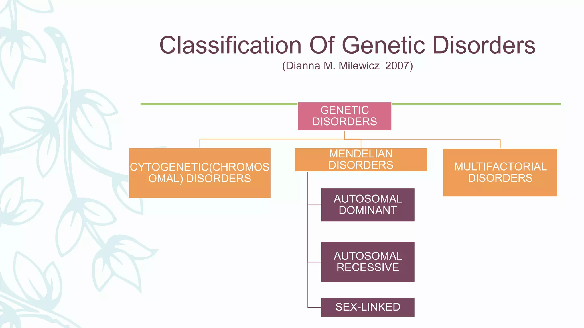
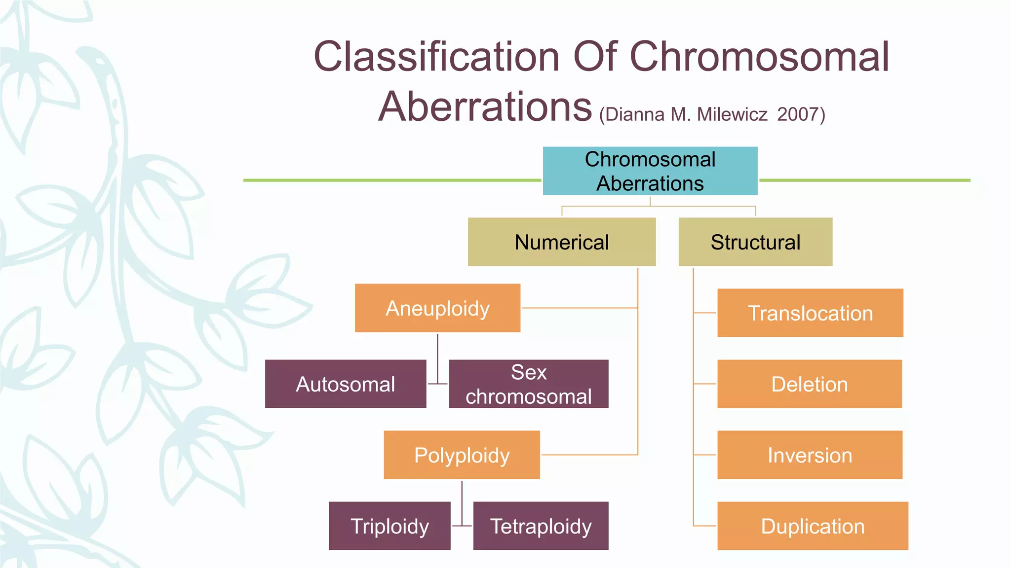
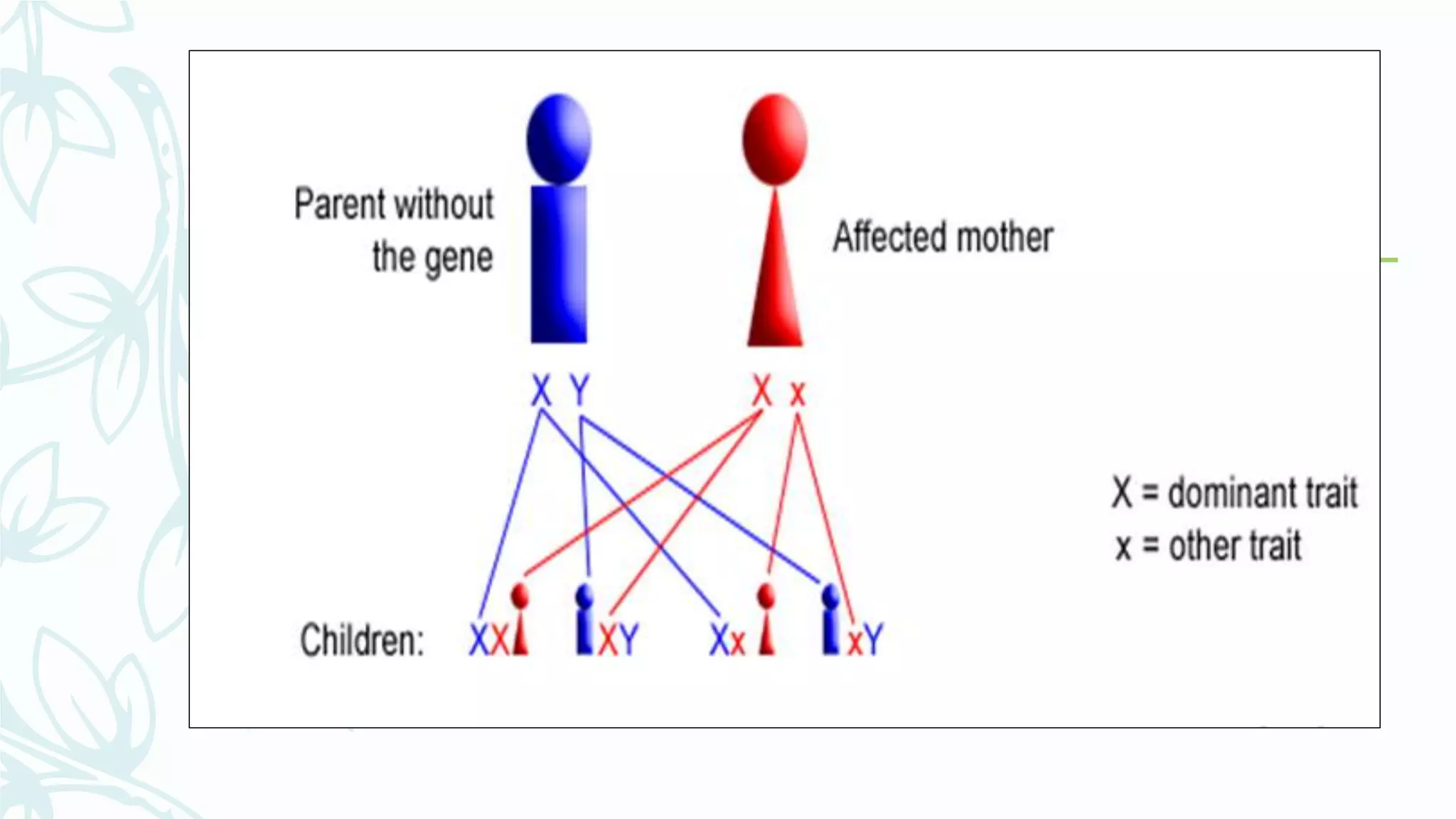
Genetic and
chromosomal
aberrations in
children
PART II POST GRADUATES
Contents
– Introduction
– Definitions
– Classification of genetic disorders
– Classification of chromosomal aberrations
– Chromosomal abnormalities
– Pattern of inheritance
– Structural abnormalities
– Autosomal dominant conditions
– Autosomal recessive conditions
– X-linked conditions
– Chromosomal syndromes
– Polygenic conditions
– Conclusion
– References
Introduction
– Genetic disorders are likely to effect everyone at some time.
– Some are of more obvious importance than others, depending on the
age of the onset of the disease, the degree of mental or physical
impairment, the numbers of affected individuals, and the cost of care
– The dentist often in a unique position to pick up a previously
unrecognized genetic or birth defect problem in a patient or family.
Definitions
– Gene : The functional unit of hereditary responsible for the transmission
of characters to the progeny or off-springs.
– Alleles : They are a pair of genes present at a specific locus in
homologous chromosomes.
– Homozygous : Condition where the paired genes are similar on
homologous chromosomes for a character.
– Heterozygous : Condition where the paired genes are dissimilar on
homologous chromosomes for a character.
– Hybrid : It is an offspring of two parents differing in one or
more characters.
– Dominant gene : It is a gene which expresses itself in the
presence of a contrasting gene.
– Recessive gene : It is a gene which fails to express itself in
the presence of a dominant gene.
Classification Of Genetic Disorders
(Dianna M. Milewicz 2007)
GENETIC
DISORDERS
CYTOGENETIC(CHROMOS
OMAL) DISORDERS
MENDELIAN
DISORDERS
AUTOSOMAL
DOMINANT
AUTOSOMAL
RECESSIVE
SEX-LINKED
MULTIFACTORIAL
DISORDERS
Classification Of Chromosomal
Aberrations(Dianna M. Milewicz 2007)
Chromosomal
Aberrations
Numerical
Aneuploidy
Autosomal
Sex
chromosomal
Polyploidy
Triploidy Tetraploidy
Structural
Translocation
Deletion
Inversion
Duplication
Chromosomal Abnormalities
NUMERICAL ABNORMALITIES
1.Polyploidy: chromosomes count exceeds the diploid number
and is also an exact multiple of its haploid number.
– e.g. Triploidy is a condition in which there are three times
the no. of haploid chromosomes. .
– Tetraploidy is a condition in which there are four times the
no. of haploid chromosomes.
2.Aneuploidy: the diploid chromosomes number of a cell is not
an exact multiple of its haploid number.
– Autosomal aneuploidy –
Trisomy 21(Down’s Syndrome)
Trisomy 13(Patau’Syndrome)
Trisomy 18(Edward Syndrome)
– Sex chromosomal aneuploidy
Kleinfelter’s (47,XXY)
Turner’s (45,X0)
Triple X Syndrome(47,XXX)
Non-Disjunction
– Nondisjunction is the failure of chromosome pairs to separate
properly during cell division. This could arise from a failure of
homologous chromosomes to separate in meiosis I, or the failure of
sister chromatids to separate during meiosis II.
– When a single chromosome is lost (2n-1), it is called a monosomy.
When a chromosome is gained, it is called trisomy,
– Examples of nondisjunction: Down syndrome, Triple-X syndrome,
Klinefelter's Syndrome, Turner's Syndrome, Edward Syndrome,
Patau’s Syndrome
Pattern Of Inheritance
– Inheritance is a term used to denote transmission of
characters from parents to off springs.
Mendelian
Multifactorial
Mendelian Inheritance
– The manner in which genes and traits are passed from
parents to their children. Disorders caused by a defect in a
single gene follow the patterns of inheritance described by
Mendel(1965).
Autosomal
dominant
Autosomal
recessive
Sex-linked
dominant
Sex-linked
recessive
– Dominant : If a trait or disease manifests itself when the
affected person carries only one copy of the gene
responsible, along with one normal allele, the mode of
inheritance of the trait is called dominant.
– Recessive : If two copies of the defective gene are required
for expression of the trait, the mode of inheritance is
called recessive.
Autosomal Dominant
 Disease can be inherited from one parent also.
 Can affect both males and females
 Can be traced through many generations of a family.
 Affected people are heterozygous for the abnormal allele
and transmit the gene for the disease to half their
offspring, whether male or female.
Autosomal Recessive
 Disease can appear only if both parents transmit it.
 Unaffected parents can transmit the trait to their
offsprings(if both parents carry recessive gene)
 Siblings may be affected but parents usually are
apparently normal
 Abnormal gene remains suppressed and does not manifest
in a heterozygous individual.
 Affected person is homozygous for the trait.
X-Linked Dominant
 The gene is located on the X chromosome, but the gene
acts in a dominant manner.
 This means that both males and females can display the
trait or disorder, by only having one copy of the gene.
X-Linked Recessive
 X-linked recessive genes are expressed in females only if there
are two copies of recessive gene (one on each X chromosome).
 In Males there only needs to be one copy of an X-linked
recessive gene in order for the trait or disorder to be expressed.
 Only males are affected, but can be transmitted through
healthy female carriers.
 A female carrier will transmit the condition to half her sons,
and half her daughters will be carriers.
 An affected male will transmit the defective gene to all his
daughters but to none of his sons.
 This absence of male to male transmission is a hallmark
of X linked inheritance.
 Trait is passed from an affected male to all of his
daughters and then to ½ of the daughter’s sons.
Multifactorial Inheritance
– Polygenic inheritance, also known as quantitative or
multifactorial inheritance refers to inheritance of a
phenotypic characteristic (trait) that is attributable to two
or more genes and their interaction with the environment.
Unlike monogenic traits, polygenic traits do not follow
patterns of Mendelian inheritance.
Structural Abnormalities
Chromosomal Translocation
– ROBERTSONIAN TRANSLOCATION: Result from
breakage of two accocenteric chromosomes with
subsequent fusion of long arms
– RECIPROCAL TRANSLOCATION: It involves breakage
of at least two chromosomes and exchange of fragments.
Chromosome Deletion
– Deletion is the loss of a portion of a chromosome
Chromosome Ring
– It is a deletion chromosome in which both ends are lost
and two broken ends unite to form a ring.
– The size of the ring, depends on how much material was
lost and which chromosomes are involved.
Chromosome Inversion
– Inversion is the separation of a portion of a chromosome
followed by rearrangement but not in the same order.
– If the inverted area includes the centromere it is called a
pericentric inversion.
– If it does not, it is called a paracentric inversion.
Chromosome Duplication
– It involves presence of extra segment of chromosome which
usually results from unequal crossing over.
– Duplications are more common and less harmful to the
individual than are deletions.
Abnormalities of Structure related to sex chromosome
– Isochromosome X – a long X chromosome – which results from
deletion of the short arms of the X chromosome and duplication
of the long arm.
Autosomal dominant
conditions
Achondroplasia(Chondrodystrophia
fetalis )
– Transmitted as an autosomal dominant trait.
– It is a genetic disorder affecting the growth and development of
bone and cartilage
– Etiology : by Mutation in Fibroblast Growth Factor Receptor - 3
gene (FGFR3)
– Result in decreased endochondral ossification , inhibited
proliferation of chondrocytes in growth plate cartilage,
decreased cellular hypertrophy and decreased cartilage matrix
production.
– Common cause of dwarfism
– Women and men are equally affected
– Global Incidence : 1 in every 25,000 births. ( Wynn J, King TM,
Gambello MJ, Waller DK, Hecht JT (2007)
– In India: 12 in 1000(Debbie Siegal,2007)
Clinical features
– The head is large and the arms and legs are short when
compared to the trunk length. (disproportionally long trunks)
– Prominent forehead (frontal bossing )
– Small midface with a flat nasal bridge and narrow nasal
passages (midface hypoplasia)
– Thoracolumbar protuberance
– Limitation of joint movement
– Normal intelligence
Oral manifestations
– Maxillary retrusion
– Mandibular prognathism (class III malocclusion)
– Missing teeth may be seen in primary as well as permanent dentition.
– Crowding
– Poor oral hygiene
Radiographic features
– Lateral skull radiographs demonstrates
– Mid face hypoplasia
– Enlarged calvarias
– Frontal prominence
– Shortening of the base of the skull
– Long bones are shorter then normal
– Thickening and mild clubbing of the ends
– Epiphyses generally appear normal but may close either early or
late
Treatment
– Growth and weight should be carefully monitored.
– Head circumference should be carefully monitored.
– Orthopedic treatment, ENT treatment and neurosurgical procedures
may be necessary.
– Growth-hormone treatment increases the rate of growth during the
first year of treatment.
– Midface deficiency in achondroplasia ideally is accomplished by a Le
Fort III osteotomy to move the entire midface forward.
– Orthodontic treatment to correct malocclusion
Treacher Collin’s Syndrome
(Mandibulofacial dysostosis)
– (also known as Franceschetti-Zwahlen-Klein syndrome ) is a genetic
disorder characterized by craniofacial deformities.
– Inheritance is autosomal dominant
– Etiology : Change in the Treacher Collins-Franceschetti -1(TCOF1)
gene on chromosome 5.
– Males = Females
– Multiple generations are affected
– It is named after Edward Treacher Collins (1862–1932), who
described its essential traits in 1900.
– Global Incidence: 1 in 25,000 births(Acc to R Madhan, Sanjna,2006)
– In India : 1 in 50,000 (Acc to Statistics By Country for Treacher
Collin Syndrome,2004)
– More than 50 families have been analyzed
– Detected mostly around 3-6 years of age
– Typical craniofacial disorder of 1st and 2nd pharyngeal arches
– Affects the development of arches and produced defects of head
and face
– Caused by fetal vascular anomaly which deprives the first
visceral arch of its blood supply between the third and fifth weeks
of gestation
Clinical features
– Bird face like or fish like appearance
– Microphthalmia
– Antimongoloid down - slanting palpebral fissures
– Notched lower eyelids (coloboma)
– Deficiency of eyelashes
– Auricle usually in low position and atresia of external auditory meatus (36%)
– Underdeveloped and malformed ears (77%)
– Hypoplasia of facial bones, Hypoplastic zygoma(28%)
– Depressed cheeks and hypoplasia of mandible
– Other anomalies
– facial clefts and skeletal deformities
– Harelip
– Deafness
– Large tongue and large irregular upper incisors
– Dwarfism
– Defects in brain development as microcephaly , mental retardation
and psychomotor delay.
Dental findings
– Hypoplasia of mandible and maxilla)
– Dysplasia of TMJ and Limited mouth opening
– Midline deviation
– Improper tongue position
– Cleft palate
– Open bite
– Impacted Supernumery teeth
– Small mouth and high palate
– High plaque index and poor efficacy of tooth-brushing
– ( acc to gisele da silva dalben lucimara teixeira das neves marcia
ribeiro gomide,2008)
Radiographic features
– Malformed auditory ossicles with fusion between malleus
and incus
– Non-fusion of the zygomatic arches as well as absence of
the palatine bones
– Hypogenesis and agenesis of the mandible
– Paranasal sinus are grossly underdeveloped
Treatment after birth
– At time of birth child with TCS concerns centers – adequacy of airway
, swallowing and feeding, hearing, vision, presence of cleft palate
– Airway may be compromised – maxillary hypoplasia and mandibular
hypoplasia
– May necessitate special infant positioning , extended hospital stay ,
pulse oximetry monitoring , immediate or delayed tracheostomy or
placement of mandibular distractor.
– If newborn with TCS have difficulty in swallowing – placement of
gastrostomy tube
Treatment
– Early childhood program for speech and language stimulation may
be recommended.
– Bone-anchored hearing aids in case of hearing problems
– People with the syndrome undergo surgeries for facial and external
ear reconstruction.
– Surgical closure of the cleft in case of cleft palate.
– Combined orthodontic therapy along with orthognathic surgery is
also sometimes required.
Cleidocranial Dysplasia
(Marie-Sainton’s Disease)
– Congenital disorder of bone formation
– Etiology : Rearrangement of long arm of chromosome 8 and 6.
Mutation in the core-binding factor alpha-1(CBFA-1) gene
– First described by Meckel and Martin in the mid 8th century.
– Global Incidence:1 in 10,00,000 (I.Golan,2003)
– In India,1 in 5100 (Dhavendra Kumar,2004)
– Reported in all ethnical group
– No sex predilection
– Manifested with
– Hypoplastic or aplastic clavicle with narrow thorax
– delayed ossification of the skull
– excessively large fontanelles(brachycephalic skull)
– delayed closing of suture
– Midface hypoplasia
– Short stature
Etiopathogenesis
– Clavicle is first bone to ossify in body in man (5th week)
– Clavicle – partially formed in membrane and partially in
cartilage
– Frontal bone , parietal bone , temporal bone , the bones of
face , part of occipital bone - formed in membrane
– CCD usually affects – bones formed from membrane
Clinical features (shoulder girdle)
– Partial or complete absence of clavicles or thinning of one or both
clavicle.
– Unusual mobility of the shoulder and Ability to bring the shoulders
together in front of the body until they meet in midline
– Chest is often found to be funnel shaped and sternum is
depressed
– Diminishing in size, origin and insertion of sternocleidomastoid,
subclavian, pactoralis major, deltoid and trapezius muscles are
found
Clinical features(skull and head)
– Delayed or incomplete closure (ossification) of the space between the bones of
the skull (fontanels) – tend to be large
– Frontal , partial and occipital bones are prominent – give the skull large
globular shape with small face
– Premature closing of the coronal suture with interposition of wormian bone.
– Relative macrocephaly , depressed nasal bridge and defects may occur at the
bregma
– Paranasal sinuses and mastoid air sinuses are underdeveloped and narrow
Other findings
– Short stature
– Scoliosis of the spine
– Wide pelvic bone
– Loose joints
– Shortened middle phalanges
– Hearing loss and/or frequent infections
Dental findings
– Maxilla is commonly micrognathous
– Mandibular prognathism
– High, narrow arched palate with median furrow
– A complete cleft of bony tissue of palate with soft parts intact
has been reported
– Lacrimal and zygomatic bones are – underdeveloped
– Prolonged retention of the primary teeth and Delayed appearance or unerupted
permanent teeth. Delayed and arrest of physiological root resorption and bone
resorption can be attributed as cause
– Unusually shaped and peg-like teeth
– Formation of supernumerary teeth can be found in all region , ectopic eruption of
these teeth can be found.
– Formation of dentigerous cyst is common.
– 1st and 2nd molar erupts normally and 3rd molar missing completely
– Enamel defects , curved roots are common
– All this leads to malocclusion and accentuates liability of caries , periodontal
problems and hypertrophied gums.
Treatment
– There is no specific treatment for the bone problems.
– Early institution of preventive care problems
– Endocrinological investigation should done
– Administration of thyroid extract and vitamin therapy to
stimulate eruption
– An otologist should check for hearing problems
– Excellent oral prophylaxis should done
– Extraction of primary teeth is contraindicated and it should be
restored
– Surgery may be performed to remove Supernumery teeth
– open the bony coverings surrounding the permanent teeth, with the
goal of promoting their eruption.
– Orthodontic procedures: required to align the teeth.
– Construction of suitable prosthetic appliance
– Bilateral osteotomy should done to reduce mandibular prognathism
Marfan’s Syndrome
– Described by Parisian professor of pediatrics, Antoine Bernard
Marfaan.
– 1896, presented a case of a 5 years old girl Gabrielle-Spider fingers,
-Slender limbs.
– Six year later, Mery and Babonneix noted a spinal curvature and
thoracic asymmetry.
– Others- Cardiovascular abnormalities.
– Generalized systemic disorder of connective tissue characterized by
– Dislocation of ocular lenses
– Long-bone overgrowth
– Long slender digits
– Proximal aortic aneurysms
– Autosomal dominant disease
– Both sexes equally
– Incidence 1:10,000
– Reviewed infrequently at 5, 10, 13 and 16 years
Etiology
– An abnormal fibrillin was identified in the skin and aorta of the
affected patients
– An abnormal fibrillin was linked in chromosome 5.
– FBN1 is a major component of extracellular microfibrils.
– Abnormal fibrillin – stiffness.
Clinical manifestations
– Phenotypically the tissues are commonly affected are:
– Growth plates of the long-bone
– Eyes
– Aortic root
– Mitral apparatus
– Overgrowth of arms and legs
– Wrist sign or Walker-Mudrock
– Steinberg or a thumb sign
According to system affected
1. Skull
• Tall stature
• Arachnodactyly
• Funnel chest, pigeon chest
• Scoliosis
• Kyphosis
• Flat feet
• Contracture of finger
• Joint laxicity
• Degenerative arthritis
2. Ocular
• Octopia lentis
• Magalocornea
• Myopia
• Retinal detachment
3. Cardiovascular
• Mitral valve prolapse
• Mitral valve regurgitation
• Aortic root dialatation
• Aortic regurgitation
• ECG abnormalities
4. Pulmonary
• Pneumothorax
5. Skin and integument
• Strai atrophicae
• Inguinal hernia
• Dural ectasia
6. CNS
• Poor motor performance
• Attention deficit disorder
• Learning disability
CRANIOFACIAL MANIFESTATIONS:
• Long narrow skull
• High arched palate
• Severe periodontitis
• Tooth crowding
• Rettrognathia
• Micrognathia
• Malar flattening
• Downward slanting of palpebral fissures
• Local hypoplastic enamel defects
• Cleft palate
• Bifid uvula
• Teeth – long and narrow
• Malocclusion
• Partial anodontia
• TMJ disorders
• Abnormal dentin formation
• Abnormal pulp shape and root deformity
Medical management
– Complete evaluation by a pediatric cardiologist and a geneticist.
– Include a systemic appraisal of the several non cardiovascular
systems
– Cardiac evaluation include a detailed general physical evaluation
and to detect signs of heart failure from mitral valve disease
– Mitral valve surgery is important
– Beta- adrenergic blockade – atenolol
– At each follow up – echocardiographic measurement of the aortic
root diameter and shape.
– Should undergo yearly assessment by ophthalmologist.
– Patient should be counselled not to engage in contact sports,
competitive athletics, or isometric exercise.
Dental management
– Daily dental care
– Consult a cardiologist for antibiotics before dental procedures
– Consider if dental or orthodontic work is needed
– Important to balance treatment of dental issues against risks to
the heart
– When an individual requires a lot of dental treatment- under
sedation.
Craniofacial Dysostosis
 Crouzon syndrome is a genetic disorder characterized by the
premature fusion of certain skull bones (craniosynostosis)which
prevents the skull from growing normally and affects the shape of the
head and face.(Harold Chen,2007)
 In 1912 Crouzon first described a woman and her daughter with this
disorder.
 Caused due to defect in FGFR2 gene (fibroblast growth factor receptor
2) in chromosome 10.
– Global incidence: 1 in 25,000 live births (Acc to Johnston in
Behrman (2004) Nelson Pediatrics, p. 1992-3)
– Detected in the newborn or infant period
Types:
1. Brachycephaly
2. Scaphocephaly
3. Plagiocephaly
4. trigonocephaly
Craniofacial manifestations
– Early synostosis of sutures mainly
coronal suture
– Patients nose resembles Parrots beak
– Ocular proptosis
– Hypertelorism and exophthalmous
– May often be mentally retarded
Dental findings
– V-shaped maxillary dental arch
– Hypoplasia of maxilla with prognathic mandible (Class III
Malocclusion)
– High arched palate
– Short hypotonic lips
– Crowding of the upper teeth
Treatment
– Mainly done for cosmetic purpose and for vision.
– Sometimes Craniectomy is done at an early age-to provide
space for rapidly developing brain
– Frontoorbital Advancement to correct ocular defects
– Midface Advancement to correct maxillary hypoplasia
– Sagittal split osteotomy with mandibular setback to correct
mandibular prognathism
Dental Management:
– Orthodontic evaluation – at an early age
– Might require extraction of some permanent teeth as well as
expansion of maxilla.
Apert Syndrome
– Apert syndrome (AS) is a severe disorder, characterized by
craniosynostosis and complex syndactyly of the hands and feet and
midfacial hypoplasia (Newfoundland Department of Education,1992).
– In 1906, Eugène Apert, first described nine people with this disorder
– In 1995, A.O.M Wilkie proved that acrocephalosyndactyly is caused
by a defect in a gene called fibroblast growth factor receptor 2, on
chromosome 10
– Occurs in approximately 1 per 160,000 to 1 per 200,000 live
births(Acc to Kaplan, L C (2004).)
In India 1 in 1923 (Acc to M.L.Kulkarni2004)
Detected in newborn period
males and females are affected equally.
Clinical features
Early closure of sutures between bones of the skull
Acrobrachycephaly(tower skull)
Ocular proptosis
Severe under-development of the mid-face
Fusion or severe webbing of the 2nd, 3rd, and 4th fingers, often
called "mitten hands"
Webbing or fusion of the toes
 slow intellectual development
Short height
Hearing loss
Frequent ear infections
Oral findings
Trapezoid like mouth with protruded lips
Delayed and/or ectopic eruption and shovel-shaped incisors.
Supernumerary teeth Or bifid uvula
Maxillary hypoplasia
Class III Malocclusion
Cleft lip
Cleft palate
Treatment
Craniectomy is performed during 1st year of life to treat
craniosynostosis
Frontofacial advancement and midface advancement to correct
proptosis and midface hypoplasia respectively.
Combined orthodontic and orthognathic surgery can help to relieve
some of the facial deformities, such as the flat or concave face.
 LeFort III, can be used to detach the midface from the rest of the
skull to reposition it appropriately.
Corrective surgery is only undertaken if the child has a
reasonable chance of living for some time as it is usually fatal
within the first six months of life.
Autosomal recessive
conditions
Cystic Fibrosis
– It is a chronic disabling disease associated with early demise.
– Caused by mutation of CFTR gene on long arm of chromosome 7.
– Gene is responsible for regulation of cellular chloride channels.
– Influence salt and water movement across cell.
Pathophysiology
Manifestations
ENT: Chronic Sinusitis, Nasal Polyps
Lungs:Cough and sputum, Airflow obstruction
Recurrent infection
GI: Pancreatic insufficiency (malnutrition)
Pancreatitis (PS), Meconium ileus,
Biliary cirrhosis and portal hypertension
Sex
organs: Obstructive azoospermia
Others: Salty tasting skin
Poor growth and poor weight gain
Oral Manifestations
– Oral mucous glands are affected
– Salivary gland enlargement
– Thick ropy saliva
– Enamel defects
– Periodontal disease
– Dental caries
– Tooth discoloration
– It can be detected by a positive history of chronic airway disease and can be confirmed using a
sweat test.
– Treatment:
– Promote clearance of secretions
– Control of lung infection
– Provide adequate nutrition
– Prevent intestinal obstruction
– oral hygiene instruction for infection control
X-Linked conditions
– Mutation in genes located on X-chromosome.
– Females are carrier and do not normally manifest the
disorder.
– Males, on other hand only have one X chromosome which is
inherited from mother, 50% chances of manifesting the
disease.
– Example: Hemophilia
X-linked hypohydrotic ectodermal dysplasia
Fragile X-syndrome.
Hemophilia
– It is a group of hereditary genetic disorders.
– Impairs the body’s ability to control blood coagulation
– Occurs only in males while females being carrier.
– Types:
Hemophilia A: Deficiency of clotting factor VIII.
Hemophilia B: Deficiency of factor IX.
Hemophilia C: Deficiency of factor XI.
Signs and symptoms:
- Internal or external bleeding episodes
Mild: 5-40%
Moderate: 1-5%
Severe: Less than 1%
- Persistent bleeding
- Hemorrhage into subcutaneous tissues, internal organs and joints.
- Massive hematomas.
– Prolonged clotting time and aPTT
– Children with mild to moderate hemophilia may not any signs or
symptoms at birth
– First symptoms are large bruises and hematomas from frequent falls
– Hemophilia A can be mimicked by von Willebrand’s disease.
– Severe cases of vitamin K deficiency can present similar symptoms to
hemophilia.
Management:
- Intravenous injection of factor VIII concentrate
Oral considerations:
- 30% factor rise is essential for infiltration anesthesia in maxillary arch
- For extractions, deep scaling or root planing, 50% factor rise is
essential.
- For surgical extractions, fractured jaw or facial bones, 100% rise may be
necessary.
– The problem of dental extractions is a difficult one
– Without proper premedication, even minor surgical procedure may result
in death.
– Tooth extraction by means of rubber bands has been used successfully.
– Tranexamic acid mouthwash 10% for 7-10 days after extraction
– Soft diet for 7 days
– Fibrin glue as local hemostatic agent
– Splints can also be used.
Fragile X Syndrome
– It is secondary to an abnormality of a gene near the end of long arm of
chromosome X.
– Males affected are moderately retarded and require a sheltered existence
throughout life.
– Carrier females are mildly affected.
– Physical alteration are not reliable indicators, although prognathism, long
face and ears are typical.
Physical characteristics:
- Long narrow face
- Prominent ears, jaw, chin
- Excessive growth rate during early childhood years
- May have intellectual disability
-Autistic like behaviors
-Strabismus and slanted eyes
- Poor muscle tone and co-ordination
- 15-20% of patients may have seizures.
It is important for the dentist to provide the parent with the information
and tools required to assess and maintain good oral hygiene and healthy
dietary practices for these children.
Ectodermal Dysplasia
– An X-linked recessive disorder
– Abnormal development of ectodermal derivatives such as nails,
teeth, hair and sweat glands.
– Rare genetic disorder with incidence of 0.00005%
– Most frequently observed is the anhidrotic X-linked form.
Ectodermal Dysplasia
Clinical features:
- Dry and rough skin and Heat intolerance
- Facial pigmentation
- Sparse hair
- Abnormal nails
-Typical face: frontal bossing, peri-orbital pigmentation, depressed nasal bridge,
protuberant lips, low set ears and scanty scalp hair.
-Moderate built and poorly nourished.
Intra-oral manifestations:
– Aberrations in number and shape of teeth
– Malformed teeth – conical shape CI and LI.
– Premature caries
– Gingivitis
– Early tooth loss of both the dentitions
– Premature exfoliation
– Dry and sticky oral mucosa
– Knife edge alveolar ridges
– Partial anodontia/ hypodontia
Radiographic manifestations:
– Altered morphology of teeth
– Aplasia of alveolar bone
– Mandibular aplasia
– Decreased lower facial height
Management:
– Dry skin: emollients
– For decreased lacrimation: artificial tears
– For dry nasal mucosa: saline sprays
– Antibiotics for any infection occurs
– For dental malalignment: orthodontic treatment
– Surgery for oral and dental rehabilitation
– Prosthesis for missing teeth
Polygenic conditions
Cleft Lip And Cleft Palate
– Most common congenital malformation occurring in man and is of a
polygenic condition.
– Results from multiple genes and gene-environmental interactions rather
than a single gene defect.
– In polygenic conditions, the incidence is 1:800 with a recurrence rate of
4%.
– Combined cleft lip and palate: More common in males
– Isolated cleft palate: More common in females
– Unilateral defect more common than bilateral defect
– In unilateral defects: left side is more common
Clinical features:
– Difficulty in speech, hearing and swallowing
– Supernumerary teeth or congenitally missing teeth
– Peg shaped teeth
– Thick curved hypoplastic incisors
– Delayed eruption of permanent teeth
– Isolated enamel defects
– Feeding difficulties
Clinical significance:
- It is customary to operate the cleft before the patient is one
month old.
- Eating and drinking are difficult because of regurgitation of
food and liquid through nose.
- Speech problem is also serious
Treatment
– Requires an experienced team including otolaryngologist, speech
pathologist, dentist and psychologist.
– Requires years of specialized care.
– At birth, predental treatment is provided which comprises feeding plate
and presurgical orthopedics.
– Timing of first lip surgery – at 3 months of age.
– Pediatricians follow rule of ‘three 10s’ as a requirement for identifying the
child’s status
Neural Tube Defects
– Multifactorial inheritance
– These conditions result from defective closure of the developing neural
tube during the first month of embryonic life.
– A defect occurring at the upper end of the developing neural tube
results in encephalocele.
– A defect occurring at the lower end leads to a spinal lesion such as
lumbo-sacral myelocele.
– Neural tube defects seldom affect the oral area directly but may cause
changes indirectly because of a resulting hydrocephalus or seizure
disorder that requires anticonvulsants.
– Repair of primary lesion is preformed early to protect CNS infections.
– Learning problems may be present in mild or more severe degrees
depending on whether a CNS infection or hydrocephalus has been
present.
– Leg bracing or wheelchair use is usual.
– For dentist, subacute bacterial endocarditis prophylaxis may be important
when a shunt tube or catheter is present, especially if the tip of the distal
end of the shunt has been placed within the ventricle of heart.
Chromosomal syndromes
Down’s Syndrome
– A chromosomal condition characterized by physical malformations and some
degree of mental retardation, concerned with a defect in the twenty-first
chromosome(presence of three representative chromosomes in a cell instead
of the usual pair)(Miller & Keane, 1972).
– Most common chromosomal disorder in humans.
– Also known as mongolism and trisomy 21(Acc to Schreiner1992)
– Dr LANGDON DOWN first described it in the clinical lecture report in
London hospital in 1866
– In 1959, Lejeune and Jacobs independently determined that Down
syndrome is caused by trisomy 21.
Incidence of Down Syndrome
– 1-in-800 overall births(global) – acc to Center for Disease Control
2006)
– In India 1 in 1139 live births(acc to M.P. Kava,2004)
Incidence extrapolations of USA for Down Syndrome:
– 340,000 per year (Association for Children with Down Syndrome2003)
Pathogenesis
- In 90% of cases down’s syndrome occur due to nondisjunction which
occurs during first and second meiotic division
- It occurs in offspring of mothers of all ages but the risk increases with the
increase in maternal age
Types
- Trisomy 21( presence of extrachromosome)
- Translocation type ( extra chromosome is not free but translocated on
any other chromosome usually 13 or 15)
- Mosaicism ( some cells have 46 and the other cells have 47
chromosomes)
Common physical signs include:
– Decreased muscle tone at birth
– Excessive skin at the nape of the neck
– Flattened nose
– Separated sutures (joints between the bones of the skull)
– Single crease in the palm of the hand(Simian Crease)
– Small ears
– Upward slanting eyes
– Wide, short hands with short fingers
– White spots on the colored part of the eye (Brushfield spots)
– Slower Physical development than normal
– Widely spaced toes
Craniofacial features
- Brachycephaly due to failure of basal segments to elongate normally
- Bony hypoplasia of midface produces ocular hypotelorism
- Flattening of nasal bridge
- Relative mandibular prognathism
- The ears are small in size
- Maxillary sinuses are hypoplastic
Many different medical conditions are seen in babies born with Down
syndrome, including
– Birth defects involving the heart such as an atrial septal defect or
ventricular septal defect
– Eye problems such as cataracts
– Hearing problems
– Sleep apnea
Oral manifestations
– Macroglossia with protrusion of tongue
– Fissured tongue
– Retardation of growth of maxilla
– Commonly have high arched palate
– Enamel Hypoplasia & microdontia
– Delayed eruption and early shedding of deciduous dentition
– Defective or absent upper incisor
• Tooth shape anomalies
• Bruxism
• Malocclusion
• Congenital absence of permanent teeth
• Prevalence and severity of periodontal diseases is much higher
• Caries susceptibility is low
Natural history
• Most children are happy ,affectionate and well behaved
• Affected child shows broad range of intellectual activity
• IQ ranges from 25-75 average being 40-45 (American Association Of
Mental Deficiency Classification)
• Adult height around 150 cms
• Average life expectancy 50-60 years
• Early death in 15- 20% with severe cardiac abnormalities
• Most people develop Alzheimer’s disease later in life
Management
– Medical management, home environment, education, and vocational
training.
– There is no specific treatment for Down syndrome, special education
and training is offered in most communities for children with delays
in mental development.
– Speech therapy may help improve language skills. Physical therapy
may be needed to teach movement skills.
Dental Management
– Abnormalities related to Down Syndrome that can affect dental
treatment are congenital heart disease and behavioural problems due
to retardation
– Provide oral care in an environment with few distractions.
– Use of fluoride should be enhanced to reduce caries
– Try to reduce unnecessary sights, sounds, or other stimuli that might
make it difficult for your patient to cooperate. Most of patients with
Down syndrome are friendly and co-operative
– Premedication may be given to allay apprehension
Systemic factors affecting dental care
- Complete medical history should be taken & reviewed
- Consult family, physician and caregivers to obtain proper medical history
- Almost 50% of these patients have cardiac abnormality so they require
antibiotic prophylaxis before dental treatment
- These pts have compromised immune system which causes higher rate of
infection and high incidence of periodontal disease
- Most of the pts have upper respiratory tract infection causing mouth
breathing
- Greater incidence of apthous ulcers, candida infections and ANUG which
should be treated aggresively
- Importance of fluoride application should be stressed to prevent caries
- Use of lip balm should be done to ease the strain on lips
- Reduced muscle tone causes inefficient cleaning and natural cleaning of
teeth
- Use of pillows should be done to stabilize the patients
- seizures are also common in these individuals can be controlled by using
anticonvulsants
– If seizure occur during treatment then instruments in the are to be
removed from the mouth
– Pt should be turned on the side to reduce the risk of aspiration
Visual & hearing impairement affecting dental care
- The pt should be assisted throughout while moving in the clinic
- Tactile feedback like a warm handshake should be given to make them
comfortable
- Dentist should talk while looking at him and should inform about each
upcoming step
- Written instructions should be given in large print
- The hearing aids should be turned off
Sleep apnea
- The decreased airway size with lowered muscle tone predisposes the pt to
sleep apnea
- Symptoms- snoring, restless sleep, unusual sleeping position
- refer the pt to the sleep disorders clinic
- Positive airway pressure and surgical correction is helpful
- Adenotonsillectomy proves helpful
Orofacial features affecting dental care
- Because of high arched palate and macroglossia there is difficulty in
speech and mastication so speech pathologist should be consulted to
teach correct tongue positioning and increase the tone of orofacial muscles
- Fissures of tongue may become food lodged leading to halitosis so regular
cleaning of dorsal surface of tongue proves helpful
- Because of delayed eruption of teeth diet has to be modified
- In case of severe crowding after eruption of all permanent teeth selective
extraction proves helpful
– Spacing by microdontia- restoration or orthodontic correction
– Severe illness causes hypoplasia and hypocalcification-primary teeth
should be preserved
– Space maintainers can be given
– Hypocalcified tooth should be observed in case of any early onset of
decay
– Pit & fissure sealants, stainless steel crowns, fluoride application can be
done
– Trauma or injury of oral cavity- tooth saving kit should be given
– Periodontal disease- chlohexidine applied using spray bottle or tooth brush
– Regular maintainence of oral hygiene and professional cleaning is
recommended
Turner’s Syndrome
– A chromosome disorder in females that is characterized by the
absence of all or part of a second sex chromosome in some or all
cells(Webster's New World Medical Dictionary2004)
– Described by Henry Turner in 1938
– Missing X-chromosome(45,X)
– Global Incidence is 1 in 2500 Births( Sybert, V.P., McCauley, E. (2004).
– In India: 1 in 3000-(M. Patil,2006)
Clinical features
– Short stature
– Defective vision
– Webbed neck
– Lower posterior hair line
– Auditory Defects
– Broad chest
– Widely spaced nipples
– Streak ovaries, infertility
– Hypoplastic nails
– Pigmented nevi
Oral Findings
– Small sized mandible
– High palate
– Narrow maxilla
– Multiple carious teeth
Treatment
– Estrogen replacement therapy from puberty onwards for of Secondary sexual
characters and prevention of osteoporosis
– Growth hormone, approved by the U.S. Food and Drug Administration used for
treatment of Turner syndrome will increases final height.
Dental management of a patient with Turner syndrome.
– In case of multiple carious teeth, necessitated full mouth dental rehabilitation
with the use of general anesthesia because of presence of cardiovascular
abnormalities and long term antibiotic prophylaxis.
– According to, National Health Institute of Child Health and Human
Development, Orthodontic evaluation should be started at 7 yrs or
more for treatment of malocclusion and other dental anomalies.
– Modifications that might be required to orthodontic treatment plans
include (1) antibiotic prophylaxis, (2) occlusal adjustments to
account for altered dental morphology, (3) altered treatment timing
because of major differences in growth and differences between
chronological and skeletal ages
Klinefelter’s Syndrome
• Klinefelter syndrome is a chromosomal disorder that affects only
males having two X chromosomes in addition to Y chromosome.
(Beers, Mark H., MD, and Robert Berkow,2004)
• This condition was first recognized by Dr. Harry Klinefelter in
1942
• Global Incidence: 1 in 1000 male live births .(M.L. Hunter, MM
Collard, T.Razavi& B. Hunter,2003)
– 1 in 800 males in India(Dada , A C Ammini , K Kucheria ,2005)
Pathogenesis
- The extra X chromosome is usually acquired through an error of
nondisjunction during gametogenesis, where a sperm or an egg
caries an extra X chromosome in addition to normal single sex
chromosome
- It may also result from an error in division during mitosis in the
zygote
Clinical manifestations
- These patients are quiet, undemanding & little passive
- As toddlers they are a bit shy and reserved in nature and
throughout the life they preserve the same temprament
- Although this they can make friends with other children but they
tend to have only few friends at a time
- As children they learn to speak later in life
- These children show difficulty to put thoughts, express their
emotions and ideas in words
Clinical features
– Tall height
– Abnormal body proportions (long legs, short trunk)
– Small, firm testicles
– Small penis
– Enlarged breasts(gynecomastia)
– Scanty growth of hair
Dental findings
– Taurodontism
– Increased tooth size in permanent dentition
– increased mesiodistal width in primary incisors and molars.
– Shovel shaped incisors
– Maxillary and mandibular prognathism
Treatment
– Once a diagnosis is made, individuals should be referred to an
endocrinologist who can monitor sexual development and
initiate testosterone treatment at the appropriate stage of
development.
– Testosterone from beginning of puberty, about age 11 to 12 years
is beneficial for secondary sexual characters.
– once the therapy is started it should continue throughout life
– This therapy will also help to produce positive changes in mood
and behaviour , muscle mass and bone integrity
– Typical dose to maintain the normal serum testosterone level in
adults is 200 mg every 2 weeks
– When started at puberty the dose is 50-100 mg initially with an
increase of 50-100 mg every 2 to 4 weeks until adult dose is
reached
– Surgical treatment is usually done to reduce gynecomastia and
can be very well treated with plastic surgery
Dental management
- Maintain good oral hygiene to reduce gingival inflammation
– Endodontic management of a taurodontic symptomatic molars
and premolar under L.A.
– Preformed SS crowns are recommended for treatment of
taurodont molars because of poor oral hygiene and recurrent
caries due to lack of patients cooperation and medication
induced xerostomia
Conclusion
As a pediatric dentist, we are the ones who might encounter
the patient at an early age and hence, enough knowledge
about the genetic abnormalities helps us to diagnose and treat
easily.
References
– EMERY’S elements of medical genetics , 11th edition, Robert F , Ian D young .
– GENES IN POPULATION-ELIOT B SPIESS(2ND EDITION)
– Textbook Of Pedodontics 2003(1st Edition):Shobha Tandon
– PRINCIPLES AND PRACTICE OF PEDODONTICS2006(1st
EDITION):ARATHI RAO
– BASIC HUMAN GENETICS,2ND EDITION,2005:V KAPUR & RK SURI
– Human Genetics, Vol1 &2,2001,-Amita Sarkar
– SHAFER’S TEXTBOOK OF ORAL PATHOLOGY(5TH EDITION)-
SHAFER,HINE,LEVY
– PEDIATRIC DENTISTRY:Infancy Through Adolescence,4th EDITION-
PINKHAM
– Mossey PA: The heritability of malocclusion. 2. The influence of
genetics in malocclusion, Br J Orthod 26:,1999
– Oral&Maxillofacial Pathology2005,2nd edition:Neville,Damm
Allen,Bouquot
– Clinical Pedodontics(4th edition)-Sidney B.Finn
– Ref:Genetic Rearrangement in Leukaemia and Lymphoma-
I.M.Goldman&D.G.Hamden
Thank
You

Genetic and chromosomal aberrations in children

  • 1.
  • 2.
    Contents – Introduction – Definitions –Classification of genetic disorders – Classification of chromosomal aberrations – Chromosomal abnormalities – Pattern of inheritance – Structural abnormalities – Autosomal dominant conditions – Autosomal recessive conditions
  • 3.
    – X-linked conditions –Chromosomal syndromes – Polygenic conditions – Conclusion – References
  • 4.
    Introduction – Genetic disordersare likely to effect everyone at some time. – Some are of more obvious importance than others, depending on the age of the onset of the disease, the degree of mental or physical impairment, the numbers of affected individuals, and the cost of care – The dentist often in a unique position to pick up a previously unrecognized genetic or birth defect problem in a patient or family.
  • 5.
    Definitions – Gene :The functional unit of hereditary responsible for the transmission of characters to the progeny or off-springs. – Alleles : They are a pair of genes present at a specific locus in homologous chromosomes. – Homozygous : Condition where the paired genes are similar on homologous chromosomes for a character. – Heterozygous : Condition where the paired genes are dissimilar on homologous chromosomes for a character.
  • 6.
    – Hybrid :It is an offspring of two parents differing in one or more characters. – Dominant gene : It is a gene which expresses itself in the presence of a contrasting gene. – Recessive gene : It is a gene which fails to express itself in the presence of a dominant gene.
  • 7.
    Classification Of GeneticDisorders (Dianna M. Milewicz 2007) GENETIC DISORDERS CYTOGENETIC(CHROMOS OMAL) DISORDERS MENDELIAN DISORDERS AUTOSOMAL DOMINANT AUTOSOMAL RECESSIVE SEX-LINKED MULTIFACTORIAL DISORDERS
  • 8.
    Classification Of Chromosomal Aberrations(DiannaM. Milewicz 2007) Chromosomal Aberrations Numerical Aneuploidy Autosomal Sex chromosomal Polyploidy Triploidy Tetraploidy Structural Translocation Deletion Inversion Duplication
  • 9.
    Chromosomal Abnormalities NUMERICAL ABNORMALITIES 1.Polyploidy:chromosomes count exceeds the diploid number and is also an exact multiple of its haploid number. – e.g. Triploidy is a condition in which there are three times the no. of haploid chromosomes. . – Tetraploidy is a condition in which there are four times the no. of haploid chromosomes. 2.Aneuploidy: the diploid chromosomes number of a cell is not an exact multiple of its haploid number.
  • 10.
    – Autosomal aneuploidy– Trisomy 21(Down’s Syndrome) Trisomy 13(Patau’Syndrome) Trisomy 18(Edward Syndrome) – Sex chromosomal aneuploidy Kleinfelter’s (47,XXY) Turner’s (45,X0) Triple X Syndrome(47,XXX)
  • 11.
    Non-Disjunction – Nondisjunction isthe failure of chromosome pairs to separate properly during cell division. This could arise from a failure of homologous chromosomes to separate in meiosis I, or the failure of sister chromatids to separate during meiosis II. – When a single chromosome is lost (2n-1), it is called a monosomy. When a chromosome is gained, it is called trisomy, – Examples of nondisjunction: Down syndrome, Triple-X syndrome, Klinefelter's Syndrome, Turner's Syndrome, Edward Syndrome, Patau’s Syndrome
  • 12.
    Pattern Of Inheritance –Inheritance is a term used to denote transmission of characters from parents to off springs. Mendelian Multifactorial
  • 13.
    Mendelian Inheritance – Themanner in which genes and traits are passed from parents to their children. Disorders caused by a defect in a single gene follow the patterns of inheritance described by Mendel(1965). Autosomal dominant Autosomal recessive Sex-linked dominant Sex-linked recessive
  • 14.
    – Dominant :If a trait or disease manifests itself when the affected person carries only one copy of the gene responsible, along with one normal allele, the mode of inheritance of the trait is called dominant. – Recessive : If two copies of the defective gene are required for expression of the trait, the mode of inheritance is called recessive.
  • 15.
    Autosomal Dominant  Diseasecan be inherited from one parent also.  Can affect both males and females  Can be traced through many generations of a family.  Affected people are heterozygous for the abnormal allele and transmit the gene for the disease to half their offspring, whether male or female.
  • 16.
    Autosomal Recessive  Diseasecan appear only if both parents transmit it.  Unaffected parents can transmit the trait to their offsprings(if both parents carry recessive gene)  Siblings may be affected but parents usually are apparently normal  Abnormal gene remains suppressed and does not manifest in a heterozygous individual.  Affected person is homozygous for the trait.
  • 17.
    X-Linked Dominant  Thegene is located on the X chromosome, but the gene acts in a dominant manner.  This means that both males and females can display the trait or disorder, by only having one copy of the gene.
  • 18.
    X-Linked Recessive  X-linkedrecessive genes are expressed in females only if there are two copies of recessive gene (one on each X chromosome).  In Males there only needs to be one copy of an X-linked recessive gene in order for the trait or disorder to be expressed.  Only males are affected, but can be transmitted through healthy female carriers.  A female carrier will transmit the condition to half her sons, and half her daughters will be carriers.
  • 19.
     An affectedmale will transmit the defective gene to all his daughters but to none of his sons.  This absence of male to male transmission is a hallmark of X linked inheritance.  Trait is passed from an affected male to all of his daughters and then to ½ of the daughter’s sons.
  • 20.
    Multifactorial Inheritance – Polygenicinheritance, also known as quantitative or multifactorial inheritance refers to inheritance of a phenotypic characteristic (trait) that is attributable to two or more genes and their interaction with the environment. Unlike monogenic traits, polygenic traits do not follow patterns of Mendelian inheritance.
  • 21.
    Structural Abnormalities Chromosomal Translocation –ROBERTSONIAN TRANSLOCATION: Result from breakage of two accocenteric chromosomes with subsequent fusion of long arms – RECIPROCAL TRANSLOCATION: It involves breakage of at least two chromosomes and exchange of fragments.
  • 22.
    Chromosome Deletion – Deletionis the loss of a portion of a chromosome Chromosome Ring – It is a deletion chromosome in which both ends are lost and two broken ends unite to form a ring. – The size of the ring, depends on how much material was lost and which chromosomes are involved.
  • 23.
    Chromosome Inversion – Inversionis the separation of a portion of a chromosome followed by rearrangement but not in the same order. – If the inverted area includes the centromere it is called a pericentric inversion. – If it does not, it is called a paracentric inversion.
  • 24.
    Chromosome Duplication – Itinvolves presence of extra segment of chromosome which usually results from unequal crossing over. – Duplications are more common and less harmful to the individual than are deletions. Abnormalities of Structure related to sex chromosome – Isochromosome X – a long X chromosome – which results from deletion of the short arms of the X chromosome and duplication of the long arm.
  • 25.
  • 26.
    Achondroplasia(Chondrodystrophia fetalis ) – Transmittedas an autosomal dominant trait. – It is a genetic disorder affecting the growth and development of bone and cartilage – Etiology : by Mutation in Fibroblast Growth Factor Receptor - 3 gene (FGFR3) – Result in decreased endochondral ossification , inhibited proliferation of chondrocytes in growth plate cartilage, decreased cellular hypertrophy and decreased cartilage matrix production.
  • 27.
    – Common causeof dwarfism – Women and men are equally affected – Global Incidence : 1 in every 25,000 births. ( Wynn J, King TM, Gambello MJ, Waller DK, Hecht JT (2007) – In India: 12 in 1000(Debbie Siegal,2007)
  • 28.
    Clinical features – Thehead is large and the arms and legs are short when compared to the trunk length. (disproportionally long trunks) – Prominent forehead (frontal bossing ) – Small midface with a flat nasal bridge and narrow nasal passages (midface hypoplasia) – Thoracolumbar protuberance – Limitation of joint movement – Normal intelligence
  • 29.
    Oral manifestations – Maxillaryretrusion – Mandibular prognathism (class III malocclusion) – Missing teeth may be seen in primary as well as permanent dentition. – Crowding – Poor oral hygiene
  • 30.
    Radiographic features – Lateralskull radiographs demonstrates – Mid face hypoplasia – Enlarged calvarias – Frontal prominence – Shortening of the base of the skull – Long bones are shorter then normal – Thickening and mild clubbing of the ends – Epiphyses generally appear normal but may close either early or late
  • 31.
    Treatment – Growth andweight should be carefully monitored. – Head circumference should be carefully monitored. – Orthopedic treatment, ENT treatment and neurosurgical procedures may be necessary. – Growth-hormone treatment increases the rate of growth during the first year of treatment. – Midface deficiency in achondroplasia ideally is accomplished by a Le Fort III osteotomy to move the entire midface forward. – Orthodontic treatment to correct malocclusion
  • 32.
    Treacher Collin’s Syndrome (Mandibulofacialdysostosis) – (also known as Franceschetti-Zwahlen-Klein syndrome ) is a genetic disorder characterized by craniofacial deformities. – Inheritance is autosomal dominant – Etiology : Change in the Treacher Collins-Franceschetti -1(TCOF1) gene on chromosome 5. – Males = Females – Multiple generations are affected
  • 33.
    – It isnamed after Edward Treacher Collins (1862–1932), who described its essential traits in 1900. – Global Incidence: 1 in 25,000 births(Acc to R Madhan, Sanjna,2006) – In India : 1 in 50,000 (Acc to Statistics By Country for Treacher Collin Syndrome,2004) – More than 50 families have been analyzed – Detected mostly around 3-6 years of age
  • 34.
    – Typical craniofacialdisorder of 1st and 2nd pharyngeal arches – Affects the development of arches and produced defects of head and face – Caused by fetal vascular anomaly which deprives the first visceral arch of its blood supply between the third and fifth weeks of gestation
  • 35.
    Clinical features – Birdface like or fish like appearance – Microphthalmia – Antimongoloid down - slanting palpebral fissures – Notched lower eyelids (coloboma) – Deficiency of eyelashes – Auricle usually in low position and atresia of external auditory meatus (36%) – Underdeveloped and malformed ears (77%) – Hypoplasia of facial bones, Hypoplastic zygoma(28%) – Depressed cheeks and hypoplasia of mandible
  • 36.
    – Other anomalies –facial clefts and skeletal deformities – Harelip – Deafness – Large tongue and large irregular upper incisors – Dwarfism – Defects in brain development as microcephaly , mental retardation and psychomotor delay.
  • 37.
    Dental findings – Hypoplasiaof mandible and maxilla) – Dysplasia of TMJ and Limited mouth opening – Midline deviation – Improper tongue position – Cleft palate – Open bite
  • 38.
    – Impacted Supernumeryteeth – Small mouth and high palate – High plaque index and poor efficacy of tooth-brushing – ( acc to gisele da silva dalben lucimara teixeira das neves marcia ribeiro gomide,2008)
  • 39.
    Radiographic features – Malformedauditory ossicles with fusion between malleus and incus – Non-fusion of the zygomatic arches as well as absence of the palatine bones – Hypogenesis and agenesis of the mandible – Paranasal sinus are grossly underdeveloped
  • 40.
    Treatment after birth –At time of birth child with TCS concerns centers – adequacy of airway , swallowing and feeding, hearing, vision, presence of cleft palate – Airway may be compromised – maxillary hypoplasia and mandibular hypoplasia – May necessitate special infant positioning , extended hospital stay , pulse oximetry monitoring , immediate or delayed tracheostomy or placement of mandibular distractor. – If newborn with TCS have difficulty in swallowing – placement of gastrostomy tube
  • 42.
    Treatment – Early childhoodprogram for speech and language stimulation may be recommended. – Bone-anchored hearing aids in case of hearing problems – People with the syndrome undergo surgeries for facial and external ear reconstruction. – Surgical closure of the cleft in case of cleft palate. – Combined orthodontic therapy along with orthognathic surgery is also sometimes required.
  • 43.
    Cleidocranial Dysplasia (Marie-Sainton’s Disease) –Congenital disorder of bone formation – Etiology : Rearrangement of long arm of chromosome 8 and 6. Mutation in the core-binding factor alpha-1(CBFA-1) gene – First described by Meckel and Martin in the mid 8th century. – Global Incidence:1 in 10,00,000 (I.Golan,2003) – In India,1 in 5100 (Dhavendra Kumar,2004) – Reported in all ethnical group – No sex predilection
  • 44.
    – Manifested with –Hypoplastic or aplastic clavicle with narrow thorax – delayed ossification of the skull – excessively large fontanelles(brachycephalic skull) – delayed closing of suture – Midface hypoplasia – Short stature
  • 45.
    Etiopathogenesis – Clavicle isfirst bone to ossify in body in man (5th week) – Clavicle – partially formed in membrane and partially in cartilage – Frontal bone , parietal bone , temporal bone , the bones of face , part of occipital bone - formed in membrane – CCD usually affects – bones formed from membrane
  • 46.
    Clinical features (shouldergirdle) – Partial or complete absence of clavicles or thinning of one or both clavicle. – Unusual mobility of the shoulder and Ability to bring the shoulders together in front of the body until they meet in midline – Chest is often found to be funnel shaped and sternum is depressed – Diminishing in size, origin and insertion of sternocleidomastoid, subclavian, pactoralis major, deltoid and trapezius muscles are found
  • 48.
    Clinical features(skull andhead) – Delayed or incomplete closure (ossification) of the space between the bones of the skull (fontanels) – tend to be large – Frontal , partial and occipital bones are prominent – give the skull large globular shape with small face – Premature closing of the coronal suture with interposition of wormian bone. – Relative macrocephaly , depressed nasal bridge and defects may occur at the bregma – Paranasal sinuses and mastoid air sinuses are underdeveloped and narrow
  • 50.
    Other findings – Shortstature – Scoliosis of the spine – Wide pelvic bone – Loose joints – Shortened middle phalanges – Hearing loss and/or frequent infections
  • 52.
    Dental findings – Maxillais commonly micrognathous – Mandibular prognathism – High, narrow arched palate with median furrow – A complete cleft of bony tissue of palate with soft parts intact has been reported – Lacrimal and zygomatic bones are – underdeveloped
  • 53.
    – Prolonged retentionof the primary teeth and Delayed appearance or unerupted permanent teeth. Delayed and arrest of physiological root resorption and bone resorption can be attributed as cause – Unusually shaped and peg-like teeth – Formation of supernumerary teeth can be found in all region , ectopic eruption of these teeth can be found. – Formation of dentigerous cyst is common. – 1st and 2nd molar erupts normally and 3rd molar missing completely – Enamel defects , curved roots are common – All this leads to malocclusion and accentuates liability of caries , periodontal problems and hypertrophied gums.
  • 56.
    Treatment – There isno specific treatment for the bone problems. – Early institution of preventive care problems – Endocrinological investigation should done – Administration of thyroid extract and vitamin therapy to stimulate eruption – An otologist should check for hearing problems
  • 57.
    – Excellent oralprophylaxis should done – Extraction of primary teeth is contraindicated and it should be restored – Surgery may be performed to remove Supernumery teeth – open the bony coverings surrounding the permanent teeth, with the goal of promoting their eruption. – Orthodontic procedures: required to align the teeth. – Construction of suitable prosthetic appliance – Bilateral osteotomy should done to reduce mandibular prognathism
  • 58.
    Marfan’s Syndrome – Describedby Parisian professor of pediatrics, Antoine Bernard Marfaan. – 1896, presented a case of a 5 years old girl Gabrielle-Spider fingers, -Slender limbs. – Six year later, Mery and Babonneix noted a spinal curvature and thoracic asymmetry. – Others- Cardiovascular abnormalities.
  • 59.
    – Generalized systemicdisorder of connective tissue characterized by – Dislocation of ocular lenses – Long-bone overgrowth – Long slender digits – Proximal aortic aneurysms – Autosomal dominant disease – Both sexes equally – Incidence 1:10,000
  • 60.
    – Reviewed infrequentlyat 5, 10, 13 and 16 years
  • 61.
    Etiology – An abnormalfibrillin was identified in the skin and aorta of the affected patients – An abnormal fibrillin was linked in chromosome 5. – FBN1 is a major component of extracellular microfibrils. – Abnormal fibrillin – stiffness.
  • 62.
    Clinical manifestations – Phenotypicallythe tissues are commonly affected are: – Growth plates of the long-bone – Eyes – Aortic root – Mitral apparatus – Overgrowth of arms and legs – Wrist sign or Walker-Mudrock – Steinberg or a thumb sign
  • 63.
    According to systemaffected 1. Skull • Tall stature • Arachnodactyly • Funnel chest, pigeon chest • Scoliosis • Kyphosis • Flat feet • Contracture of finger • Joint laxicity • Degenerative arthritis
  • 64.
    2. Ocular • Octopialentis • Magalocornea • Myopia • Retinal detachment 3. Cardiovascular • Mitral valve prolapse • Mitral valve regurgitation • Aortic root dialatation • Aortic regurgitation • ECG abnormalities
  • 65.
    4. Pulmonary • Pneumothorax 5.Skin and integument • Strai atrophicae • Inguinal hernia • Dural ectasia 6. CNS • Poor motor performance • Attention deficit disorder • Learning disability
  • 66.
    CRANIOFACIAL MANIFESTATIONS: • Longnarrow skull • High arched palate • Severe periodontitis • Tooth crowding • Rettrognathia • Micrognathia • Malar flattening • Downward slanting of palpebral fissures • Local hypoplastic enamel defects
  • 67.
    • Cleft palate •Bifid uvula • Teeth – long and narrow • Malocclusion • Partial anodontia • TMJ disorders • Abnormal dentin formation • Abnormal pulp shape and root deformity
  • 68.
    Medical management – Completeevaluation by a pediatric cardiologist and a geneticist. – Include a systemic appraisal of the several non cardiovascular systems – Cardiac evaluation include a detailed general physical evaluation and to detect signs of heart failure from mitral valve disease – Mitral valve surgery is important – Beta- adrenergic blockade – atenolol – At each follow up – echocardiographic measurement of the aortic root diameter and shape.
  • 69.
    – Should undergoyearly assessment by ophthalmologist. – Patient should be counselled not to engage in contact sports, competitive athletics, or isometric exercise.
  • 70.
    Dental management – Dailydental care – Consult a cardiologist for antibiotics before dental procedures – Consider if dental or orthodontic work is needed – Important to balance treatment of dental issues against risks to the heart – When an individual requires a lot of dental treatment- under sedation.
  • 71.
    Craniofacial Dysostosis  Crouzonsyndrome is a genetic disorder characterized by the premature fusion of certain skull bones (craniosynostosis)which prevents the skull from growing normally and affects the shape of the head and face.(Harold Chen,2007)  In 1912 Crouzon first described a woman and her daughter with this disorder.  Caused due to defect in FGFR2 gene (fibroblast growth factor receptor 2) in chromosome 10.
  • 72.
    – Global incidence:1 in 25,000 live births (Acc to Johnston in Behrman (2004) Nelson Pediatrics, p. 1992-3) – Detected in the newborn or infant period
  • 73.
    Types: 1. Brachycephaly 2. Scaphocephaly 3.Plagiocephaly 4. trigonocephaly
  • 74.
    Craniofacial manifestations – Earlysynostosis of sutures mainly coronal suture – Patients nose resembles Parrots beak – Ocular proptosis – Hypertelorism and exophthalmous – May often be mentally retarded
  • 75.
    Dental findings – V-shapedmaxillary dental arch – Hypoplasia of maxilla with prognathic mandible (Class III Malocclusion) – High arched palate – Short hypotonic lips – Crowding of the upper teeth
  • 76.
    Treatment – Mainly donefor cosmetic purpose and for vision. – Sometimes Craniectomy is done at an early age-to provide space for rapidly developing brain – Frontoorbital Advancement to correct ocular defects – Midface Advancement to correct maxillary hypoplasia – Sagittal split osteotomy with mandibular setback to correct mandibular prognathism
  • 77.
    Dental Management: – Orthodonticevaluation – at an early age – Might require extraction of some permanent teeth as well as expansion of maxilla.
  • 78.
    Apert Syndrome – Apertsyndrome (AS) is a severe disorder, characterized by craniosynostosis and complex syndactyly of the hands and feet and midfacial hypoplasia (Newfoundland Department of Education,1992). – In 1906, Eugène Apert, first described nine people with this disorder – In 1995, A.O.M Wilkie proved that acrocephalosyndactyly is caused by a defect in a gene called fibroblast growth factor receptor 2, on chromosome 10 – Occurs in approximately 1 per 160,000 to 1 per 200,000 live births(Acc to Kaplan, L C (2004).)
  • 79.
    In India 1in 1923 (Acc to M.L.Kulkarni2004) Detected in newborn period males and females are affected equally. Clinical features Early closure of sutures between bones of the skull Acrobrachycephaly(tower skull) Ocular proptosis
  • 80.
    Severe under-development ofthe mid-face Fusion or severe webbing of the 2nd, 3rd, and 4th fingers, often called "mitten hands" Webbing or fusion of the toes  slow intellectual development Short height Hearing loss Frequent ear infections
  • 81.
    Oral findings Trapezoid likemouth with protruded lips Delayed and/or ectopic eruption and shovel-shaped incisors. Supernumerary teeth Or bifid uvula Maxillary hypoplasia Class III Malocclusion Cleft lip Cleft palate
  • 82.
    Treatment Craniectomy is performedduring 1st year of life to treat craniosynostosis Frontofacial advancement and midface advancement to correct proptosis and midface hypoplasia respectively. Combined orthodontic and orthognathic surgery can help to relieve some of the facial deformities, such as the flat or concave face.  LeFort III, can be used to detach the midface from the rest of the skull to reposition it appropriately.
  • 83.
    Corrective surgery isonly undertaken if the child has a reasonable chance of living for some time as it is usually fatal within the first six months of life.
  • 84.
  • 86.
    Cystic Fibrosis – Itis a chronic disabling disease associated with early demise. – Caused by mutation of CFTR gene on long arm of chromosome 7. – Gene is responsible for regulation of cellular chloride channels. – Influence salt and water movement across cell.
  • 87.
  • 89.
    Manifestations ENT: Chronic Sinusitis,Nasal Polyps Lungs:Cough and sputum, Airflow obstruction Recurrent infection GI: Pancreatic insufficiency (malnutrition) Pancreatitis (PS), Meconium ileus, Biliary cirrhosis and portal hypertension Sex organs: Obstructive azoospermia Others: Salty tasting skin Poor growth and poor weight gain
  • 90.
    Oral Manifestations – Oralmucous glands are affected – Salivary gland enlargement – Thick ropy saliva – Enamel defects – Periodontal disease – Dental caries – Tooth discoloration
  • 91.
    – It canbe detected by a positive history of chronic airway disease and can be confirmed using a sweat test. – Treatment: – Promote clearance of secretions – Control of lung infection – Provide adequate nutrition – Prevent intestinal obstruction – oral hygiene instruction for infection control
  • 92.
  • 93.
    – Mutation ingenes located on X-chromosome. – Females are carrier and do not normally manifest the disorder. – Males, on other hand only have one X chromosome which is inherited from mother, 50% chances of manifesting the disease. – Example: Hemophilia X-linked hypohydrotic ectodermal dysplasia Fragile X-syndrome.
  • 94.
    Hemophilia – It isa group of hereditary genetic disorders. – Impairs the body’s ability to control blood coagulation – Occurs only in males while females being carrier. – Types: Hemophilia A: Deficiency of clotting factor VIII. Hemophilia B: Deficiency of factor IX. Hemophilia C: Deficiency of factor XI.
  • 95.
    Signs and symptoms: -Internal or external bleeding episodes Mild: 5-40% Moderate: 1-5% Severe: Less than 1% - Persistent bleeding - Hemorrhage into subcutaneous tissues, internal organs and joints. - Massive hematomas.
  • 97.
    – Prolonged clottingtime and aPTT – Children with mild to moderate hemophilia may not any signs or symptoms at birth – First symptoms are large bruises and hematomas from frequent falls – Hemophilia A can be mimicked by von Willebrand’s disease. – Severe cases of vitamin K deficiency can present similar symptoms to hemophilia.
  • 98.
    Management: - Intravenous injectionof factor VIII concentrate Oral considerations: - 30% factor rise is essential for infiltration anesthesia in maxillary arch - For extractions, deep scaling or root planing, 50% factor rise is essential. - For surgical extractions, fractured jaw or facial bones, 100% rise may be necessary.
  • 99.
    – The problemof dental extractions is a difficult one – Without proper premedication, even minor surgical procedure may result in death. – Tooth extraction by means of rubber bands has been used successfully. – Tranexamic acid mouthwash 10% for 7-10 days after extraction – Soft diet for 7 days – Fibrin glue as local hemostatic agent – Splints can also be used.
  • 100.
    Fragile X Syndrome –It is secondary to an abnormality of a gene near the end of long arm of chromosome X. – Males affected are moderately retarded and require a sheltered existence throughout life. – Carrier females are mildly affected. – Physical alteration are not reliable indicators, although prognathism, long face and ears are typical.
  • 101.
    Physical characteristics: - Longnarrow face - Prominent ears, jaw, chin - Excessive growth rate during early childhood years - May have intellectual disability -Autistic like behaviors -Strabismus and slanted eyes - Poor muscle tone and co-ordination - 15-20% of patients may have seizures.
  • 102.
    It is importantfor the dentist to provide the parent with the information and tools required to assess and maintain good oral hygiene and healthy dietary practices for these children.
  • 103.
    Ectodermal Dysplasia – AnX-linked recessive disorder – Abnormal development of ectodermal derivatives such as nails, teeth, hair and sweat glands. – Rare genetic disorder with incidence of 0.00005% – Most frequently observed is the anhidrotic X-linked form.
  • 104.
    Ectodermal Dysplasia Clinical features: -Dry and rough skin and Heat intolerance - Facial pigmentation - Sparse hair - Abnormal nails -Typical face: frontal bossing, peri-orbital pigmentation, depressed nasal bridge, protuberant lips, low set ears and scanty scalp hair. -Moderate built and poorly nourished.
  • 105.
    Intra-oral manifestations: – Aberrationsin number and shape of teeth – Malformed teeth – conical shape CI and LI. – Premature caries – Gingivitis – Early tooth loss of both the dentitions – Premature exfoliation – Dry and sticky oral mucosa – Knife edge alveolar ridges – Partial anodontia/ hypodontia
  • 107.
    Radiographic manifestations: – Alteredmorphology of teeth – Aplasia of alveolar bone – Mandibular aplasia – Decreased lower facial height
  • 108.
    Management: – Dry skin:emollients – For decreased lacrimation: artificial tears – For dry nasal mucosa: saline sprays – Antibiotics for any infection occurs – For dental malalignment: orthodontic treatment – Surgery for oral and dental rehabilitation – Prosthesis for missing teeth
  • 109.
  • 110.
    Cleft Lip AndCleft Palate – Most common congenital malformation occurring in man and is of a polygenic condition. – Results from multiple genes and gene-environmental interactions rather than a single gene defect. – In polygenic conditions, the incidence is 1:800 with a recurrence rate of 4%. – Combined cleft lip and palate: More common in males – Isolated cleft palate: More common in females
  • 111.
    – Unilateral defectmore common than bilateral defect – In unilateral defects: left side is more common
  • 112.
    Clinical features: – Difficultyin speech, hearing and swallowing – Supernumerary teeth or congenitally missing teeth – Peg shaped teeth – Thick curved hypoplastic incisors – Delayed eruption of permanent teeth – Isolated enamel defects – Feeding difficulties
  • 114.
    Clinical significance: - Itis customary to operate the cleft before the patient is one month old. - Eating and drinking are difficult because of regurgitation of food and liquid through nose. - Speech problem is also serious
  • 115.
    Treatment – Requires anexperienced team including otolaryngologist, speech pathologist, dentist and psychologist. – Requires years of specialized care. – At birth, predental treatment is provided which comprises feeding plate and presurgical orthopedics. – Timing of first lip surgery – at 3 months of age. – Pediatricians follow rule of ‘three 10s’ as a requirement for identifying the child’s status
  • 116.
    Neural Tube Defects –Multifactorial inheritance – These conditions result from defective closure of the developing neural tube during the first month of embryonic life. – A defect occurring at the upper end of the developing neural tube results in encephalocele. – A defect occurring at the lower end leads to a spinal lesion such as lumbo-sacral myelocele.
  • 117.
    – Neural tubedefects seldom affect the oral area directly but may cause changes indirectly because of a resulting hydrocephalus or seizure disorder that requires anticonvulsants. – Repair of primary lesion is preformed early to protect CNS infections. – Learning problems may be present in mild or more severe degrees depending on whether a CNS infection or hydrocephalus has been present. – Leg bracing or wheelchair use is usual.
  • 118.
    – For dentist,subacute bacterial endocarditis prophylaxis may be important when a shunt tube or catheter is present, especially if the tip of the distal end of the shunt has been placed within the ventricle of heart.
  • 119.
  • 120.
    Down’s Syndrome – Achromosomal condition characterized by physical malformations and some degree of mental retardation, concerned with a defect in the twenty-first chromosome(presence of three representative chromosomes in a cell instead of the usual pair)(Miller & Keane, 1972). – Most common chromosomal disorder in humans. – Also known as mongolism and trisomy 21(Acc to Schreiner1992) – Dr LANGDON DOWN first described it in the clinical lecture report in London hospital in 1866 – In 1959, Lejeune and Jacobs independently determined that Down syndrome is caused by trisomy 21.
  • 121.
    Incidence of DownSyndrome – 1-in-800 overall births(global) – acc to Center for Disease Control 2006) – In India 1 in 1139 live births(acc to M.P. Kava,2004) Incidence extrapolations of USA for Down Syndrome: – 340,000 per year (Association for Children with Down Syndrome2003)
  • 123.
    Pathogenesis - In 90%of cases down’s syndrome occur due to nondisjunction which occurs during first and second meiotic division - It occurs in offspring of mothers of all ages but the risk increases with the increase in maternal age
  • 125.
    Types - Trisomy 21(presence of extrachromosome) - Translocation type ( extra chromosome is not free but translocated on any other chromosome usually 13 or 15) - Mosaicism ( some cells have 46 and the other cells have 47 chromosomes)
  • 127.
    Common physical signsinclude: – Decreased muscle tone at birth – Excessive skin at the nape of the neck – Flattened nose – Separated sutures (joints between the bones of the skull) – Single crease in the palm of the hand(Simian Crease)
  • 128.
    – Small ears –Upward slanting eyes – Wide, short hands with short fingers – White spots on the colored part of the eye (Brushfield spots) – Slower Physical development than normal – Widely spaced toes
  • 129.
    Craniofacial features - Brachycephalydue to failure of basal segments to elongate normally - Bony hypoplasia of midface produces ocular hypotelorism - Flattening of nasal bridge - Relative mandibular prognathism - The ears are small in size - Maxillary sinuses are hypoplastic
  • 131.
    Many different medicalconditions are seen in babies born with Down syndrome, including – Birth defects involving the heart such as an atrial septal defect or ventricular septal defect – Eye problems such as cataracts – Hearing problems – Sleep apnea
  • 132.
    Oral manifestations – Macroglossiawith protrusion of tongue – Fissured tongue – Retardation of growth of maxilla – Commonly have high arched palate – Enamel Hypoplasia & microdontia – Delayed eruption and early shedding of deciduous dentition – Defective or absent upper incisor
  • 133.
    • Tooth shapeanomalies • Bruxism • Malocclusion • Congenital absence of permanent teeth • Prevalence and severity of periodontal diseases is much higher • Caries susceptibility is low
  • 135.
    Natural history • Mostchildren are happy ,affectionate and well behaved • Affected child shows broad range of intellectual activity • IQ ranges from 25-75 average being 40-45 (American Association Of Mental Deficiency Classification) • Adult height around 150 cms • Average life expectancy 50-60 years • Early death in 15- 20% with severe cardiac abnormalities • Most people develop Alzheimer’s disease later in life
  • 136.
    Management – Medical management,home environment, education, and vocational training. – There is no specific treatment for Down syndrome, special education and training is offered in most communities for children with delays in mental development. – Speech therapy may help improve language skills. Physical therapy may be needed to teach movement skills.
  • 137.
    Dental Management – Abnormalitiesrelated to Down Syndrome that can affect dental treatment are congenital heart disease and behavioural problems due to retardation – Provide oral care in an environment with few distractions. – Use of fluoride should be enhanced to reduce caries – Try to reduce unnecessary sights, sounds, or other stimuli that might make it difficult for your patient to cooperate. Most of patients with Down syndrome are friendly and co-operative – Premedication may be given to allay apprehension
  • 138.
    Systemic factors affectingdental care - Complete medical history should be taken & reviewed - Consult family, physician and caregivers to obtain proper medical history - Almost 50% of these patients have cardiac abnormality so they require antibiotic prophylaxis before dental treatment - These pts have compromised immune system which causes higher rate of infection and high incidence of periodontal disease - Most of the pts have upper respiratory tract infection causing mouth breathing
  • 139.
    - Greater incidenceof apthous ulcers, candida infections and ANUG which should be treated aggresively - Importance of fluoride application should be stressed to prevent caries - Use of lip balm should be done to ease the strain on lips - Reduced muscle tone causes inefficient cleaning and natural cleaning of teeth - Use of pillows should be done to stabilize the patients - seizures are also common in these individuals can be controlled by using anticonvulsants
  • 140.
    – If seizureoccur during treatment then instruments in the are to be removed from the mouth – Pt should be turned on the side to reduce the risk of aspiration
  • 141.
    Visual & hearingimpairement affecting dental care - The pt should be assisted throughout while moving in the clinic - Tactile feedback like a warm handshake should be given to make them comfortable - Dentist should talk while looking at him and should inform about each upcoming step - Written instructions should be given in large print - The hearing aids should be turned off
  • 142.
    Sleep apnea - Thedecreased airway size with lowered muscle tone predisposes the pt to sleep apnea - Symptoms- snoring, restless sleep, unusual sleeping position - refer the pt to the sleep disorders clinic - Positive airway pressure and surgical correction is helpful - Adenotonsillectomy proves helpful
  • 143.
    Orofacial features affectingdental care - Because of high arched palate and macroglossia there is difficulty in speech and mastication so speech pathologist should be consulted to teach correct tongue positioning and increase the tone of orofacial muscles - Fissures of tongue may become food lodged leading to halitosis so regular cleaning of dorsal surface of tongue proves helpful - Because of delayed eruption of teeth diet has to be modified - In case of severe crowding after eruption of all permanent teeth selective extraction proves helpful
  • 144.
    – Spacing bymicrodontia- restoration or orthodontic correction – Severe illness causes hypoplasia and hypocalcification-primary teeth should be preserved – Space maintainers can be given – Hypocalcified tooth should be observed in case of any early onset of decay – Pit & fissure sealants, stainless steel crowns, fluoride application can be done – Trauma or injury of oral cavity- tooth saving kit should be given
  • 145.
    – Periodontal disease-chlohexidine applied using spray bottle or tooth brush – Regular maintainence of oral hygiene and professional cleaning is recommended
  • 146.
    Turner’s Syndrome – Achromosome disorder in females that is characterized by the absence of all or part of a second sex chromosome in some or all cells(Webster's New World Medical Dictionary2004) – Described by Henry Turner in 1938 – Missing X-chromosome(45,X) – Global Incidence is 1 in 2500 Births( Sybert, V.P., McCauley, E. (2004). – In India: 1 in 3000-(M. Patil,2006)
  • 147.
    Clinical features – Shortstature – Defective vision – Webbed neck – Lower posterior hair line – Auditory Defects – Broad chest
  • 148.
    – Widely spacednipples – Streak ovaries, infertility – Hypoplastic nails – Pigmented nevi Oral Findings – Small sized mandible – High palate – Narrow maxilla – Multiple carious teeth
  • 149.
    Treatment – Estrogen replacementtherapy from puberty onwards for of Secondary sexual characters and prevention of osteoporosis – Growth hormone, approved by the U.S. Food and Drug Administration used for treatment of Turner syndrome will increases final height. Dental management of a patient with Turner syndrome. – In case of multiple carious teeth, necessitated full mouth dental rehabilitation with the use of general anesthesia because of presence of cardiovascular abnormalities and long term antibiotic prophylaxis.
  • 150.
    – According to,National Health Institute of Child Health and Human Development, Orthodontic evaluation should be started at 7 yrs or more for treatment of malocclusion and other dental anomalies. – Modifications that might be required to orthodontic treatment plans include (1) antibiotic prophylaxis, (2) occlusal adjustments to account for altered dental morphology, (3) altered treatment timing because of major differences in growth and differences between chronological and skeletal ages
  • 151.
    Klinefelter’s Syndrome • Klinefeltersyndrome is a chromosomal disorder that affects only males having two X chromosomes in addition to Y chromosome. (Beers, Mark H., MD, and Robert Berkow,2004) • This condition was first recognized by Dr. Harry Klinefelter in 1942 • Global Incidence: 1 in 1000 male live births .(M.L. Hunter, MM Collard, T.Razavi& B. Hunter,2003) – 1 in 800 males in India(Dada , A C Ammini , K Kucheria ,2005)
  • 153.
    Pathogenesis - The extraX chromosome is usually acquired through an error of nondisjunction during gametogenesis, where a sperm or an egg caries an extra X chromosome in addition to normal single sex chromosome - It may also result from an error in division during mitosis in the zygote
  • 154.
    Clinical manifestations - Thesepatients are quiet, undemanding & little passive - As toddlers they are a bit shy and reserved in nature and throughout the life they preserve the same temprament - Although this they can make friends with other children but they tend to have only few friends at a time - As children they learn to speak later in life - These children show difficulty to put thoughts, express their emotions and ideas in words
  • 155.
    Clinical features – Tallheight – Abnormal body proportions (long legs, short trunk) – Small, firm testicles – Small penis – Enlarged breasts(gynecomastia) – Scanty growth of hair
  • 156.
    Dental findings – Taurodontism –Increased tooth size in permanent dentition – increased mesiodistal width in primary incisors and molars. – Shovel shaped incisors – Maxillary and mandibular prognathism
  • 157.
    Treatment – Once adiagnosis is made, individuals should be referred to an endocrinologist who can monitor sexual development and initiate testosterone treatment at the appropriate stage of development. – Testosterone from beginning of puberty, about age 11 to 12 years is beneficial for secondary sexual characters. – once the therapy is started it should continue throughout life – This therapy will also help to produce positive changes in mood and behaviour , muscle mass and bone integrity
  • 158.
    – Typical doseto maintain the normal serum testosterone level in adults is 200 mg every 2 weeks – When started at puberty the dose is 50-100 mg initially with an increase of 50-100 mg every 2 to 4 weeks until adult dose is reached – Surgical treatment is usually done to reduce gynecomastia and can be very well treated with plastic surgery
  • 159.
    Dental management - Maintaingood oral hygiene to reduce gingival inflammation – Endodontic management of a taurodontic symptomatic molars and premolar under L.A. – Preformed SS crowns are recommended for treatment of taurodont molars because of poor oral hygiene and recurrent caries due to lack of patients cooperation and medication induced xerostomia
  • 160.
    Conclusion As a pediatricdentist, we are the ones who might encounter the patient at an early age and hence, enough knowledge about the genetic abnormalities helps us to diagnose and treat easily.
  • 161.
    References – EMERY’S elementsof medical genetics , 11th edition, Robert F , Ian D young . – GENES IN POPULATION-ELIOT B SPIESS(2ND EDITION) – Textbook Of Pedodontics 2003(1st Edition):Shobha Tandon – PRINCIPLES AND PRACTICE OF PEDODONTICS2006(1st EDITION):ARATHI RAO – BASIC HUMAN GENETICS,2ND EDITION,2005:V KAPUR & RK SURI – Human Genetics, Vol1 &2,2001,-Amita Sarkar – SHAFER’S TEXTBOOK OF ORAL PATHOLOGY(5TH EDITION)- SHAFER,HINE,LEVY
  • 162.
    – PEDIATRIC DENTISTRY:InfancyThrough Adolescence,4th EDITION- PINKHAM – Mossey PA: The heritability of malocclusion. 2. The influence of genetics in malocclusion, Br J Orthod 26:,1999 – Oral&Maxillofacial Pathology2005,2nd edition:Neville,Damm Allen,Bouquot – Clinical Pedodontics(4th edition)-Sidney B.Finn – Ref:Genetic Rearrangement in Leukaemia and Lymphoma- I.M.Goldman&D.G.Hamden
  • 163.

Editor's Notes

  • #27 Non lethal form of chondrodysplasia
  • #28 inappropriate appearance is contrast to pituitary dwarf. And dis discrepancy become more pronounced as he approaches to adulthood
  • #29 They have unusual strength and agility – professional wrestler.
  • #30 Maxillary retrusion because of restriction of growth of the base of the skull Produce malocclusion
  • #32 Middle ear infections are common and dental crowding is common
  • #36 Wide variations in clinical expression Microphtlmia – small and abnormal eyes
  • #41 Maxillary hypoplasia – tends to constrict the nasal passage Mandibular hypoplasia – retropositioned tongue
  • #44 Aftern then 700 case has been reported
  • #45 Fontanelles may remain open till adulthood Refer as Arnold face – named after progeny of Chinese who settled in south Africa
  • #53 Absence of cellular cementum Crypt formation pver imapacted or ectopic teeth
  • #54 Decidious teeth are slow in erupting and slow in exfoliating