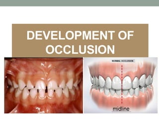
This document discusses the development of occlusion from the neonatal period through adulthood. It covers the following key points:
1. Occlusion develops through four periods: neonatal, primary dentition, mixed dentition, and permanent dentition. Each period has characteristic features and relationships between the teeth.
2. During the neonatal period, the gum pads have an anterior open bite relationship. As the primary teeth erupt, different molar relationships can form that influence the permanent dentition.
3. In the mixed dentition period, the first permanent molars erupt and can cause early or late shifts to a class I molar relationship through mechanisms like leeway space. Anterior teeth are also exchanged.
The Indian Dental Academy is the Leader in continuing dental education , training dentists in all aspects of dentistry and
offering a wide range of dental certified courses in different formats.for more details please visit
www.indiandentalacademy.com
The Indian Dental Academy is the Leader in continuing dental education , training dentists in all aspects of dentistry and
offering a wide range of dental certified courses in different formats.for more details please visit
www.indiandentalacademy.com
DEVELOPMENT OF OCCLUSION IN PEDIATRIC DENTISTRYChsaiteja3
HELLO VISITERS, IAM SAITEJA , BDS 3RD YEAR STUDENT FROM MNR DENTAL COLLEGE , SANGAREDDY. I AND MY BATCH HAS DEVELOPED A PPT ON DEVELOPMENT OF OCCLUSION IN PEDIATRIC DENTISTRY. PLEASE GO THROUGH THE PPT. EVERY TOPIC IS CLEARLY EXPLAINED IN THIS PPT ALONG WITH DIAGRAMS.
Differences between primary and permanent teeth and importanceKarishma Sirimulla
This is a small brief presentation and contains basic differences between primary and permanent dentition an also an added note on importance of young permanent molar and its management clinically
India Clinical Trials Market: Industry Size and Growth Trends [2030] Analyzed...Kumar Satyam
According to TechSci Research report, "India Clinical Trials Market- By Region, Competition, Forecast & Opportunities, 2030F," the India Clinical Trials Market was valued at USD 2.05 billion in 2024 and is projected to grow at a compound annual growth rate (CAGR) of 8.64% through 2030. The market is driven by a variety of factors, making India an attractive destination for pharmaceutical companies and researchers. India's vast and diverse patient population, cost-effective operational environment, and a large pool of skilled medical professionals contribute significantly to the market's growth. Additionally, increasing government support in streamlining regulations and the growing prevalence of lifestyle diseases further propel the clinical trials market.
Growing Prevalence of Lifestyle Diseases
The rising incidence of lifestyle diseases such as diabetes, cardiovascular diseases, and cancer is a major trend driving the clinical trials market in India. These conditions necessitate the development and testing of new treatment methods, creating a robust demand for clinical trials. The increasing burden of these diseases highlights the need for innovative therapies and underscores the importance of India as a key player in global clinical research.
Health Education on prevention of hypertensionRadhika kulvi
Hypertension is a chronic condition of concern due to its role in the causation of coronary heart diseases. Hypertension is a worldwide epidemic and important risk factor for coronary artery disease, stroke and renal diseases. Blood pressure is the force exerted by the blood against the walls of the blood vessels and is sufficient to maintain tissue perfusion during activity and rest. Hypertension is sustained elevation of BP. In adults, HTN exists when systolic blood pressure is equal to or greater than 140mmHg or diastolic BP is equal to or greater than 90mmHg. The
The dimensions of healthcare quality refer to various attributes or aspects that define the standard of healthcare services. These dimensions are used to evaluate, measure, and improve the quality of care provided to patients. A comprehensive understanding of these dimensions ensures that healthcare systems can address various aspects of patient care effectively and holistically. Dimensions of Healthcare Quality and Performance of care include the following; Appropriateness, Availability, Competence, Continuity, Effectiveness, Efficiency, Efficacy, Prevention, Respect and Care, Safety as well as Timeliness.
One of the most developed cities of India, the city of Chennai is the capital of Tamilnadu and many people from different parts of India come here to earn their bread and butter. Being a metropolitan, the city is filled with towering building and beaches but the sad part as with almost every Indian city
Leading the Way in Nephrology: Dr. David Greene's Work with Stem Cells for Ki...Dr. David Greene Arizona
As we watch Dr. Greene's continued efforts and research in Arizona, it's clear that stem cell therapy holds a promising key to unlocking new doors in the treatment of kidney disease. With each study and trial, we step closer to a world where kidney disease is no longer a life sentence but a treatable condition, thanks to pioneers like Dr. David Greene.
Medical Technology Tackles New Health Care Demand - Research Report - March 2...pchutichetpong
M Capital Group (“MCG”) predicts that with, against, despite, and even without the global pandemic, the medical technology (MedTech) industry shows signs of continuous healthy growth, driven by smaller, faster, and cheaper devices, growing demand for home-based applications, technological innovation, strategic acquisitions, investments, and SPAC listings. MCG predicts that this should reflects itself in annual growth of over 6%, well beyond 2028.
According to Chris Mouchabhani, Managing Partner at M Capital Group, “Despite all economic scenarios that one may consider, beyond overall economic shocks, medical technology should remain one of the most promising and robust sectors over the short to medium term and well beyond 2028.”
There is a movement towards home-based care for the elderly, next generation scanning and MRI devices, wearable technology, artificial intelligence incorporation, and online connectivity. Experts also see a focus on predictive, preventive, personalized, participatory, and precision medicine, with rising levels of integration of home care and technological innovation.
The average cost of treatment has been rising across the board, creating additional financial burdens to governments, healthcare providers and insurance companies. According to MCG, cost-per-inpatient-stay in the United States alone rose on average annually by over 13% between 2014 to 2021, leading MedTech to focus research efforts on optimized medical equipment at lower price points, whilst emphasizing portability and ease of use. Namely, 46% of the 1,008 medical technology companies in the 2021 MedTech Innovator (“MTI”) database are focusing on prevention, wellness, detection, or diagnosis, signaling a clear push for preventive care to also tackle costs.
In addition, there has also been a lasting impact on consumer and medical demand for home care, supported by the pandemic. Lockdowns, closure of care facilities, and healthcare systems subjected to capacity pressure, accelerated demand away from traditional inpatient care. Now, outpatient care solutions are driving industry production, with nearly 70% of recent diagnostics start-up companies producing products in areas such as ambulatory clinics, at-home care, and self-administered diagnostics.
2. INTRODUCTION
• Latin word, “occlusio” means relationship between all
components of the masticatory system.
• Ideal occlusion is perfect interdigitation of the upper
and lower teeth.
• The occlusion is regarded as a ‘dynamic’ rather than
‘static’ interrelation between the facial structures.
3. Periods of Occlusal Development
Occlusal development can be divided into the following
development periods:
• Neo-natal period.
• Primary dentition period.
• Mixed dentition period.
• Permanent dentition period.
4. NEONATAL PERIOD
➢Lasts upto 6 months after birth.
GUM PADS- alveolar processes at time of birth
➢Pink in color, firm. Covered by dense layer of fibrous
periosteum.
➢Segmented by groove- transverse groove & each segment is
a developing tooth site.
➢Pads get divided into labiobuccal & lingual portion by dental
groove.
➢Groove between canine & 1st molar region- lateral sulcus,
useful for judging inter-arch relationship at early stage.
5. Upper gum pad- horse
shoe shaped & shows:
• Gingival groove: separates
gum pad from the palate.
• Dental groove: starts at the
incisive papilla, backward till
gingival groove in canine
region & then moves laterally
to end in the molar region.
• Lateral sulcus
6. Lower gum pad- U‟
shaped and rectangular,
characterized by:
• Gingival groove: lingual
extension of the gum pads.
• Dental groove
• Lateral sulcus
7. Relationship of Gum
Pads
• Anterior open bite is seen at rest
with contact only at the molar
region.
• Complete overjet.
• Class II pattern with maxillary
gum pad being more prominent.
• Mandible is distal to the maxilla
by 2.7 mm- males & 2.5- females.
(Sillman JH 1938)
8. • The range of variation of this distal relationship is from
0 - 7 mm. (Sillman JH 1938)
• Mandibular lateral sulci lies posterior to maxillary lateral
sulci.
• Mandibular functional movements are mainly vertical,
and to a little extent antero-posterior.
• Lateral movements are absent.
9. NEONATAL JAW RELATIONSHIP
• A “precise bite” or jaw relationship is not yet seen.
• Therefore, neonatal jaw relationship cannot be
used as a diagnostic criterion for reliable
prediction of subsequent occlusion in the primary
dentition.
10. Precociously Erupted Primary Teeth
• Pre-erupted teeth or Early Infansive teeth are teeth that
erupt during the 2nd or 3rd month.
Natal tooth Neonatal teeth
11. Natal/neonatal teeth Classification
Hebling (1997) classified natal teeth into 4 clinical
categories:
1) Shell-shaped crown poorly fixed to the alveolus by
gingival tissue and absence of a root;
2) Solid crown poorly fixed to the alveolus by gingival
tissue and little or no root;
3) Eruption of the incisal margin of the crown through
gingival tissue
4) Edema of gingival tissue with an unerupted but palpable
tooth.
12. Gender - Predilection for females 66% to males 31%
(Kates)
Etiology- several factors are there-
• Superficial position of the germ
• Infection or malnutrition
• Eruption accelerated by febrile incidents or hormonal
stimulation
• Hereditary transmission of a dominant autosomal gene
• Osteoblastic activity inside the germ area related to the
remodeling phenomenon and hypovitaminosis
14. Natal/neonatal teeth Complications
• Interfere with feeding
• Risk of aspiration
• Traumatic injury to baby’s tongue or maternal breast
• Riga-Fede disease- oral condition, found rarely in
newborns manifests as ulceration on ventral surface of
tongue or inner surface of the lower lip. Caused by trauma
to the soft tissue from erupted baby teeth.
15. Diagnosis-
• Radiographic verification of relationship between natal or
neonatal tooth and adjacent structures, presence or absence of
a germ in primary tooth area- determines tooth belongs to the
normal dentition or not (Almeida CM)
• Mostly natal & neonatal teeth - primary teeth of normal
dentition (95%) & not supernumerary teeth (Brandt)
Treatment-
• If diagnosed as tooth of normal dentition - maintenance of teeth
in mouth - first treatment option (Chow MH, Roberts MW)
• Reasons for removal -- Risk of dislocation, aspiration,
traumatic injury to baby’s tongue, maternal breast (Kates GA)
16. If the treatment option is extraction, certain
precautions to be taken -
• Avoiding extraction up to the 10th day of life to prevent
hemorrhage.
• Assessing the need to administer vitamin K before
extraction (0.5-1.0 mg IM)
• Considering the general health condition of the baby
• Avoiding unnecessary injury to the gingiva
• Being alert to the risk of aspiration during removal.
17.
18. • Around 6 months to 6 yrs
• Features Of Primary Dentition
• Spacing- 2 types of dentition are seen:
• A) Spaced dentition - seen in
deciduous dentition to accommodate
the larger permanent teeth in the jaws.
• More in anterior region-‘physiological
spacing’ or ‘developmental spacing’.
• Absence of spaces in primary dentition-
crowding of permanent teeth.
PRIMARY DENTITION PERIOD
19. Non- spaced dentition
• Teeth without any spaces, due to
narrow dental arches or if teeth are
wider than usual.
• Most sub-human primates have it
throughout life and use it for
interdigitation of the opposing canines
called primate spaces’, ‘simian
spaces’ or ‘anthropoid spaces’.
• Space is used for early mesial shift.
20. Molar Relationship-
In the primary dentition classified into 3 types:
1. Straight/flush terminal plane.
2. Mesial step.
3. Distal step.
21. Flush Terminal Plane
• If the distal surface of maxillary and mandibular deciduous
second molars are in the same vertical plane; then it is
called a flush terminal plane
• Normal molar relationship in the primary dentition,
because the mesiodistal width of the mandibular molar is
greater than the mesiodistal width of the maxillary molar.
22. Mesial Step
• Distal surface of mandibular
deciduous second molar is
mesial to the distal surface of
maxillary deciduous second
molar.
Distal Step
• Distal surface of mandibular
second deciduous molar is
more distal to the distal
surface of the maxillary
second deciduous molar
23. Canine relationship
• Relationship of maxillary & mandibular deciduous canines
is one of the most stable in primary dentition
• Classified as:
Class 1 Class 2
24. MIXED DENTITION PERIOD
Around 6 years- 12 years
• The mixed dentition period can be divided into three
phases:
1. First transitional period.
2. Inter-transitional period.
3. Second transitional period.
25. First transitional period
• Eruption of 1st Permanent Molar
• The location & relation of the 1st permanent molar
depends much upon the distal surface of the upper &
lower 2nd deciduous molar.
26. Transition to Class I Molar Relation
• The shift in lower molar from a flush terminal plane to a
class I relation can occur in two ways:
• Early Shift- occurs during the early mixed dentition
period. Since this occurs early-early shift.
• Late Shift- occurs in the late mixed dentition period and is
thus called late shift.
Early shift Late shift
27. Leeway Space of Nance
• Nance (1947)
• Maxilla: 0.9 mm/segment = 1.8 mm.
• Mandible: 1.7 mm/segment = 3.4mm.
• Maxillary incisors, as a group in one
quadrant– 3.2to 3.5 mm larger
• Mandibular incisors, as a group in
one quadrant – 2.4 to 2.5 mm larger
• The latter figures balance out or
cancel the 1.7 mm of so called
leeway space
28. Distal Step
• When deciduous second molars are in distal
step, permanent first molar will erupt into
class II relation.
• This molar configuration is not self correcting
and will cause class II malocclusion despite
Leeway space and differential growth.
Mesial Step
• Primary second molars in mesial
step relationship lead to a class I
molar relation in mixed dentition.
• This may remain or progress to a
half or full cusp class III with
continued mandibular growth
29. • Influence of terminal
plane on the position of
1st permanent molar
• Distal Step – 23.3% incidence,
abnormal, Class II- 38.6%
• Straight terminal plane –
49.2% incidence, Class I or II
• Mesial Step - <2mm 26.7%,
class I 58.9% >2mm 0.8%.
Class III- 2.5%
30. Exchange of Incisors-
• During the first transitional period the deciduous incisors
are replaced by the permanent incisors..
• This difference between the amount of space needed for
the accommodation of the incisors and the amount of
space available for this, is called ‘Incisor liability’.
• It is roughly about 7.6 mm in the maxillary arch and
about 6mm in the mandibular arch. (Wayne)
31. Transition of Incisors
• Incisal liability is overcome :
• Interdental physiological
spacing in the primary incisor
region. (4mm, maxilla & 3mm,
mandibula)
• Increase in inter-canine arch
width.
Increase in anterior length of the
dental arches: Permanent incisors
erupt labial to the primary incisors
to obtain an added space of around
2-3 mm.
32. Inter-Transitional Period
• This is a stable phase where little changes take place in
the dentition.
• Root formation of emerged incisors, and molars
continues, along with concomitant increase in alveolar
process height.
• Resorption of roots of deciduous canines and molars.
33. Second Transitional Period
• Characterized by replacement of deciduous molars &
canines by premolars and permanent canines.
• At around 10 years of age the deciduous canine shed, but
just before the shedding a transient or self correcting
malocclusion is seen between the age of 8-9 years.
34. Ugly Duckling Stage (Broadbent’s
phenomenon)
• Around the age of 8 - 9 years, a midline diastema is
commonly seen in the upper arch, looks as malocclusion.
• Flaring of the lateral incisors.
• Maxillary midline diastema.
• Crowns of canines impinge on developing lateral incisor
roots.
35. • With eruption of the canines, the impingement from the
roots shift incisally thus driving the incisor crowns
medially, resulting in closure of the diastema as well as
the correction of the flared lateral incisors.
• Hence this unaesthetic metamorphosis, eventually leads
to an aesthetic result.
36. Second Transitional Period contd…
Eruption of permanent second molars
• Before emergence- second molars, oriented in a mesial &
lingual direction
• Teeth- formed palatally, guided into occlusion by Cone
Funnel mechanism , upper palatal cusps (cone) slides
into the lower occlusal fossa (funnel)
• Arch length is reduced by mesial eruptive forces
• Thereby, crowding if present is accentuated
37. SELF CORRECTING ANOMALIES
• 1. PREDENTATE
PERIOD:
• A) Retrognathic mandible
• B) Anterior open bite
• C) Infantile swallowing
pattern
• CORRECTION
• Corrects with differential
and forward growth of the
mandible
• Eruption of primary
incisors
• During the first year of life
with introduction of solid
foods
38. • PRIMARY DENTITION
• A) Anterior deep bite
• B) Flush terminal plane
• C) Spacing
• D) Edge to edge
• CORRECTION
• Eruption of deciduous
molars
• Attrition of incisal edges
• Forward and downward
growth of mandible (early
shift)
• Eruption of first permanent
molar
• Late shift (Leeway space)
• Eruption of first permanent
molars
• Eruption of permanent
incisors
39. • MIXED DENTITION
• A) Anterior deep bite
• B) Mandibular anterior crowding
• C) Ugly duckling stage
• D) End-on relation
• CORRECTION
• Proprioceptive response
condition of patient (with the
eruption of 1st permanent molars
and premature contact of the
pad of tissue overlying them as
natural bite opener)
• Tongue pressure
• Increase in intercanine width
• Maxillary canine eruption
• With eruption of first permanent
molars
• Late mesial shift in non-spaced
dentition
40. • PERMANENT DENTITION
• A) Overjet and overbite
• CORRECTION
• Decreases with eruption of
all permanent molars
• Differential growth of
mandible
41. PERMANENT DENTITION PERIOD
• Marked by eruption of the four permanent second molars.
• Calcification begins at birth with the calcification of the
cusps of the first permanent molar and extends as late as
the 25th year of life.
• Complete calcification of incisor crowns take place by 4 –
5 years and of the other permanent teeth by 6 – 8 years
except for third molars.
42. Features of Permanent
Dentition
• Coinciding midline.
• Class I molar relationship
• Vertical overbite of about one third the clinical crown
height of the mandibular central incisors.
• Overjet and over bite decreases throughout the second
decade of life due to greater forward growth of the
mandible.
43. ANDREWS KEYS TO
NORMAL OCCLUSION
• Key I – Molar relationship- MB cusp
of the max 1st molar falls into the
mesiobuccal groove of the mand 1st
molar.
• Key II Crown angulation (Tip)
Angulation of facial axis should be
positive
• Key III Crown inclination- upper
incisors the gingival portion of the
crown’s labial surface is lingual to the
incisal portion.
• Lower incisors and other crowns,
gingival portion of the labial or buccal
surface is labial or buccal to the
incisal or occlusal portion
44. • Key IV – Rotations- teeth should
be free of undesirable rotations
• Key V – Tight contacts- Contact
points should be tight (no spaces).
Key VI – Occlusal plane or curve
of spee - should have no more
than a slight arch.
Key VII – Correct tooth size or the
bolton’s ratio
(Bennett and McLaughlin, 1993).
45. Abnormalities in dental arch
1. Arch Length Discrepancy
1. Crowding
2. Spacing
2. Deviation in no. of teeth-
1. Absence of teeth (Agenesis)
2. Supernumerary teeth
Sequence of agenesis is –
• 3rd molar > Mand. 2nd premolars >
Max Lateral Incisors > Max. 2nd
Premolar
Supernumerary teeth
46. 3. Deviation in tooth size
• Its relative in nature
• All teeth combined > or < relative to size of jaws or head.
• Crowding
• Spacing
4. Ankylosis
• Frequent in mand deciduous molars.
• In permanent 2 types-
• Due to abnormal position within jaw, Max perm. Canine
• Due to lack of space, Mand 3rd molar
47.
48. Centric relation
• Definition: Relationship of mandible to maxilla when properly
aligned condyle–disk assemblies are in most superior
position against the eminentiae.
• The centric relation refers to the fully seated condylar
position.
• It is the universally accepted jaw position because it is
physiologically and biomechanically correct and is the only
jaw position that permits an interference-free occlusion.
Maximum intercuspation position
• The tooth-to-tooth relationship in maximum contact is called
as maximum intercuspation position (MIP).
49. Neutral zone
• Definition: The potential space between the lips and cheeks on
one side and the tongue on the other.
• Any part of the dentition out of harmony with the neutral zone
will result in instability, interference with function, or some
degree of discomfort.
50. CONCLUSION
Occlusion is the way the maxillary and mandibular teeth
articulate, but in reality dental occlusion is a much more
complex relationship, because it not only involves the study
of the teeth, but also their morphology and angulations, the
muscles of mastication, the skeletal structures, the
temperomandibular joint, and the functional jaw
movements.