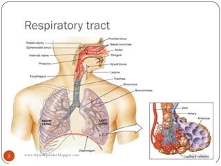
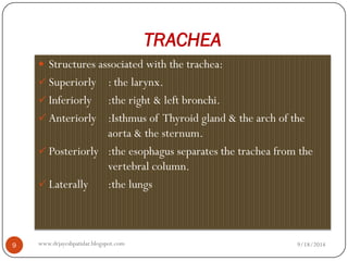
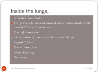
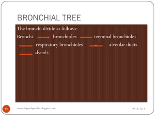
Dr. JayeshPatidar 
www.drjayeshpatidar.blogspot.com 
RESPIRATORY SYSTEM
Lower respiratory tract 
It includes: 
A.Trachea, 
B.Lungs, 
C.Bronchi. 
9/18/2014 
2 
www.drjayeshpatidar.blogspot.com
Respiratory tract 
9/18/2014 
3 
www.drjayeshpatidar.blogspot.com
TRACHEA 
Structures associated with the trachea: 
Superiorly: the larynx. 
Inferiorly:the right & left bronchi. 
Anteriorly:Isthmus of Thyroid gland & the arch of the aorta & the sternum. 
Posteriorly:the esophagus separates the trachea from the vertebral column. 
Laterally:the lungs 
9/18/2014 
9 
www.drjayeshpatidar.blogspot.com
STRUCTURE OF THE TRACHEA 
It is composed of 3 layers of tissues & held by between 16-20 incomplete rings of hyaline cartilage. 
Rings are incomplete posteriorly. 
Connective tissues & involuntary muscle join the cartilages & form the posterior wall which is in contact with the esophagus. 
9/18/2014 
10 
www.drjayeshpatidar.blogspot.com
Three layers of tissue: 
I.The outer layer: consists of fibrous & elastic tissue & encloses the cartilages. 
II.The middle layer:consists of bands of smooth muscles in a helical manner. There is some areolar tissue. 
III.The inner lining:consists of ciliated columnar epithelium, containing mucus secreting Goblet cells. 
9/18/2014 
11 
www.drjayeshpatidar.blogspot.com
LUNGS 
There are 2 lungs, each lying on each side of the midline in the thoracic cavity. 
They have- 
oApex, 
oBase, 
oCostal surface, 
oMedial surface-Hilum at 5th, 6th, 7ththoracic vertebrae. 
•The area between the lungs: Mediastinum. 
•Lobes of the right & left Lungs. 
9/18/2014 
12 
www.drjayeshpatidar.blogspot.com
PLEURA & PLEURAL CAVITY: 
Pleura consists of a closed sac of serous membrane which contains a small amount of serous fluid. 
Each lung is pushed into this sac so that 2 layers are formed: 
a.One adherent to the lungs -visceral 
b.Another adherent to the wall of the thoracic cavity-parietal 
9/18/2014 
13 
www.drjayeshpatidar.blogspot.com
Inside the lungs.. 
Bronchi & bronchioles: 
Two primary bronchi are formed when trachea divides at the level of 5ththoracic vertebra. 
The right bronchus: 
-wider, shorter & more vertical than the left one. 
-Approx 2.5 cm 
The left bronchus: 
-About 5cm long 
-Narrower. 
9/18/2014 
14 
www.drjayeshpatidar.blogspot.com
BRONCHIAL TREE 
The bronchi divide as follows: 
Bronchi bronchioles terminal bronchioles 
respiratory bronchioles alveolar ducts 
alveoli. s 
9/18/2014 
15 
www.drjayeshpatidar.blogspot.com
Thank you 
9/18/2014 
16 
www.drjayeshpatidar.blogspot.com

Respiratory system

  • 1.
  • 2.
    Lower respiratory tract It includes: A.Trachea, B.Lungs, C.Bronchi. 9/18/2014 2 www.drjayeshpatidar.blogspot.com
  • 3.
    Respiratory tract 9/18/2014 3 www.drjayeshpatidar.blogspot.com
  • 4.
    TRACHEA Structures associatedwith the trachea: Superiorly: the larynx. Inferiorly:the right & left bronchi. Anteriorly:Isthmus of Thyroid gland & the arch of the aorta & the sternum. Posteriorly:the esophagus separates the trachea from the vertebral column. Laterally:the lungs 9/18/2014 9 www.drjayeshpatidar.blogspot.com
  • 5.
    STRUCTURE OF THETRACHEA It is composed of 3 layers of tissues & held by between 16-20 incomplete rings of hyaline cartilage. Rings are incomplete posteriorly. Connective tissues & involuntary muscle join the cartilages & form the posterior wall which is in contact with the esophagus. 9/18/2014 10 www.drjayeshpatidar.blogspot.com
  • 6.
    Three layers oftissue: I.The outer layer: consists of fibrous & elastic tissue & encloses the cartilages. II.The middle layer:consists of bands of smooth muscles in a helical manner. There is some areolar tissue. III.The inner lining:consists of ciliated columnar epithelium, containing mucus secreting Goblet cells. 9/18/2014 11 www.drjayeshpatidar.blogspot.com
  • 7.
    LUNGS There are2 lungs, each lying on each side of the midline in the thoracic cavity. They have- oApex, oBase, oCostal surface, oMedial surface-Hilum at 5th, 6th, 7ththoracic vertebrae. •The area between the lungs: Mediastinum. •Lobes of the right & left Lungs. 9/18/2014 12 www.drjayeshpatidar.blogspot.com
  • 8.
    PLEURA & PLEURALCAVITY: Pleura consists of a closed sac of serous membrane which contains a small amount of serous fluid. Each lung is pushed into this sac so that 2 layers are formed: a.One adherent to the lungs -visceral b.Another adherent to the wall of the thoracic cavity-parietal 9/18/2014 13 www.drjayeshpatidar.blogspot.com
  • 9.
    Inside the lungs.. Bronchi & bronchioles: Two primary bronchi are formed when trachea divides at the level of 5ththoracic vertebra. The right bronchus: -wider, shorter & more vertical than the left one. -Approx 2.5 cm The left bronchus: -About 5cm long -Narrower. 9/18/2014 14 www.drjayeshpatidar.blogspot.com
  • 10.
    BRONCHIAL TREE Thebronchi divide as follows: Bronchi bronchioles terminal bronchioles respiratory bronchioles alveolar ducts alveoli. s 9/18/2014 15 www.drjayeshpatidar.blogspot.com
  • 11.
    Thank you 9/18/2014 16 www.drjayeshpatidar.blogspot.com