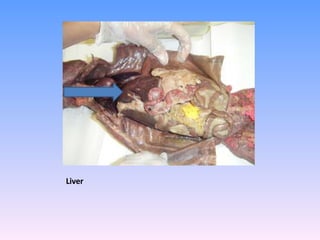
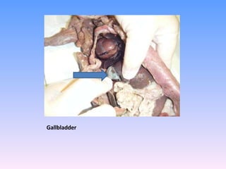
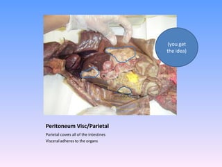
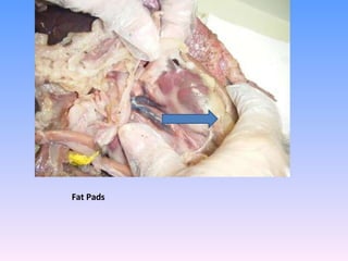
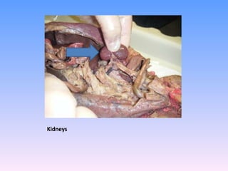
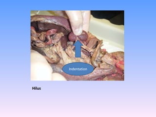
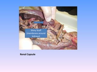
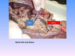
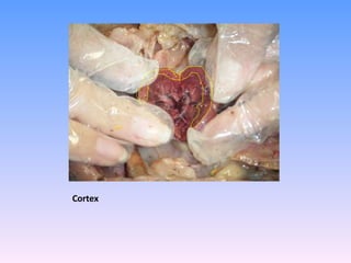
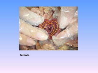
This document provides labels and brief descriptions for various anatomical structures and organs in a dissected mink specimen. Key structures identified and summarized include:
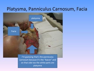
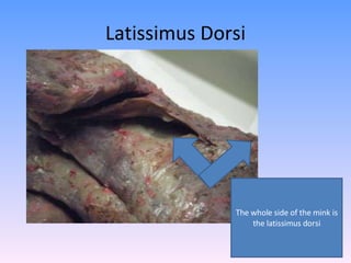
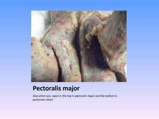
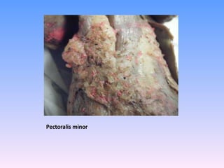
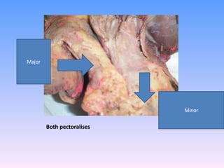
- The panniculus carnosum, platysma, latissimus dorsi, pectoralis major, and pectoralis minor muscles.
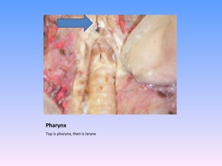
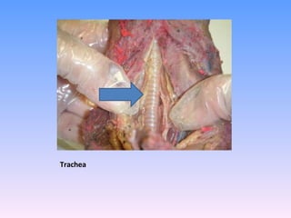
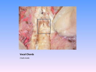
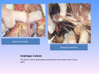
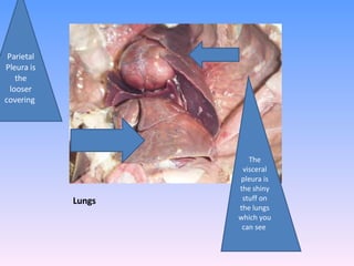
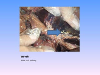
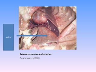
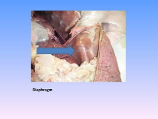
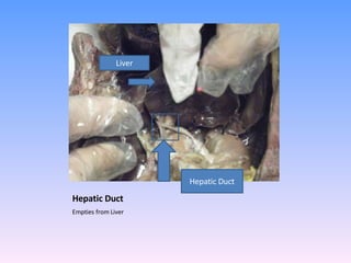
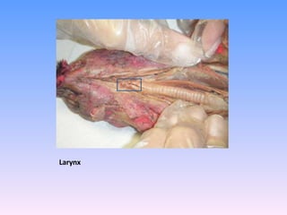
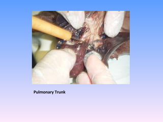
- The pharynx, larynx, trachea, esophagus, lungs, bronchi, pulmonary veins and arteries.
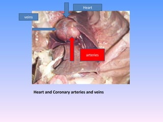
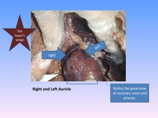
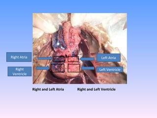
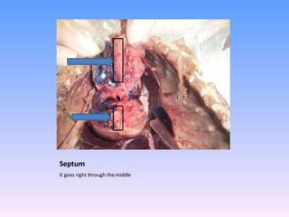
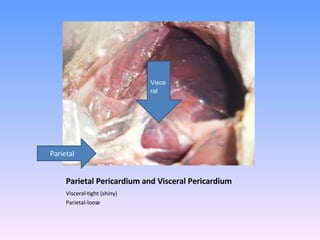
- The heart, coronary arteries and veins, right and left atria and ventricles, septum, and pericardium.
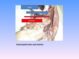
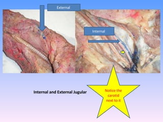
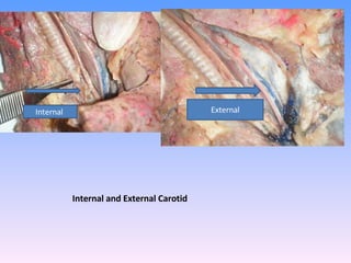
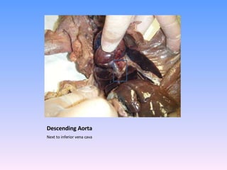
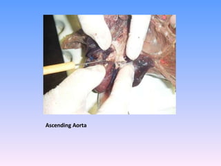
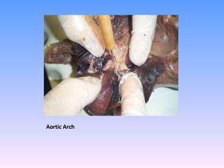
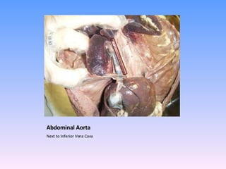
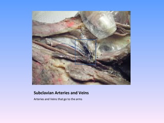
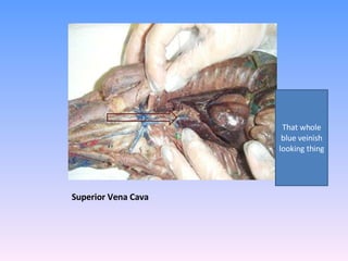
- Major blood vessels including the internal and external jugular veins, internal and external carotid arteries, descending and ascending aorta


![Thyroid final [part 1]](https://cdn.slidesharecdn.com/ss_thumbnails/thyroidfinalpart1-161126043454-thumbnail.jpg?width=640&height=640&fit=bounds)


![Anatomy introduction[1]](https://cdn.slidesharecdn.com/ss_thumbnails/9rhvq4jzrwacyv5bjs6b-signature-460517c25b85fc4e63c8080c3e27df73c8dfae9e0c6544cc7ea6d9e8b5e79cc7-poli-180213064029-thumbnail.jpg?width=640&height=640&fit=bounds)


