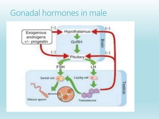
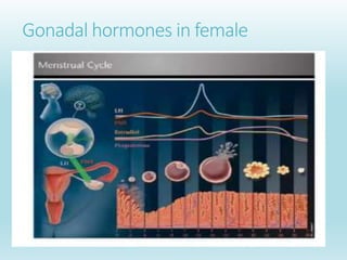
What is Gonad?
The gonad is the organ that makes gametes.
The gonads in males are the testes and the gonads in females are the ovaries.
The product- gametes, are haploid germ cells. For example, spermatozoon and egg
cells are gametes.
Reproduction – hormones of the reproductive system
Brain-hypothalamic hormones
 Gonadotropin-releasing hormone (GnRH or LHRH).
 Gonadotropin release-inhibitory factors (GnRIF)
Pituitary hormones: Gonadotropins
 Follicle-stimulating Hormone (FSH): controls gonadal growth
 Luteinizing Hormone (LH): controls gamete maturation & release
Gonadal hormones (steroids)
 Testosterone (androgen): important for spermatogenesis
 Oestrogen: important for oogenesis
 Progesterone or its derivatives (progestins): induce gamete
maturation in males and females and ovulation in females
Gonadal hormones in male
Gonadal hormones in female
Structure of LH
•LH is a glycoprotein hormone.
•Each monomeric unit is a glycoprotein molecule One alpha and one
beta subunit make the full, functional protein.
• The alpha subunits of LH contain 92 amino acids in human.
•LH beta subunit have 120 amino acids that confers its specific
biologic action and is responsible for the specificity of the
interaction with the LH receptor.
•The biologic half-life of LH is 20 minutes, shorter than that of FSH
(3–4 hours) and hCG (24 hours).
Action
•In both males and females, LH is essential for
reproduction.
•In the male, LH acts upon the Leydig cells of the testis
and is responsible for the production of testosterone, an
androgen that exerts both endocrine activity and
intratesticular activity on spermatogenesis.
Contd…
 In females LH is necessary to maintain luteal function for the first two weeks of the
menstrual cycle. If pregnancy occurs, LH levels will decrease, and luteal function will
instead be maintained by the action of hCG (a hormone very similar to LH but secreted
from the new placenta).
 In females, LH supports theca cells in the ovary that provide androgens and hormonal
precursors for estradiol production.
 At the time of menstruation, FSH initiates follicular growth, specifically affecting
granulosa cells.
 With the rise in estrogens, LH receptors are also expressed on the maturing follicle,
which causes it to produce more estradiol.
 When the follicle has fully matured, a spike in estrogen production by the follicle
stimulates a positive feedback loop in the hypothalamus that stimulates the release of
LH from the anterior pituitary. This increase in LH production only lasts for 24 to 48
hours.
Contd…
 This "LH surge" triggers ovulation, thereby not only releasing the
egg from the follicle, but also initiating the conversion of the
residual follicle into a corpus luteum that, in turn, produces
progesterone to prepare the endometrium for a possible
implantation.
 LH is necessary to maintain luteal function for the first two weeks
of menstrual cycle. If pregnancy occurs LH level will decrease and
luteal function will instead be maintained by hCG ( an hormone
similar in action but produced by developing placenta).
Follicle-stimulating hormone (FSH)
 It is synthesized and secreted by gonadotrophs of the
anterior pituitary gland.
 FSH regulates the development, growth, pubertal
maturation, and reproductive processes of the body.
 FSH and luteinizing hormone (LH) act synergistically
in reproduction. Specifically, an increase in FSH
secretion by the anterior pituitary causes ovulation.
Structure of FSH
 FSH is a glycoprotein.
 Its structure is similar to that of LH, TSH, and hCG.
 The protein dimer contains 2 polypeptide units - alpha and
beta subunits. The alpha subunits of LH, FSH, TSH, and hCG
are identical, and contain 92 amino acids. The beta subunits
vary. FSH has a beta subunit of 111amino acids (FSH β),
which confers its specific biologic action and is responsible
for interaction with the FSH- receptor.
 The half-life of FSH is 3–4 hours.
Action
FSH regulates the development, growth, pubertal maturation, and
reproductive processes of the human body.
Effects in males
 FSH enhances the production of androgen-binding protein by the
Sertoli cells of the testes by binding to FSH receptors on their
basolateral membranes, and is critical for the initiation of
spermatogenesis.
 FSH stimulates primary spermatocytes to undergo the first
division of meiosis, to form secondary spermatocytes.
Contd…
Effects in females
 FSH stimulates the growth and recruitment of immature
ovarian follicles in the ovary.
 In early (small) antral follicles, FSH is the major survival
factor that rescues the small antral follicles (2–5 mm in
diameter for humans) from apoptosis (programmed cell
death).
Gonadotropin-releasing hormone (GnRH)
 Also known as Luteinizing-hormone-releasing hormone (LHRH), is a
peptide / protein hormone responsible for the release of follicle-stimulating
hormone (FSH) and luteinizing hormone (LH) from the anterior pituitary.
 GnRH is synthesized and released from neurons within the hypothalamus.
GnRH is considered a neurohormone, a hormone produced in a specific
neural cell and released at its neural terminal.
 At the pituitary, GnRH stimulates the synthesis and secretion of the
gonadotropins, follicle- stimulating hormone (FSH), and luteinizing
hormone (LH).
 Low-frequency GnRH pulses lead to FSH release, whereas high-frequency
GnRH pulses stimulate LH release.
Gonadal Steroid hormones:Testosterone
 Testosterone is a steroid hormone from the androgen group.
 In mammals, testosterone is primarily secreted in the testicles
of males and the ovaries of females, although small amounts
are also secreted by the adrenal glands.
 It is the principal male sex hormone and an anabolic steroid.
 In men, testosterone plays a key role in the development of
male reproductive tissues such as the testis and prostate as well
as promoting secondary sexual characteristics such as
increased muscle, bone mass, and the growth of body hair.
Biosynthesis of testosterone
 The largest amounts of testosterone (>95%) are produced by
the testes in men.
 It is also synthesized in far smaller quantities in women by the
thecal cells of the ovaries, and by the placenta,
 The zona reticularis of the adrenal cortex and skin in both
sexes also produces in small quantities.
 In the testes, testosterone is produced by the Leydig cells. The
male gonads also contain Sertoli cells which require
testosterone for spermatogenesis.
Regulation of biosynthesis of testosterone
 Testosterone is primarily synthesized in Leydig cells.
 The number of Leydig cells in turn is regulated by luteinizing hormone (LH) and follicle
stimulating hormone (FSH).
 The amount of testosterone produced by existing Leydig cells is under the control of LH
which regulates the expression of 17-β hydroxysteroid dehydrogenase (HSD).
 When testosterone levels are low, gonadotropin-releasing hormone (GnRH) is released by
the hypothalamus which in turn stimulates the pituitary gland to release FSH and LH.
These later two hormones stimulate the testis to synthesize testosterone.
 Finally increasing levels of testosterone through a negative feedback loop act on the
hypothalamus and pituitary to inhibit the release of GnRH and FSH/LH respectively.
Biosynthesis of Estrogen:
 Oestrogens, in females, are produced primarily by the ovaries,
and during pregnancy, the placenta.
 Follicle-stimulating hormone (FSH) stimulates the ovarian
production of oestrogens by the granulosa cells of the ovarian
follicles and corpora lutea.
 Some oestrogens are also produced in smaller amounts by other
tissues such as the liver, adrenal glands, and the breasts.
 In females, synthesis of oestrogens starts in theca interna cells in
the ovary, by the synthesis of androstenedione from cholesterol.
Oestrogen
 They are the primary female sex hormones.
 Like all steroid hormones, oestrogens readily diffuse across the cell
membrane. Once inside the cell, they bind to and activate oestrogen
receptors which in turn modulate the expression of many genes.
 Oestradiol is the predominant (90%) and most potent form oestrogen
during reproductive years.
 During menopause, Oestrone(weak form of oestrogen) is the
predominant circulating oestrogen.
 Oestriol is degradation form of oestradiol and oestrone.
 During pregnancy Oestriol is the predominant circulating oestrogen.
Function of Oestrogen
 Promote development of female secondary sex characteristics
 Accelerate metabolism
 Increase fat stores
 Stimulate endometrial growth
 Increase uterine growth
 Increase vaginal lubrication
 Thicken the vaginal wall
 Maintenance of vessel and skin
 Reduce bone resorption, increase bone formation
 Reduce muscle mass
Progesterone
 Progesterone is a steroid hormone involved in the female
menstrual cycle, pregnancy and embryogenesis of humans
and other species.
 Progesterone belongs to a class of hormones called
progestogens, and is the major naturally occurring human
progestogen.
 Progesterone is produced in the ovaries (by corpus luteum),
the adrenal glands and, by the placenta during pregnancy.
 Progesterone is also stored in adipose (fat) tissue.
Progesterone Level
 In women, progesterone levels are relatively low during
the preovulatory phase of the menstrual cycle, rise after
ovulation, and are elevated during the luteal phase.
 Progesterone levels tend to be < 2 ng/ml prior to
ovulation, and > 5 ng/ml after ovulation.
 If pregnancy occurs, human chorionic gonadotropin is
released maintaining the corpus luteum allowing it to
maintain levels of progesterone.
Hormonal regulation in nonpregnant female
 Hypothalamus releases gonadotropin-releasing hormone (GnRH). This
stimulates the anterior pituitary to release FSH and LH.
 FSH stimulates maturation of primary oocyte in an immature follicle.
 Follicle produces oestrogen. Oestrogen: (A) builds the uterine wall /
endometrium (B) inhibits secretion of FSH.
 High levels of oestrogen further stimulate secretion of LH by anterior
pituitary. This plus FSH also causes ovulation of the secondary oocyte
– leaving follicle without egg (the corpus luteum).
Contd…
 Corpus luteum secretes oestrogen and progesterone. This
maintains the endometrium for 15-16 days and inhibits LH.
 (If oocyte is not fertilized and implanted in the uterine wall),
corpus degenerates to corpus albicans and stops producing
oestrogen and progesterone.
 Without oestrogen and progesterone, endometrium breaks down –
menstruation occurs. Menstruation is the sloughing off of the
enlarged endometrial wall along with blood and mucous.
 Decrease in progesterone and LH. Low LH causes secretion of
FSH by pituitary again. The cycle repeats.
https://www.slideshare.net/binayatamang/gonadal-hormone-
235917843

Reproductive endocrinology.pptx

  • 2.
    What is Gonad? Thegonad is the organ that makes gametes. The gonads in males are the testes and the gonads in females are the ovaries. The product- gametes, are haploid germ cells. For example, spermatozoon and egg cells are gametes.
  • 3.
    Reproduction – hormonesof the reproductive system Brain-hypothalamic hormones  Gonadotropin-releasing hormone (GnRH or LHRH).  Gonadotropin release-inhibitory factors (GnRIF) Pituitary hormones: Gonadotropins  Follicle-stimulating Hormone (FSH): controls gonadal growth  Luteinizing Hormone (LH): controls gamete maturation & release Gonadal hormones (steroids)  Testosterone (androgen): important for spermatogenesis  Oestrogen: important for oogenesis  Progesterone or its derivatives (progestins): induce gamete maturation in males and females and ovulation in females
  • 4.
  • 5.
  • 6.
    Structure of LH •LHis a glycoprotein hormone. •Each monomeric unit is a glycoprotein molecule One alpha and one beta subunit make the full, functional protein. • The alpha subunits of LH contain 92 amino acids in human. •LH beta subunit have 120 amino acids that confers its specific biologic action and is responsible for the specificity of the interaction with the LH receptor. •The biologic half-life of LH is 20 minutes, shorter than that of FSH (3–4 hours) and hCG (24 hours).
  • 7.
    Action •In both malesand females, LH is essential for reproduction. •In the male, LH acts upon the Leydig cells of the testis and is responsible for the production of testosterone, an androgen that exerts both endocrine activity and intratesticular activity on spermatogenesis.
  • 8.
    Contd…  In femalesLH is necessary to maintain luteal function for the first two weeks of the menstrual cycle. If pregnancy occurs, LH levels will decrease, and luteal function will instead be maintained by the action of hCG (a hormone very similar to LH but secreted from the new placenta).  In females, LH supports theca cells in the ovary that provide androgens and hormonal precursors for estradiol production.  At the time of menstruation, FSH initiates follicular growth, specifically affecting granulosa cells.  With the rise in estrogens, LH receptors are also expressed on the maturing follicle, which causes it to produce more estradiol.  When the follicle has fully matured, a spike in estrogen production by the follicle stimulates a positive feedback loop in the hypothalamus that stimulates the release of LH from the anterior pituitary. This increase in LH production only lasts for 24 to 48 hours.
  • 9.
    Contd…  This "LHsurge" triggers ovulation, thereby not only releasing the egg from the follicle, but also initiating the conversion of the residual follicle into a corpus luteum that, in turn, produces progesterone to prepare the endometrium for a possible implantation.  LH is necessary to maintain luteal function for the first two weeks of menstrual cycle. If pregnancy occurs LH level will decrease and luteal function will instead be maintained by hCG ( an hormone similar in action but produced by developing placenta).
  • 10.
    Follicle-stimulating hormone (FSH) It is synthesized and secreted by gonadotrophs of the anterior pituitary gland.  FSH regulates the development, growth, pubertal maturation, and reproductive processes of the body.  FSH and luteinizing hormone (LH) act synergistically in reproduction. Specifically, an increase in FSH secretion by the anterior pituitary causes ovulation.
  • 11.
    Structure of FSH FSH is a glycoprotein.  Its structure is similar to that of LH, TSH, and hCG.  The protein dimer contains 2 polypeptide units - alpha and beta subunits. The alpha subunits of LH, FSH, TSH, and hCG are identical, and contain 92 amino acids. The beta subunits vary. FSH has a beta subunit of 111amino acids (FSH β), which confers its specific biologic action and is responsible for interaction with the FSH- receptor.  The half-life of FSH is 3–4 hours.
  • 12.
    Action FSH regulates thedevelopment, growth, pubertal maturation, and reproductive processes of the human body. Effects in males  FSH enhances the production of androgen-binding protein by the Sertoli cells of the testes by binding to FSH receptors on their basolateral membranes, and is critical for the initiation of spermatogenesis.  FSH stimulates primary spermatocytes to undergo the first division of meiosis, to form secondary spermatocytes.
  • 13.
    Contd… Effects in females FSH stimulates the growth and recruitment of immature ovarian follicles in the ovary.  In early (small) antral follicles, FSH is the major survival factor that rescues the small antral follicles (2–5 mm in diameter for humans) from apoptosis (programmed cell death).
  • 14.
    Gonadotropin-releasing hormone (GnRH) Also known as Luteinizing-hormone-releasing hormone (LHRH), is a peptide / protein hormone responsible for the release of follicle-stimulating hormone (FSH) and luteinizing hormone (LH) from the anterior pituitary.  GnRH is synthesized and released from neurons within the hypothalamus. GnRH is considered a neurohormone, a hormone produced in a specific neural cell and released at its neural terminal.  At the pituitary, GnRH stimulates the synthesis and secretion of the gonadotropins, follicle- stimulating hormone (FSH), and luteinizing hormone (LH).  Low-frequency GnRH pulses lead to FSH release, whereas high-frequency GnRH pulses stimulate LH release.
  • 15.
    Gonadal Steroid hormones:Testosterone Testosterone is a steroid hormone from the androgen group.  In mammals, testosterone is primarily secreted in the testicles of males and the ovaries of females, although small amounts are also secreted by the adrenal glands.  It is the principal male sex hormone and an anabolic steroid.  In men, testosterone plays a key role in the development of male reproductive tissues such as the testis and prostate as well as promoting secondary sexual characteristics such as increased muscle, bone mass, and the growth of body hair.
  • 16.
    Biosynthesis of testosterone The largest amounts of testosterone (>95%) are produced by the testes in men.  It is also synthesized in far smaller quantities in women by the thecal cells of the ovaries, and by the placenta,  The zona reticularis of the adrenal cortex and skin in both sexes also produces in small quantities.  In the testes, testosterone is produced by the Leydig cells. The male gonads also contain Sertoli cells which require testosterone for spermatogenesis.
  • 17.
    Regulation of biosynthesisof testosterone  Testosterone is primarily synthesized in Leydig cells.  The number of Leydig cells in turn is regulated by luteinizing hormone (LH) and follicle stimulating hormone (FSH).  The amount of testosterone produced by existing Leydig cells is under the control of LH which regulates the expression of 17-β hydroxysteroid dehydrogenase (HSD).  When testosterone levels are low, gonadotropin-releasing hormone (GnRH) is released by the hypothalamus which in turn stimulates the pituitary gland to release FSH and LH. These later two hormones stimulate the testis to synthesize testosterone.  Finally increasing levels of testosterone through a negative feedback loop act on the hypothalamus and pituitary to inhibit the release of GnRH and FSH/LH respectively.
  • 18.
    Biosynthesis of Estrogen: Oestrogens, in females, are produced primarily by the ovaries, and during pregnancy, the placenta.  Follicle-stimulating hormone (FSH) stimulates the ovarian production of oestrogens by the granulosa cells of the ovarian follicles and corpora lutea.  Some oestrogens are also produced in smaller amounts by other tissues such as the liver, adrenal glands, and the breasts.  In females, synthesis of oestrogens starts in theca interna cells in the ovary, by the synthesis of androstenedione from cholesterol.
  • 19.
    Oestrogen  They arethe primary female sex hormones.  Like all steroid hormones, oestrogens readily diffuse across the cell membrane. Once inside the cell, they bind to and activate oestrogen receptors which in turn modulate the expression of many genes.  Oestradiol is the predominant (90%) and most potent form oestrogen during reproductive years.  During menopause, Oestrone(weak form of oestrogen) is the predominant circulating oestrogen.  Oestriol is degradation form of oestradiol and oestrone.  During pregnancy Oestriol is the predominant circulating oestrogen.
  • 20.
    Function of Oestrogen Promote development of female secondary sex characteristics  Accelerate metabolism  Increase fat stores  Stimulate endometrial growth  Increase uterine growth  Increase vaginal lubrication  Thicken the vaginal wall  Maintenance of vessel and skin  Reduce bone resorption, increase bone formation  Reduce muscle mass
  • 21.
    Progesterone  Progesterone isa steroid hormone involved in the female menstrual cycle, pregnancy and embryogenesis of humans and other species.  Progesterone belongs to a class of hormones called progestogens, and is the major naturally occurring human progestogen.  Progesterone is produced in the ovaries (by corpus luteum), the adrenal glands and, by the placenta during pregnancy.  Progesterone is also stored in adipose (fat) tissue.
  • 22.
    Progesterone Level  Inwomen, progesterone levels are relatively low during the preovulatory phase of the menstrual cycle, rise after ovulation, and are elevated during the luteal phase.  Progesterone levels tend to be < 2 ng/ml prior to ovulation, and > 5 ng/ml after ovulation.  If pregnancy occurs, human chorionic gonadotropin is released maintaining the corpus luteum allowing it to maintain levels of progesterone.
  • 23.
    Hormonal regulation innonpregnant female  Hypothalamus releases gonadotropin-releasing hormone (GnRH). This stimulates the anterior pituitary to release FSH and LH.  FSH stimulates maturation of primary oocyte in an immature follicle.  Follicle produces oestrogen. Oestrogen: (A) builds the uterine wall / endometrium (B) inhibits secretion of FSH.  High levels of oestrogen further stimulate secretion of LH by anterior pituitary. This plus FSH also causes ovulation of the secondary oocyte – leaving follicle without egg (the corpus luteum).
  • 24.
    Contd…  Corpus luteumsecretes oestrogen and progesterone. This maintains the endometrium for 15-16 days and inhibits LH.  (If oocyte is not fertilized and implanted in the uterine wall), corpus degenerates to corpus albicans and stops producing oestrogen and progesterone.  Without oestrogen and progesterone, endometrium breaks down – menstruation occurs. Menstruation is the sloughing off of the enlarged endometrial wall along with blood and mucous.  Decrease in progesterone and LH. Low LH causes secretion of FSH by pituitary again. The cycle repeats.
  • 25.