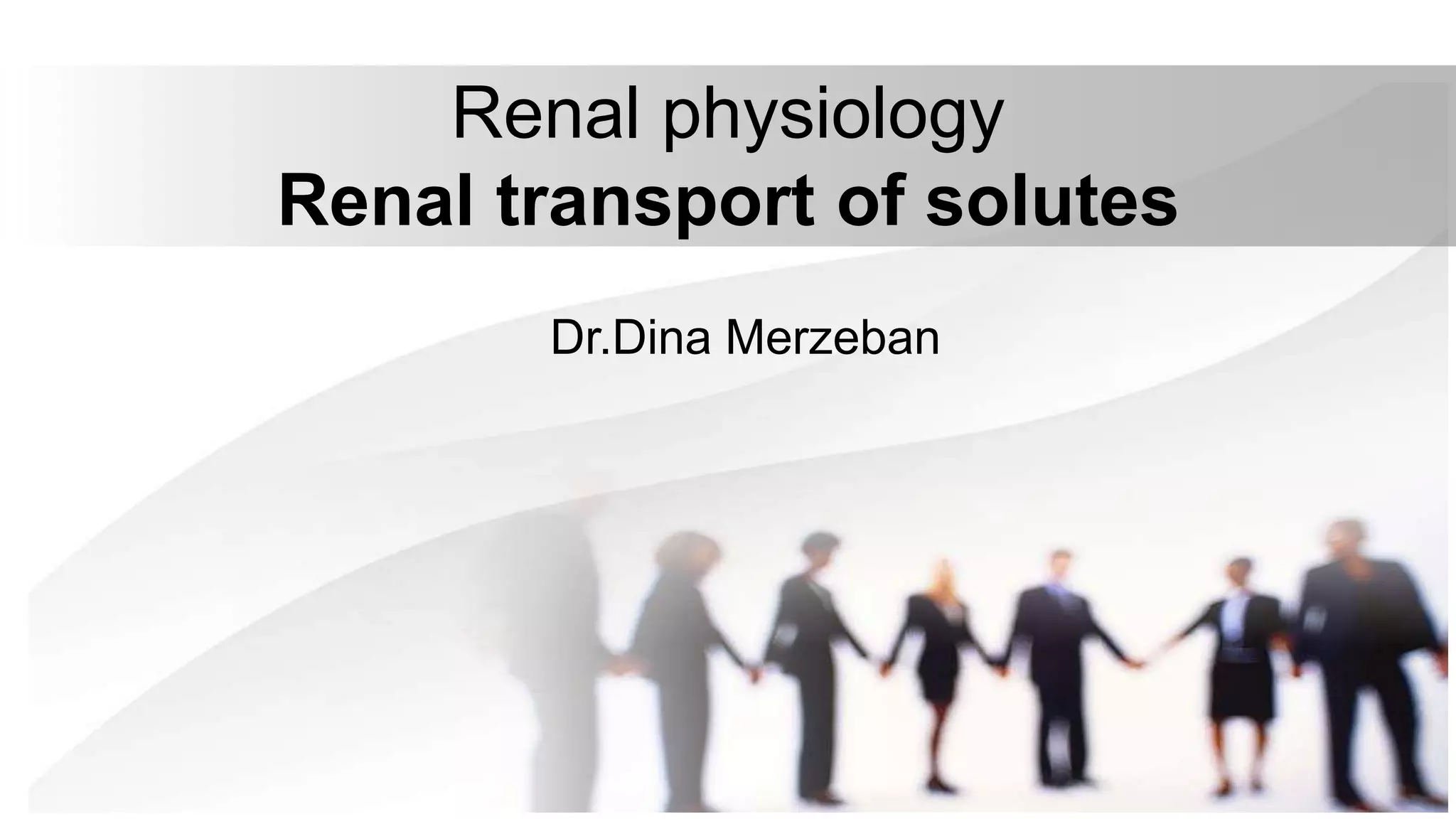
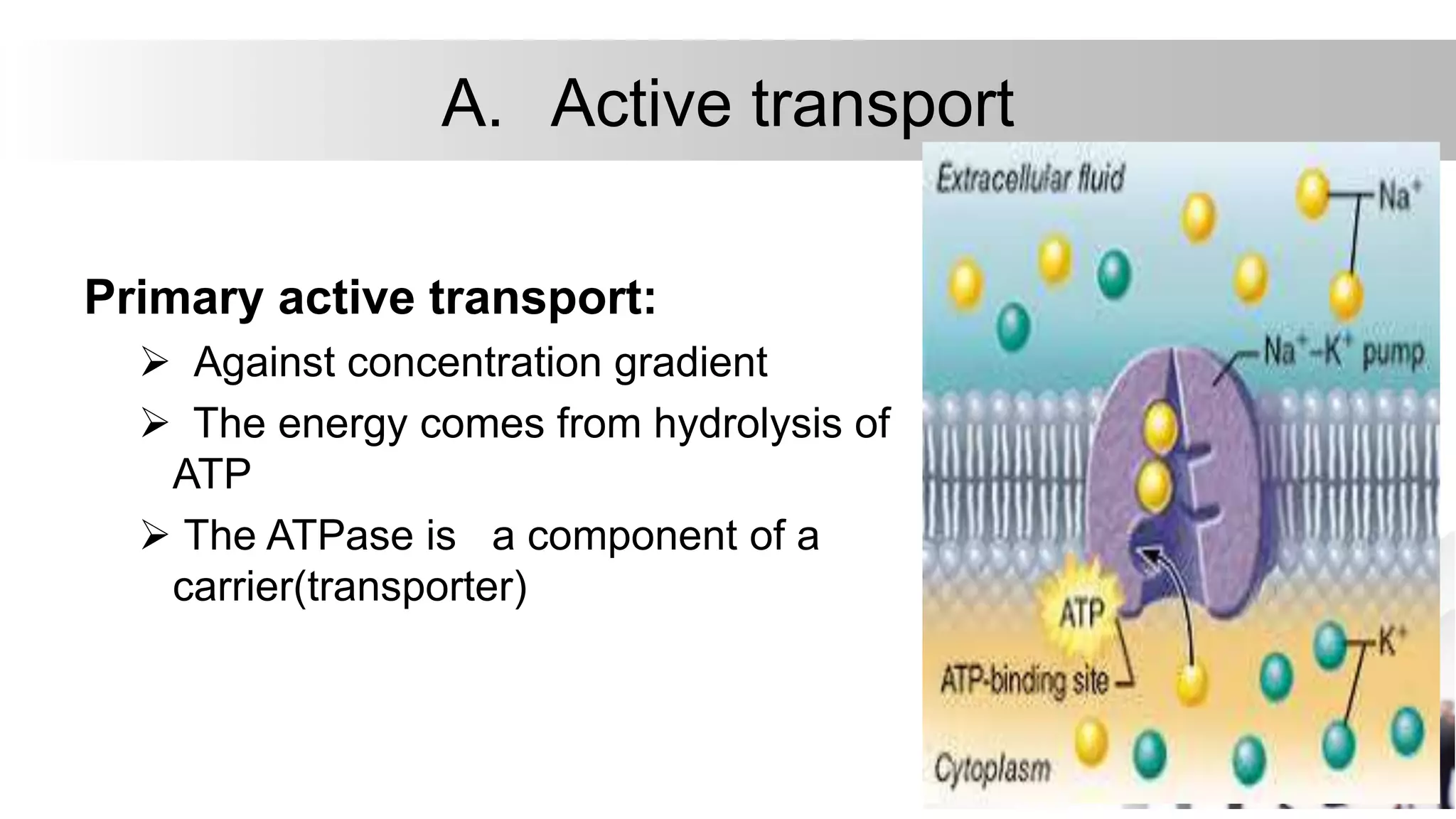
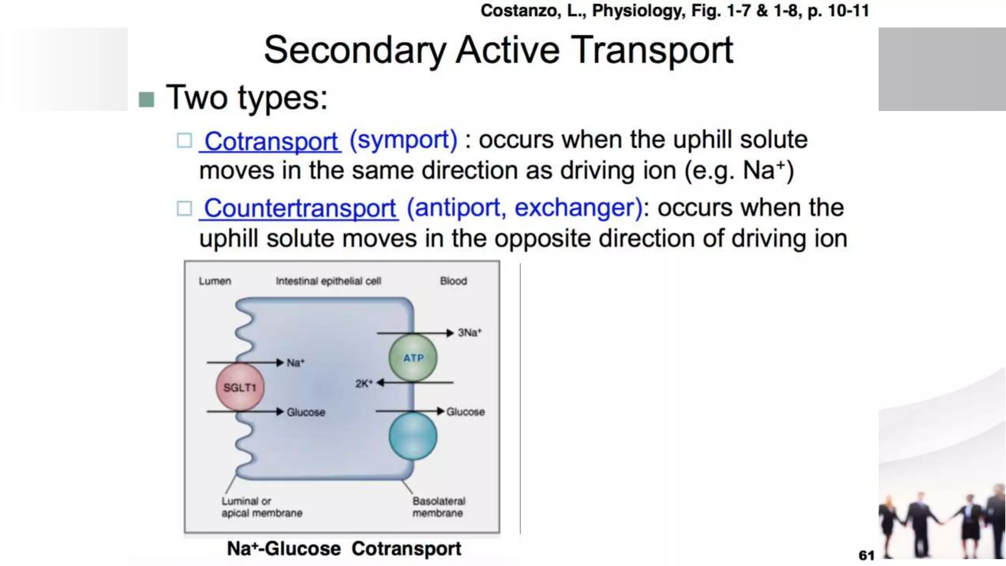
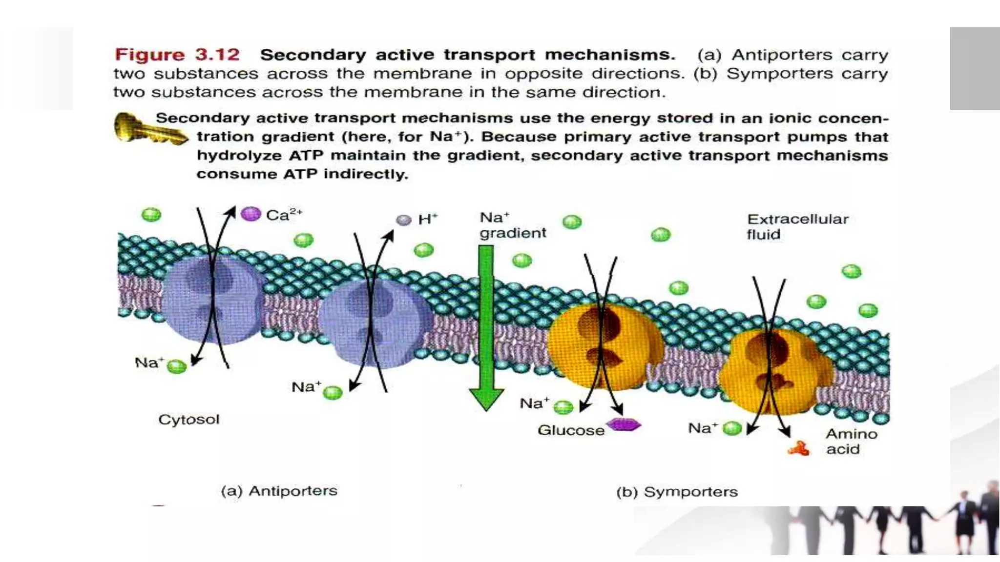
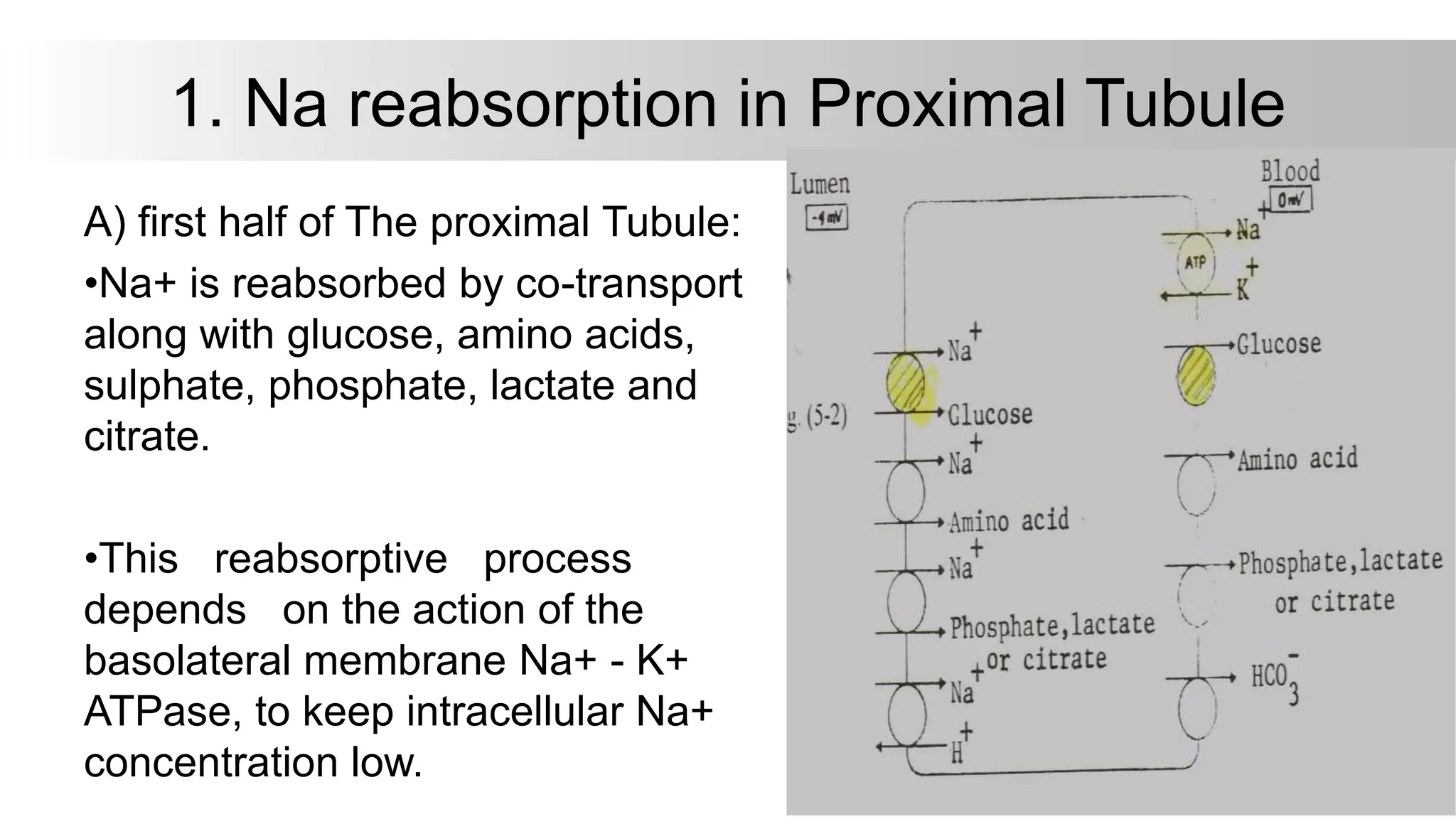
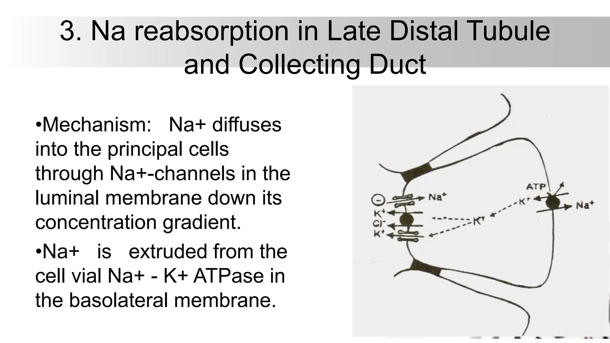
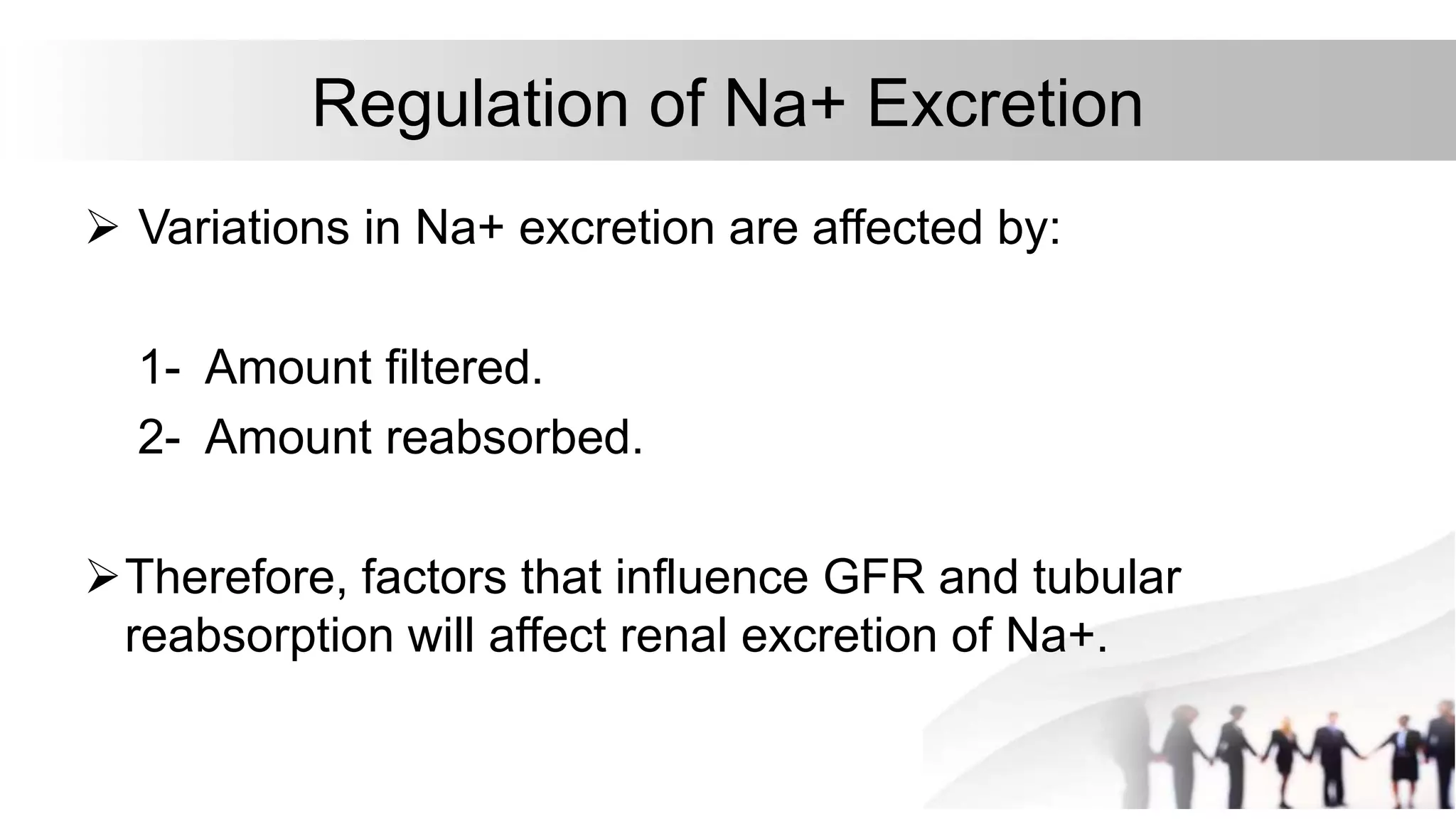
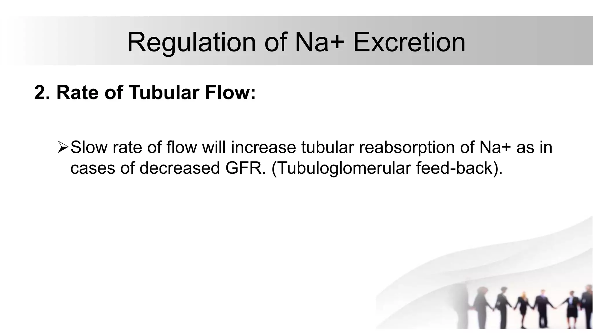
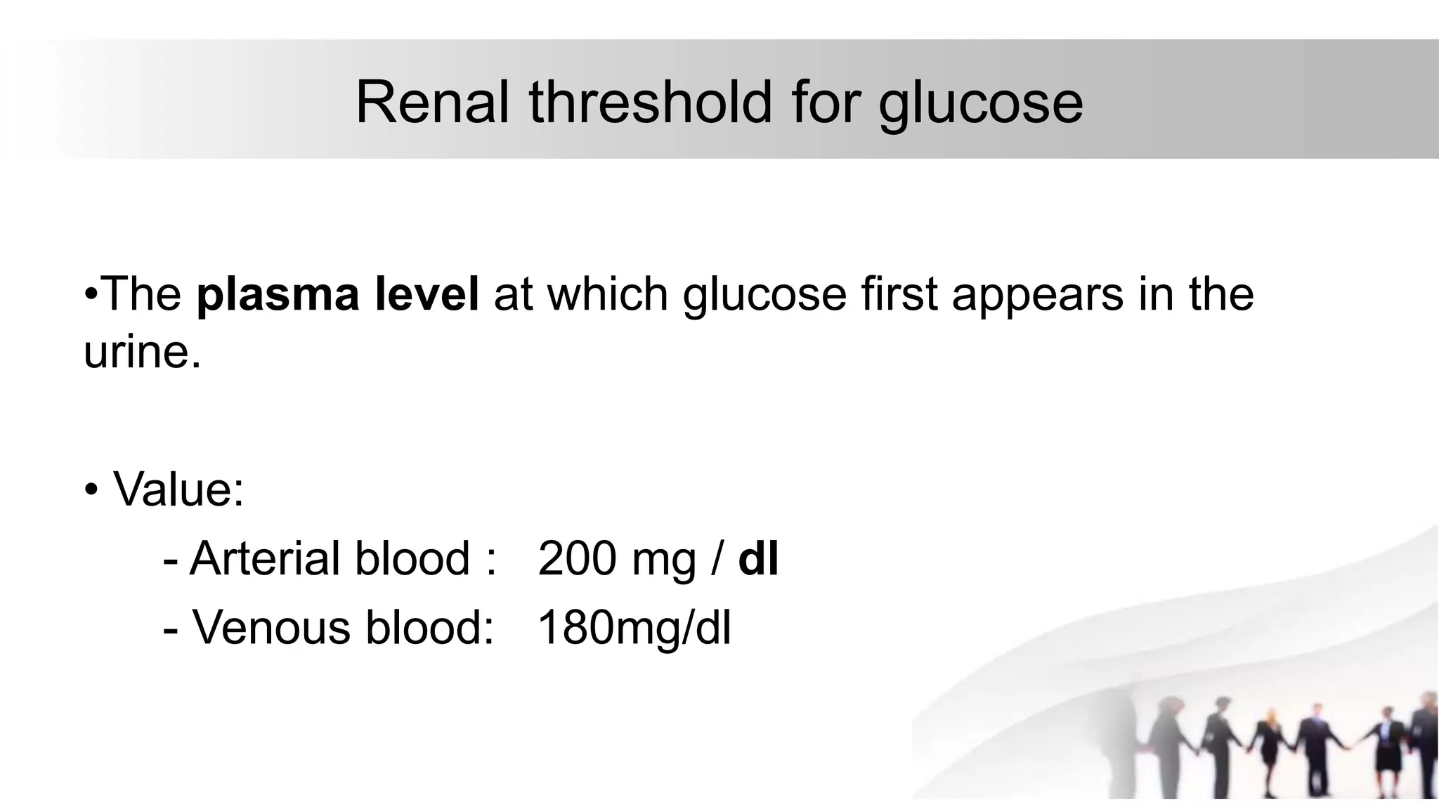
Renal physiology
Renal transport of solutes
Dr.Dina Merzeban
Mechanism of tubular transport
 Transport across the tubular membrane:
A. Active transport:
- Primary active transport.
- Secondary active transport
a) Co-transport.
b) Counter-transport.
B. Passive transport.
C. Pinocytosis.
PROPERTIES OF ACTIVE
TRANSPORT
 Active
 Needs carrier
 Against concentration gradient
„CARRIER PROTEINS OF
ACTIVE TRANSPORT
1. Uniport
2. Symport or antiport.
TYPES ACTIVE TRANSPORT:
It is further devided into:
A- Primary active transport
B- Secondary active transport
A. Active transport
Primary active transport:
 Against concentration gradient
 The energy comes from hydrolysis of
ATP
 The ATPase is a component of a
carrier(transporter)
Primary active transport
 The energy is derived directly from the breakdown of ATP.
A. Active transport
• Primary active transporters
includes:
 Sodium - Potassium ATPase.
 Hydrogen ATPase.
 Hydrogen - Potassium ATPase.
 Calcium ATPase.
SECONDARY ACTIVE TRANSPORT
In the secondary active transport, the energy is derived
secondarily from energy that has been stored in the form of ionic
concentration differences between the two sides of a membrane.
Secondary active transport is the transport of a substance with
sodium ion, by means of a common carrier protein. When sodium is
transported by a carrier protein, another substance is also
transported by the same protein simultaneously, either in the same
direction (of sodium movement) or in the opposite direction. Thus,
the transport of sodium is coupled with transport of another
substance.
Types of secondary active transport :
1- Cotransport, 2-Countertransport
B. Passive Reabsorption
1. Passive Reabsorption of Chloride
2. Osmosis of Water
3. Passive Reabsorption of Urea
C. Pinocytosis
•It is an active transport
mechanism for reabsorption of
proteins and peptides in the
proximal convoluted tubule.
Proteins in the tubular fluid
attach to the luminal membrane
of the epithelial cells.
C. Pinocytosis
•This portion of the membrane then
invaginates to the interior of the cell until
its completely pinched off. A vesicle is
formed containing the protein, which is
digested into amino acids.
•These amino acids are reabsorbed
through the basolateral membrane into
the interstitial fluid.
Tubular Transport Maximum
• The transport maximum (Tm) for the substance:
Is the maximum rate a which actively transported substances
can be transported .
• Substances that are actively reabsorbed or secreted
require a specific transport systems i.e. specific carriers
and enzymes in the tubular epithelial cells.
• this is because the carrier system becomes saturated
as the tubular load increases.
Solutes that exhibit Tm - reabsorption :Glucose, amino
acids, phosphates and sulphates
Tm- reabsorption continues so long as the transport
system is unsaturated.
For example, reabsorption of glucose and amino acids is
completed if the filtered load does not saturate the
transport system.
Renal threshold
•Substances that have a reabsorptive maximum have a
threshold concentration in the plasma below which none
of the substance appears in the urine and above which
progressively large quantities appear.
Na+ Handling by the Renal Tubule
Na+ is filtered in large amounts through the glomeruli
Na+ is actively reabsorbed out of all portions of the tubule
except the thin descending segment of the loop of Henle.
96% to 99% of the filtered Na+ is reabsorbed.
• The Figure shows Na+ handling in the nephron, and the percentages of the filtered load
reabsorbed by different segments.
1. Na reabsorption in Proximal Tubule
A) first half of The proximal Tubule:
•Na+ is reabsorbed by co-transport
along with glucose, amino acids,
sulphate, phosphate, lactate and
citrate.
•This reabsorptive process
depends on the action of the
basolateral membrane Na+ - K+
ATPase, to keep intracellular Na+
concentration low.
1. Na reabsorption in Proximal Tubule
B) Late half of The Proximal Tubule:
•Na+ is reabsorbed with chloride ion (CI-).
The late proximal tubule reabsorbs
primarily NaCI.
•Reabsorption of Na+ across the luminal
membrane is accompanied by H+
secretion via Na+ - H+.
•H+ secretion is accompanied by HCO3
reabsorption.
1. Na reabsorption in Proximal Tubule
2. Na reabsorption in Loop of Henle and
early distal tubule
Thin descending limb:
 Reabsorb water, but has no capacity to reabsorb Na+
 as the Na+ transport proteins or channels are absent from luminal
membrane.
2. Na reabsorption in Loop of Henle and
early distal tubule
•Thick ascending limb and early
distal tubule:
 30% (25% + 5%) of the filtered load of Na+,
K+, and CI- are reabsorbed by co-transport
mechanism
 that co-transports one Na+, one K+, and two
CI- from the lumen into the cells.
 Reabsorption of NaCI in the thin ascending
limb is passive by concentration gradient.
3. Na reabsorption in Late Distal Tubule
and Collecting Duct
•3% of the filtered Na+
•The principal cells
•The rate of reabsorption of Na+ is
controlled by aldosterone:
3. Na reabsorption in Late Distal Tubule
and Collecting Duct
•Mechanism: Na+ diffuses
into the principal cells
through Na+-channels in the
luminal membrane down its
concentration gradient.
•Na+ is extruded from the
cell vial Na+ - K+ ATPase in
the basolateral membrane.
Regulation of Na+ Excretion
 Na+ is the main cation in ECF. Sodium salts accounts for over
90% of the osmotically active solutes in the plasma and interstitial
fluid.
 The amount of Na+ excreted is adjusted to the amount ingested
over a wide range of dietary intake.
 Thus, the urinary Na+ output ranges from 1 mEq/day on a low-salt
diet to 400 mEq/day or more when the dietary Na+ intake is high.
Regulation of Na+ Excretion
 Variations in Na+ excretion are affected by:
1- Amount filtered.
2- Amount reabsorbed.
Therefore, factors that influence GFR and tubular
reabsorption will affect renal excretion of Na+.
Regulation of Na+ Excretion
1. Glomerular Filtration Rate:
"Glomerulotubular Balance"
Increased GFR increases the amount of Na+ filtered and
this increases the amount reabsorbed (67% of the filtered
load of Na+ and water) and Na+ excretion.
(constant percentage)
Regulation of Na+ Excretion
2. Rate of Tubular Flow:
Slow rate of flow will increase tubular reabsorption of Na+ as in
cases of decreased GFR. (Tubuloglomerular feed-back).
Regulation of Na+ Excretion
3. Pressure Natriuresis:
An increase in Na+ excretion
with rise of ABP with consequent
decrease of the ECF volume.
This is primarily a compensatory
mechanism for regulation of ABP
independent of nervous or
hormonal influence.
Regulation of Na+ Excretion
4. Hormonal Control:
a) Mineralo-corticoids:
 Aldosterone increases Na+ reabsorption in exchange
with K+ or H+.
 It acts mainly on distal tubule and collecting duct.
4. Hormonal Control of Na excretion
Aldosterone acts through:
i) Increase number of Na+
channels in the luminal
membrane
ii) Increase number of Na+ -
K+ ATPase molecules in the
basal membrane.
4. Hormonal Control of Na excretion
B) Glucocorticoids:
Cortisol has weak mineralocorticoid activity.
c) Sex Hormones:
•Estrogen increases Na+ reabsorption by renal tubule.
4. Hormonal Control of Na excretion
D) Angiotensin II:
- It is the most powerful sodium-retaining hormone, it
increases Na+ reabsorption:
i) Angiotensin II stimulates aldosterone secretion.
ii) Direct action on PCT cells:
- Stimulates Na+ - K+ ATPase pump.
- Stimulates Na+ - H+ pump.
4. Hormonal Control of Na excretion
E) Atrial natriuretic peptide (ANP):decreases Na+ reabsorption
•Facilitates the excretion of NaCI and water under conditions
of marked expansion of ECF.
i) Inhibits renin secretion: This in turn reduces the levels of
angiotensin II and the levels of aldosterone.
ii) Inhibits Na+ reabsorption by collecting ducts by direct effect:
- Inhibit Na+ - channels in the apical membrane.
- Inhibit Na+ - K+ ATPase in the basolateral membrane.
5. Sympathetic stimulation:
Decreases Na+ excretion:
 Increases Na+ reabsorption by the proximal tubule and
thick ascending limb of Loop of Henle.
 Increases renin secretion and angiotensin II formation
increases aldosterone→ increases Na+ reabsorption.
 Reduces GFR by constricting renal vessel (afferent).
6. Diuretics:
•Increase Na+ excretion.
Glucose reabsorption by the renal
tubules
Glucose reabsorption by the renal tubules
•Site: - Normally all of the freely filtered glucose is
reabsorbed in the early portion of the proximal convoluted
tubule.
•Mechanism: Secondary active transport,
i.e. secondary to the primary active transport of Na+
Glucose reabsorption by the renal tubules
At the luminal border:
•Glucose and Na+ bind to a common carrier
SGLT-2 (Sodium-dependent glucose
transporter) in the luminal membrane. As Na+
moves down its chemical gradient, glucose is
carried into the cells.
•This transport is dependent on Na+.
At the basolateral border:
•Glucose is carried into the interstitium by
facilitated diffusion. The carrier is GLUT-2
(glucose transporter).
Tubular Transport Maximum for glucose (TmG)
•TmG : is defined as the maximum amount of glucose (in
mg) that can be reabsorbed by the renal tubules per
minute.
•It is an indication of the reabsorptive capacity of the kidney
and is determined by the number of glucose carriers in the
proximal tubule.
•Value: TmG : 300 mg / min in female.
375 mg / min in male.
Renal threshold for glucose
•The plasma level at which glucose first appears in the
urine.
• Value:
- Arterial blood : 200 mg / dl
- Venous blood: 180mg/dl
Glycosuria (Glucosuria)
•It is excretion of glucose in urine in considerable amounts.
•Causes:
1) Diabetes Mellitus:
Glycosuria occurs when the blood glucose level is
elevated and exceeds renal threshold.
Glycosuria (Glucosuria)
2) Renal glycosuria:
Glycosuria occurs at normal plasma glucose level.
The renal threshold for glucose is lowered below180 mg % due
to congenital defect in the glucose transport mechanism in the
renal tubules.
TmG is markedly decreased in renal glycosuria.
Water reabsorption
•Normally 180 L of fluid is filtered through the glomeruli
each day, while the average daily urine volume is about 1
L.
•Water reabsorption is a passive process (osmosis)
throughout the whole nephron.
•It is of two types:
I. Obligatory water reabsorption
Water reabsorption
I. Obligatory water reabsorption
•It comprises 87% of the filtered water, which is reabsorbed
independent of ADH.
65% from PCT: following the reabsorbed solutes.
15% from descending limb of the loop of Henle.
The ascending limb is impermeable to water.
Early distal tubule: is relatively impermeable to water.
Water reabsorption
II. Facultative water reabsorption
•It comprises about 13% of the filtered water, which is controlled by
ADH in the following segments:
1- Late Distal Convoluted Tubules.
2- Collecting ducts
•This facultative water reabsorption can produce concentrated urine.
ADH increases the permeability of these segments to water by causing
insertion of water channels into the luminal membrane of the principal
cells.
ADH increases the permeability of these segments to water by causing insertion of water channels into the luminal membrane of
the principal cells.
Urine concentration and dilution
•The kidneys can excrete either concentrated or diluted
urine according to the water balance of the body.
•In dehydration: water is conserved in the body and
concentrated urine having high osmolarity is excreted.
•On the other hand, in case of hydration, excess water is
eliminated from the body and diluted urine is excreted.
Urine concentration and dilution
Since water reabsorption is obligatory in the proximal
tubule and loop of Henle,
it is clear that the final adjustment of the urine volume and
osmolarity depends only on the extent of facultative water
reabsorption (ADH).
Thank you

Renal physiology lecture 2 dina

  • 1.
    Renal physiology Renal transportof solutes Dr.Dina Merzeban
  • 2.
    Mechanism of tubulartransport  Transport across the tubular membrane: A. Active transport: - Primary active transport. - Secondary active transport a) Co-transport. b) Counter-transport. B. Passive transport. C. Pinocytosis.
  • 3.
    PROPERTIES OF ACTIVE TRANSPORT Active  Needs carrier  Against concentration gradient
  • 4.
    „CARRIER PROTEINS OF ACTIVETRANSPORT 1. Uniport 2. Symport or antiport.
  • 5.
    TYPES ACTIVE TRANSPORT: Itis further devided into: A- Primary active transport B- Secondary active transport
  • 6.
    A. Active transport Primaryactive transport:  Against concentration gradient  The energy comes from hydrolysis of ATP  The ATPase is a component of a carrier(transporter)
  • 7.
    Primary active transport The energy is derived directly from the breakdown of ATP.
  • 8.
    A. Active transport •Primary active transporters includes:  Sodium - Potassium ATPase.  Hydrogen ATPase.  Hydrogen - Potassium ATPase.  Calcium ATPase.
  • 9.
    SECONDARY ACTIVE TRANSPORT Inthe secondary active transport, the energy is derived secondarily from energy that has been stored in the form of ionic concentration differences between the two sides of a membrane. Secondary active transport is the transport of a substance with sodium ion, by means of a common carrier protein. When sodium is transported by a carrier protein, another substance is also transported by the same protein simultaneously, either in the same direction (of sodium movement) or in the opposite direction. Thus, the transport of sodium is coupled with transport of another substance. Types of secondary active transport : 1- Cotransport, 2-Countertransport
  • 12.
    B. Passive Reabsorption 1.Passive Reabsorption of Chloride 2. Osmosis of Water 3. Passive Reabsorption of Urea
  • 13.
    C. Pinocytosis •It isan active transport mechanism for reabsorption of proteins and peptides in the proximal convoluted tubule. Proteins in the tubular fluid attach to the luminal membrane of the epithelial cells.
  • 14.
    C. Pinocytosis •This portionof the membrane then invaginates to the interior of the cell until its completely pinched off. A vesicle is formed containing the protein, which is digested into amino acids. •These amino acids are reabsorbed through the basolateral membrane into the interstitial fluid.
  • 15.
    Tubular Transport Maximum •The transport maximum (Tm) for the substance: Is the maximum rate a which actively transported substances can be transported . • Substances that are actively reabsorbed or secreted require a specific transport systems i.e. specific carriers and enzymes in the tubular epithelial cells. • this is because the carrier system becomes saturated as the tubular load increases.
  • 16.
    Solutes that exhibitTm - reabsorption :Glucose, amino acids, phosphates and sulphates Tm- reabsorption continues so long as the transport system is unsaturated. For example, reabsorption of glucose and amino acids is completed if the filtered load does not saturate the transport system.
  • 17.
    Renal threshold •Substances thathave a reabsorptive maximum have a threshold concentration in the plasma below which none of the substance appears in the urine and above which progressively large quantities appear.
  • 18.
    Na+ Handling bythe Renal Tubule Na+ is filtered in large amounts through the glomeruli Na+ is actively reabsorbed out of all portions of the tubule except the thin descending segment of the loop of Henle. 96% to 99% of the filtered Na+ is reabsorbed.
  • 19.
    • The Figureshows Na+ handling in the nephron, and the percentages of the filtered load reabsorbed by different segments.
  • 20.
    1. Na reabsorptionin Proximal Tubule A) first half of The proximal Tubule: •Na+ is reabsorbed by co-transport along with glucose, amino acids, sulphate, phosphate, lactate and citrate. •This reabsorptive process depends on the action of the basolateral membrane Na+ - K+ ATPase, to keep intracellular Na+ concentration low.
  • 21.
    1. Na reabsorptionin Proximal Tubule B) Late half of The Proximal Tubule: •Na+ is reabsorbed with chloride ion (CI-). The late proximal tubule reabsorbs primarily NaCI. •Reabsorption of Na+ across the luminal membrane is accompanied by H+ secretion via Na+ - H+. •H+ secretion is accompanied by HCO3 reabsorption.
  • 22.
    1. Na reabsorptionin Proximal Tubule
  • 23.
    2. Na reabsorptionin Loop of Henle and early distal tubule Thin descending limb:  Reabsorb water, but has no capacity to reabsorb Na+  as the Na+ transport proteins or channels are absent from luminal membrane.
  • 24.
    2. Na reabsorptionin Loop of Henle and early distal tubule •Thick ascending limb and early distal tubule:  30% (25% + 5%) of the filtered load of Na+, K+, and CI- are reabsorbed by co-transport mechanism  that co-transports one Na+, one K+, and two CI- from the lumen into the cells.  Reabsorption of NaCI in the thin ascending limb is passive by concentration gradient.
  • 25.
    3. Na reabsorptionin Late Distal Tubule and Collecting Duct •3% of the filtered Na+ •The principal cells •The rate of reabsorption of Na+ is controlled by aldosterone:
  • 26.
    3. Na reabsorptionin Late Distal Tubule and Collecting Duct •Mechanism: Na+ diffuses into the principal cells through Na+-channels in the luminal membrane down its concentration gradient. •Na+ is extruded from the cell vial Na+ - K+ ATPase in the basolateral membrane.
  • 27.
    Regulation of Na+Excretion  Na+ is the main cation in ECF. Sodium salts accounts for over 90% of the osmotically active solutes in the plasma and interstitial fluid.  The amount of Na+ excreted is adjusted to the amount ingested over a wide range of dietary intake.  Thus, the urinary Na+ output ranges from 1 mEq/day on a low-salt diet to 400 mEq/day or more when the dietary Na+ intake is high.
  • 28.
    Regulation of Na+Excretion  Variations in Na+ excretion are affected by: 1- Amount filtered. 2- Amount reabsorbed. Therefore, factors that influence GFR and tubular reabsorption will affect renal excretion of Na+.
  • 29.
    Regulation of Na+Excretion 1. Glomerular Filtration Rate: "Glomerulotubular Balance" Increased GFR increases the amount of Na+ filtered and this increases the amount reabsorbed (67% of the filtered load of Na+ and water) and Na+ excretion. (constant percentage)
  • 30.
    Regulation of Na+Excretion 2. Rate of Tubular Flow: Slow rate of flow will increase tubular reabsorption of Na+ as in cases of decreased GFR. (Tubuloglomerular feed-back).
  • 31.
    Regulation of Na+Excretion 3. Pressure Natriuresis: An increase in Na+ excretion with rise of ABP with consequent decrease of the ECF volume. This is primarily a compensatory mechanism for regulation of ABP independent of nervous or hormonal influence.
  • 32.
    Regulation of Na+Excretion 4. Hormonal Control: a) Mineralo-corticoids:  Aldosterone increases Na+ reabsorption in exchange with K+ or H+.  It acts mainly on distal tubule and collecting duct.
  • 33.
    4. Hormonal Controlof Na excretion Aldosterone acts through: i) Increase number of Na+ channels in the luminal membrane ii) Increase number of Na+ - K+ ATPase molecules in the basal membrane.
  • 34.
    4. Hormonal Controlof Na excretion B) Glucocorticoids: Cortisol has weak mineralocorticoid activity. c) Sex Hormones: •Estrogen increases Na+ reabsorption by renal tubule.
  • 35.
    4. Hormonal Controlof Na excretion D) Angiotensin II: - It is the most powerful sodium-retaining hormone, it increases Na+ reabsorption: i) Angiotensin II stimulates aldosterone secretion. ii) Direct action on PCT cells: - Stimulates Na+ - K+ ATPase pump. - Stimulates Na+ - H+ pump.
  • 36.
    4. Hormonal Controlof Na excretion E) Atrial natriuretic peptide (ANP):decreases Na+ reabsorption •Facilitates the excretion of NaCI and water under conditions of marked expansion of ECF. i) Inhibits renin secretion: This in turn reduces the levels of angiotensin II and the levels of aldosterone. ii) Inhibits Na+ reabsorption by collecting ducts by direct effect: - Inhibit Na+ - channels in the apical membrane. - Inhibit Na+ - K+ ATPase in the basolateral membrane.
  • 37.
    5. Sympathetic stimulation: DecreasesNa+ excretion:  Increases Na+ reabsorption by the proximal tubule and thick ascending limb of Loop of Henle.  Increases renin secretion and angiotensin II formation increases aldosterone→ increases Na+ reabsorption.  Reduces GFR by constricting renal vessel (afferent).
  • 38.
  • 39.
    Glucose reabsorption bythe renal tubules
  • 40.
    Glucose reabsorption bythe renal tubules •Site: - Normally all of the freely filtered glucose is reabsorbed in the early portion of the proximal convoluted tubule. •Mechanism: Secondary active transport, i.e. secondary to the primary active transport of Na+
  • 41.
    Glucose reabsorption bythe renal tubules At the luminal border: •Glucose and Na+ bind to a common carrier SGLT-2 (Sodium-dependent glucose transporter) in the luminal membrane. As Na+ moves down its chemical gradient, glucose is carried into the cells. •This transport is dependent on Na+. At the basolateral border: •Glucose is carried into the interstitium by facilitated diffusion. The carrier is GLUT-2 (glucose transporter).
  • 42.
    Tubular Transport Maximumfor glucose (TmG) •TmG : is defined as the maximum amount of glucose (in mg) that can be reabsorbed by the renal tubules per minute. •It is an indication of the reabsorptive capacity of the kidney and is determined by the number of glucose carriers in the proximal tubule. •Value: TmG : 300 mg / min in female. 375 mg / min in male.
  • 43.
    Renal threshold forglucose •The plasma level at which glucose first appears in the urine. • Value: - Arterial blood : 200 mg / dl - Venous blood: 180mg/dl
  • 45.
    Glycosuria (Glucosuria) •It isexcretion of glucose in urine in considerable amounts. •Causes: 1) Diabetes Mellitus: Glycosuria occurs when the blood glucose level is elevated and exceeds renal threshold.
  • 46.
    Glycosuria (Glucosuria) 2) Renalglycosuria: Glycosuria occurs at normal plasma glucose level. The renal threshold for glucose is lowered below180 mg % due to congenital defect in the glucose transport mechanism in the renal tubules. TmG is markedly decreased in renal glycosuria.
  • 47.
    Water reabsorption •Normally 180L of fluid is filtered through the glomeruli each day, while the average daily urine volume is about 1 L. •Water reabsorption is a passive process (osmosis) throughout the whole nephron. •It is of two types: I. Obligatory water reabsorption
  • 48.
    Water reabsorption I. Obligatorywater reabsorption •It comprises 87% of the filtered water, which is reabsorbed independent of ADH. 65% from PCT: following the reabsorbed solutes. 15% from descending limb of the loop of Henle. The ascending limb is impermeable to water. Early distal tubule: is relatively impermeable to water.
  • 49.
    Water reabsorption II. Facultativewater reabsorption •It comprises about 13% of the filtered water, which is controlled by ADH in the following segments: 1- Late Distal Convoluted Tubules. 2- Collecting ducts •This facultative water reabsorption can produce concentrated urine. ADH increases the permeability of these segments to water by causing insertion of water channels into the luminal membrane of the principal cells.
  • 51.
    ADH increases thepermeability of these segments to water by causing insertion of water channels into the luminal membrane of the principal cells.
  • 52.
    Urine concentration anddilution •The kidneys can excrete either concentrated or diluted urine according to the water balance of the body. •In dehydration: water is conserved in the body and concentrated urine having high osmolarity is excreted. •On the other hand, in case of hydration, excess water is eliminated from the body and diluted urine is excreted.
  • 53.
    Urine concentration anddilution Since water reabsorption is obligatory in the proximal tubule and loop of Henle, it is clear that the final adjustment of the urine volume and osmolarity depends only on the extent of facultative water reabsorption (ADH).
  • 54.