![Sodium transport in the kidney
Page 2 of 5
~
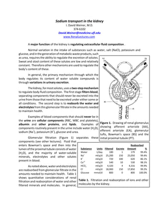
3 Na+
2 K+
[Na+] ~10-20
mEq/L
-70
mV
Figure 3. Effect of basolateral Na+
‐
K+
‐ATPase on intracellular sodium
concentration and membrane
potential.
Figure 4. Sodium reabsorption
along the nephron. PT ‐ Proximal
tubule; TAL ‐ thick ascending limb of
the loop of Henle; DT ‐ distal
connecting tubule; CCD ‐ cortical
collecting duct; IMCD ‐ inner
medullary collecting duct.
the kidneys adjust reabsorption so that excretion balances the
difference between intake, typically oral intake, and extra‐renal
excretion. For example, if someone is dehydrated, GFR and thus the
filtered amount of water is constant, but renal water reabsorption
increases to decrease net urinary water excretion. Conversely, if
someone is studying and drinking lots of iced tea, which is
predominantly H2O, the amount of H2O filtered at the glomerulus
does not change substantially, but H2O reabsorption decreases
resulting in more H2O excretion in the urine and resultant
maintenance of water homeostasis. The sodium transporters used
to maintain sodium balance are the primary focus of this lecture.
The kidney generally, uses a multi‐step process to reabsorb
filtered solutes. The first step is bulk reabsorption of large
quantities of solute. Although some regulation occurs at this stage,
the large quantities of solute reabsorbed preclude the exact
regulation necessary for long‐term health. The second step is
intermediate quantity reabsorption associated with intermediate
level regulation, and the third and final step, which mediates the
final regulation of the urine composition, has low rates of solute
transport but with high level regulation. Typically, these three
steps occur in the proximal tubule, loop of Henle and the collecting
duct (please give me some artistic
license to simplify these functions
and their anatomic location).
Throughout the nephron,
sodium reabsorption is due to the
effects of basolateral Na+
‐K+
‐
ATPase. As shown in Figure 3, a
ubiquitous basolateral Na+
‐K+
‐
ATPase, present in all renal tubular
cells, generates both intracellular
electronegativity and a low intra‐
cellular sodium concentration, both
of which provide a gradient for
sodium entry into cells.
Figure 4 summarizes the
relative roles of sodium reabsorption
along the nephron. The proximal
tubule is the primary site of bulk reabsorption, while the thick
ascending limb of the loop of Henle reabsorbs most of the
remaining sodium, and the distal tubule and the collecting duct
accounts for the remaining sodium reabsorption.
Figure 2. Diagram showing
intimate contact between
peritubular capillaries and nephron.
This enables solute reabsorbed by
renal epithelial cells to diffuse into
peritubular capillaries and be
returned to systemic circulation via
renal vein.](data:image/gif;base64,R0lGODlhAQABAIAAAAAAAP///yH5BAEAAAAALAAAAAABAAEAAAIBRAA7)
Recommended
More Related Content
What's hot
What's hot (20)
Similar to Sodium Transport Mechanism in Regulating ECF Fluid Osmolarity.
Similar to Sodium Transport Mechanism in Regulating ECF Fluid Osmolarity. (20)
More from meducationdotnet
More from meducationdotnet (20)
Sodium Transport Mechanism in Regulating ECF Fluid Osmolarity.
- 1. Figure 1. Drawing of renal glomerulus showing afferent arteriole (AA), efferent arteriole (EA), glomerulur tufts, Bowman’s space (BS) and the initial proximal tubule (PT). Sodium transport in the kidney I. David Weiner, M.D. 374‐6102 David.Weiner@medicine.ufl.edu www.RenalLectures.com A major function of the kidney is regulating extracellular fluid composition . Normal variation in the intake of substances such as water, salt (NaCl), potassium and glucose, and in the generation of metabolic waste products, such as urea, requires the ability to regulate the excretion of solutes. Sweat and stool content of these solutes are low and relatively constant. Therefore other mechanisms are used to regulate the body's content of these. In general, the primary mechanism through which the body regulates its content of water soluble compounds is through variations in urinary excretion. The kidney, for most solutes, uses a two‐step mechanism to regulate body fluid composition. The first stage filters blood, separating components that should never be excreted into the urine from those that need to be excreted under either some or all conditions. The second step is to reabsorb the water and electrolytes from this glomerular filtrate in the amounts needed to maintain health. Examples of blood components that should never be in the urine are cellular components (RBC, WBC and platelets), albumin and other proteins, and lipids. Examples of components routinely present in the urine include water (H2O), sodium (Na+ ), potassium (K+ ), glucose and urea. Glomerular filtration (Figure 1) separates these components (see other lectures). Fluid that enters Bowman’s space and then into the lumen of the proximal tubule consists of water (H2O), and the majority of water‐soluble minerals, electrolytes and other solutes present in blood. As noted above, water and electrolytes are reabsorbed from glomerular filtrate in the amounts needed to maintain health. Table 1 shows quantitative considerations of renal filtration and reabsorption of water and other filtered minerals and molecules. In general, Substance Units Filtered Excrete Reabsorbed Amount % Water L/day 180 1 179 99.4% Na+ mEq/d 25,200 150 25,050 99.4% K+ mEq/d 720 100 620 86.1% Ca+2 mEq/d 540 10 530 98.1% HCO3 ‐ mEq/d 4,320 4 4,316 99.9% Cl‐ mEq/d 18,000 150 17,850 99.2% Glucose mmol/d 800 0 800 100.0% Table 1. Filtration and reabsorption of ions and other molecules by the kidney.
- 2. Sodium transport in the kidney Page 2 of 5 ~ 3 Na+ 2 K+ [Na+] ~10-20 mEq/L -70 mV Figure 3. Effect of basolateral Na+ ‐ K+ ‐ATPase on intracellular sodium concentration and membrane potential. Figure 4. Sodium reabsorption along the nephron. PT ‐ Proximal tubule; TAL ‐ thick ascending limb of the loop of Henle; DT ‐ distal connecting tubule; CCD ‐ cortical collecting duct; IMCD ‐ inner medullary collecting duct. the kidneys adjust reabsorption so that excretion balances the difference between intake, typically oral intake, and extra‐renal excretion. For example, if someone is dehydrated, GFR and thus the filtered amount of water is constant, but renal water reabsorption increases to decrease net urinary water excretion. Conversely, if someone is studying and drinking lots of iced tea, which is predominantly H2O, the amount of H2O filtered at the glomerulus does not change substantially, but H2O reabsorption decreases resulting in more H2O excretion in the urine and resultant maintenance of water homeostasis. The sodium transporters used to maintain sodium balance are the primary focus of this lecture. The kidney generally, uses a multi‐step process to reabsorb filtered solutes. The first step is bulk reabsorption of large quantities of solute. Although some regulation occurs at this stage, the large quantities of solute reabsorbed preclude the exact regulation necessary for long‐term health. The second step is intermediate quantity reabsorption associated with intermediate level regulation, and the third and final step, which mediates the final regulation of the urine composition, has low rates of solute transport but with high level regulation. Typically, these three steps occur in the proximal tubule, loop of Henle and the collecting duct (please give me some artistic license to simplify these functions and their anatomic location). Throughout the nephron, sodium reabsorption is due to the effects of basolateral Na+ ‐K+ ‐ ATPase. As shown in Figure 3, a ubiquitous basolateral Na+ ‐K+ ‐ ATPase, present in all renal tubular cells, generates both intracellular electronegativity and a low intra‐ cellular sodium concentration, both of which provide a gradient for sodium entry into cells. Figure 4 summarizes the relative roles of sodium reabsorption along the nephron. The proximal tubule is the primary site of bulk reabsorption, while the thick ascending limb of the loop of Henle reabsorbs most of the remaining sodium, and the distal tubule and the collecting duct accounts for the remaining sodium reabsorption. Figure 2. Diagram showing intimate contact between peritubular capillaries and nephron. This enables solute reabsorbed by renal epithelial cells to diffuse into peritubular capillaries and be returned to systemic circulation via renal vein.
- 3. Sodium transport in the kidney Page 3 of 5 Na+ K+ 2 Cl- Na+ H+ Cl- ~ Na+ K+ Lumen Peritubular Figure 6. Sodium reabsorption in the thick ascending limb of the loop of Henle. Na+ H+ Glucose, Phosphate, Amino acids, or lactate Na+ Na+ K+ Lumen (Urine) Peritubular (Blood) Figure 5. Sodium reabsorption in the early proximal tubule. Na+ Cl- ~ Na+ K+ Cl- Lumen Peritubular H2O Figure 7. Sodium reabsorption in the distal convoluted tubule. The specific mechanism of sodium reabsorption differs in different segments. The majority of sodium reabsorption occurs in the proximal tubule. Sodium reabsorption is linked to reabsorption of bicarbonate, glucose, phosphate, amino acids and chloride. Figure 5 summarizes sodium reabsorption in the early regions of the proximal tubule. The importance of sodium‐linked proton secretion will be discussed in the lecture on acid‐base transport. Sodium‐linked glucose, phosphate, amino acid and lactate reabsorption are critical to reabsorbing these essential solutes, and occurs predominantly in the proximal tubule. Little‐to‐no sodium is reabsorbed in the descending thin limb or the ascending thin limb of the loop of Henle. The thick ascending limb of the loop of Henle is the next major region of sodium reabsorption. Approximately 25% of filtered sodium is reabsorbed here. Figure 6 summarizes sodium reabsorption in this region. The primary mechanism of sodium reabsorption in this region is an apical Na+ ‐K+ ‐2Cl‐ cotransporter. Because this protein transports equal numbers of cations and anions, it does not move net charge. Instead the low intracellular sodium and chloride concentrations “drive” the transport, and result in potassium moving into the cell despite the high intracellular potassium concentration. This potassium recycles into the luminal fluid through a potassium channel. The movement of potassium back into the luminal fluid causes the luminal fluid to become positively charged. This voltage is important for reabsorption of calcium in this segment. An apical Na+ /H+ exchanger is also present, but is quantitatively less important in sodium reabsorption. The distal convoluted tubule is the next site for sodium reabsorption. Its sodium transport mechanism is shown in Figure 7. Sodium is reabsorbed by an apical sodium‐chloride cotransporter that transports equal amounts of sodium and chloride. Because water does not cross the apical membrane in this segment hypotonic luminal fluid with a low sodium concentration is generated (the importance of this will be seen in the lecture on water transport). The last segment involved in sodium transport is the collecting duct. In this segment sodium is reabsorbed by a specific cell, the principal cell. Figure 8 summarizes sodium transport in the collecting duct. Sodium enters the cell via an apical protein, the
- 4. Sodium transport in the kidney Page 4 of 5 Na+ K+ ~ Na+ K+ Lumen Peritubular Figure 8. Sodium transport in the collecting duct principal cell. epithelial Na channel (ENaC), that transports sodium in the form of Na+ . Na+ moves into the cell because of the low intracellular Na+ concentration and the intracellular electronegativity that are generated by the basolateral Na+ ‐K+ ‐ATPase. Because sodium enters the cell without an associated anion, its movement out of the luminal fluid results in loss of positive charge and the subsequent development of a net negative charge in the luminal fluid. Potassium can move out of the cell into the luminal fluid partially as a result of luminal electronegativity and partially because intracellular potassium concentrations are high (due to basolateral Na+ ‐K+ ‐ATPase). Conditions that regulate sodium transport in the collecting duct also regulate potassium secretion. Regulation of sodium transport Because sodium balance determines the circulating plasma volume, any condition that alters plasma volume will alter sodium transport by the renal tubules. These effects are mediated via hormones released by other cells. In general, conditions associated with plasma volume expansion inhibit stimuli that increase sodium reabsorption, whereas the opposite occurs in response to plasma volume contraction. Table 2 summarizes important sodium transport regulators. Pharmacologic inhibition of sodium transport Many clinical conditions are associated with relatively inadequate sodium excretion by the kidney. Examples include hypertension, heart failure, liver disease and kidney disease. Medications that inhibit renal sodium transport are very effective for treating these conditions. Effective inhibitors of proximal tubule sodium transport mechanisms are not available. Inhibitors of the thick ascending limb of loop of Henle Na+ ‐K+ ‐2Cl‐ cotransporter are termed “loop diuretics” and are potent medications that increase sodium excretion. Distal convoluted tubule sodium‐chloride cotransport inhibitors, known as “thiazide diuretics” because of their chemical structure, are the second most potent drugs for increasing urinary sodium excretion. Both loop diuretics and thiazide diuretics increase sodium delivery to the collecting duct. The collecting duct does not have adequate sodium transport capacity, so the net effect is increased sodium excretion into the urine. However, the collecting duct will “attempt” to correct Hormone Stimulus Site of action Effect Angiotensin II Renin PT, TAL, CD 8 Aldosterone AII, 8[K+ ] DT, CD 8 Sympathetic nerves 9BP, 9ECV PT, TAL 8 ANP 8BP, 8ECV CD 9 NO PT, CD 9 Table 2. Hormonal regulation of renal Na+ transport. Abbreviations: BP, blood pressure; ECV, extracellular volume; PT, proximal tubule; TAL, thick ascending limb of the loop of Henle; DT, distal tubule; CD, collecting duct.
- 5. Sodium transport in the kidney Page 5 of 5 this increase sodium excretion by increasing sodium reabsorption. Although this “attempt” is insufficient to correct the sodium excretion, it does cause collecting duct potassium secretion to increase, and can lead to potassium deficiency (this is covered in more detail in the potassium lecture). Inhibitors of collecting duct ENaC have less effect on net sodium transport because so little sodium is reabsorbed in the collecting duct. However, sodium reabsorption is critical for potassium secretion, and the collecting duct is the primary site regulating renal potassium excretion. Accordingly, collecting duct sodium transport inhibitors decrease potassium excretion. Consequently, they are often termed “potassium‐sparing diuretics.”