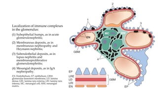
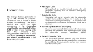
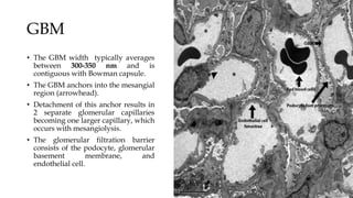
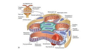
RENAL PATHOLOGY
STRUCTURE OF GLOMERULUS
Glomerulus
• Mesangial Cells
• Mesangial cells are modified smooth muscle cells with
phagocytic capability that can influence intracapillary
blood pressure.
• Endothelial Cells
• Endothelial cell nuclei protrude into the glomerular
capillary lumen and these should not be included in the
assessment for mesangial cellularity. The endothelial cells
are fenestrated with fenestrae that measure 70-100 nm in
diameter.
• Visceral Epithelial Cells (Podocytes)
• Podocytes are a terminally differentiated cell with complex
processes that intertwine with adjacent podocytes along
the glomerular basement membranes (GBM).
• Parietal Epithelial Cells
• The 4th cell type (parietal epithelial cell) lines Bowman
capsules, but the lesser known parietal podocyte can also
be found lining Bowman capsules and are more common
near the vascular pole.
The average human glomerulus is
up to 200 microns in diameter.
Glomerular size is harder to assess
without morphometry. One quick
method that works for most
microscopes – the glomerulus
should not be more than half of the
field of view when viewing with
the 40X objective lens.
The glomerular tuft consists of three
cells types (mesangial, endothelial,
and visceral epithelial cells – also
known as podocytes).
GBM
• The GBM width typically averages
between 300-350 nm and is
contiguous with Bowman capsule.
• The GBM anchors into the mesangial
region (arrowhead).
• Detachment of this anchor results in
2 separate glomerular capillaries
becoming one larger capillary, which
occurs with mesangiolysis.
• The glomerular filtration barrier
consists of the podocyte, glomerular
basement membrane, and
endothelial cell.
Scanning electron micrograph (400×) showing a vascular
cast of two juxtamedullary glomeruli (rat). Each capillary
tuft is supplied by an afferent arteriole (AA) which, on
the surface of the tuft, immediately divides into several
branches. Efferent arterioles (EA) emerge out of the
centre of the tuft.
• Part of a glomerular lobule (rat), showing the
arrangement of structures in the glomerular tuft.
• The capillary (C) is outlined by a flat fenestrated
endothelium (E). The podocyte layer (PO) and the
glomerular basement membrane (GBM) do not
encircle the individual capillary completely, they
form a common surface cover around the lobule.
• In the peripheral portion of the capillary the
filtration barrier is formed.
• Two subdomains of the GBM are delineated from
each other by mesangial angles (arrows): the
pericapillary GBM (cGBM) faced by the podocyte
layer and the endothelial layer, and the
perimesangial GBM (mGBM) bordered by the
podocyte layer and the mesangium.
• Within the mesangium two types of cells are
shown: contractile mesangial cells (M) and a cell (*)
which is probably a macrophage that has invaded
the mesangium.
• Note the intimate relationships between the
endothelium and the mesangium (arrowheads).
US = urinary space. 6100×.
• Longitudinal section through the
glomerular vascular pole showing the
juxtaglomerular apparatus with both
arterioles (rat).
• At the entrance into the glomerulus, the
afferent arteriole (AA) immediately
branches into capillaries (C).
• The efferent arteriole (EA) usually arises
deeper in the tuft and can be identified by
the high number of endothelial cells (E) at
the exit from the glomerulus.
• The macula densa (MD) of the thick
ascending limb is in contact with the
extraglomerular mesangium (EGM) and the
glomerular arterioles. The media of the AA
contains granular cells (G). M, mesangial
cells; PE, parietal epithelium; PO,
podocytes; US, urinary space.
• Transmission electron micrograph (1300×).
• Scanning electron micrograph (3300×)
of rat glomerular capillaries.
• The urinary side of the capillary is
covered by the highly branched
podocytes.
• The interdigitating system of primary
(P) and secondary (F) processes lines
the entire surface of the glomerular
basement membrane an proceeds also
beneath the cell bodies.
• In between the interdigitating foot
processes (F) of neighbouring cells the
filtration slits are spared
.
• Filtration barrier.
• The peripheral part of the glomerular
capillary wall comprises the fenestrated
endothelial layer (E), the glomerular basemen
membrane, and the interdigitating foot
processes (F).
• The filtration slits between the foot processes
are bridged by thin diaphragms (long
arrows).
• Arrowhead point to the endothelial pores.
• The glomerular basement membrane shows
lamina densa (2) bounded by the lamina rara
interna (l) and the lamina rar externa (3).
• In this picture, tannic acid staining allows
discrimination between the alternating foot
processes of two neighbouring podocytes: the
more densely stained processes belong to one
cell, and the others to the neighbouring cell C
= capillary lumen. 60,000×.
Different Microscopy Technique
Optical Microscope
• It is a type of microscope that commonly uses visible
light and a system of lenses to generate magnified
images of small objects.
• All modern optical microscopes designed for viewing
samples by transmitted light share the same basic
components of the light path.
• Components:
• Eyepiece (ocular lens) (1)
• Objective turret, revolver, or revolving nose piece (to
hold multiple objective lenses) (2)
• Objective lenses (3)
• Focus knobs (to move the stage)
• Coarse adjustment (4)
• Fine adjustment (5)
• Stage (to hold the specimen) (6)
• Light source (a light or a mirror) (7)
• Diaphragm and condenser(8)
• Mechanical stage (9)
Immunofluorescence
• Immunofluorescence is a technique
used for light microscopy with
a fluorescence microscope and is used
primarily on microbiological samples.
• This technique uses the specificity
of antibodies to their antigen to
target fluorescent dyes to
specific biomolecule targets within a
cell, and therefore allows visualization
of the distribution of the target
molecule through the sample.
Immunofluorescence
• In order to probe the tissue with antibodies, two techniques are available:
immunofluorescence uses labelled antisera or antibodies (which require native tissue
without fixation) and immunohistochemistry (which can be done with formalin-fixed
tissue, whilst more aggressive fixatives destroy the epitopes and preclude
immunohistochemical investigations.
• The workhorses for immunofluorescence/ immunohistochemistry are antisera or
monoclonal antibodies against immunoglobulins (IgA, IgG and IgM) and components of
the classical or alternative complement pathway (C1q, C3c and C4) as well as - and -light
chains, albumin and fibrinogen.
• The pathologist should not only report whether the reaction is positive, but should also
comment on the pattern of staining, e.g. mesangial vs capillary staining pattern, linear (or
pseudolinear) vs granular staining.
• If possible, he should also describe where the deposits are located, e.g. in a
subendothelial, intramembranous or subepithelial position.
• In renal transplant biopsies, immunostaining for the C4d fragment of the complement
pathway has become extremely popular.
Electron Microscopy
• An electron microscope is a microscope that uses
a beam of accelerated electrons as a source of
illumination.
• As the wavelength of an electron can be up to
100,000 times shorter than that of visible
light photons, electron microscopes have a
higher resolving power than light
microscopes and can reveal the structure of
smaller objects.
• A scanning transmission electron microscope has
achieved better than 50 pm resolution in annular
dark-field imaging mode and magnifications of
up to about 10,000,000× whereas most light
microscopes are limited by diffraction to about
200 nm resolution and useful magnifications
below 2000×.
Electron Microscopy
• EM is valuable for defining the morphology of
• The basement membranes, which is abnormal in some forms of hereditary nephropathy (e.g.,
Alport syndrome and thin basement membrane nephropathy),
• For identifying fibrils (e.g., in amyloidosis)
• Tubuloreticular intracellular structures (e.g., in lupus nephritis).
• EM is also useful for localizing the site of immune deposits, which are usually
homogeneous and electron dense.
• Electron-densevdeposits are seen in the mesangium or along the capillary wall on the
subepithelial or subendothelial side of the GBM.
• Infrequently, the electron-dense material follows a linear pattern within the GBM.
• The sites of immune deposits are helpful in the classification of the types of GN.
• For some renal diseases, the definite diagnosis requires electron microscopy, such as
Alport’s disease, thin basement disease, immunotactoid disease, minimal change
nephropathy.
Sample Handling and
Transportation
Clinical Information
• The absence of clinical information is a sore point in many partnerships between
clinicians and pathologists.
• In an ideal world, the pathologist obtains information on the clinical history, recent
laboratory values in particular urine (proteinuria, haematuria, leukocyturia, cylindruria)
and serum [urea, creatinine, cholesterol, total protein, creatine clearance, C3, C4, ANA,
ANCA, Anti GBM], presence of diabetes mellitus or hypertension or other systemic
diseases, other parameters of interest (if available) and current therapy
Light Microscopy
• Fixative
• The most commonly used fixative for LM is buffered, 10% aqueous formaldehyde solution
(formalin).
• Formalin is stable at room temperature, provides acceptable morphology, and allows IHC or
molecular studies to be performed.
• Some laboratories prefer alcoholic Bouin’s, Duboscq-Brasil or Zenker’s fixatives that provide
better preservation of certain morphologic details. However, these fixatives limit recovery of
material for EM, IP or molecular studies, and require additional precautions and handling.
• Bouin’s and Duboscq-Brasil fluid contain picric acid that is highly explosive when dry and can
be a disposal problem.
• Processing and sectioning
• To avoid specimen loss during processing, the specimen for LM should be gently wrapped in
lens paper that has been prewetted with fixative.
• Neither sponges nor plastic embedding bags should be used because mechanical artefact
almost always occurs.
• Tissues can be processed overnight using a protocol appropriate for small biopsy samples.
Light Microscopy
• Sectioning and staining
• Serial sections of 2 µm thickness are cut and at least two sections should be placed on each
slide.
• There are many acceptable staining protocols; most include staining alternating slides with
hematoxylin and eosin stain (H&E) periodic acid–Schiff reaction (PAS), silver methenamine
and trichrome stains.
Immunofluorescence
• Preparation
• IF is best performed on unfixed, frozen sections.
• Tissues can be transported to the laboratory fresh on saline-soaked gauze or in Michel’s fixative
(Zeus medium’).
• 15 Serial sections are cut at 2–4 µm in a cryostat.
• IP staining requires no special tissue preparation in that the same formalin-fixed, paraffin-
embedded material used for LM is also used for IP.
• Staining
• The antigens that should be routinely examined include: immunoglobulins (primarily IgG,
IgM and IgA), complement components (primarily C3, C1q, and C4), fibrin, and kappa and
lambda light chains.
• Additional antibodies may be required in specific circumstances, for example, amyloid
speciation, collagen IV alpha chains in hereditary nephritis,IgG subclasses, virus identification,
lymphocyte phenotyping in allografts in suspected cases of PTLD, C4d in allograft biopsies,
etc.
• In the absence of appropriate tissue in the IF sample, paraffin-embedded material can be
examined using IP techniques.
Immunofluorescence
• Controls
• IHC controls should include a negative control (no antibody applied) and a positive control
(albumin can serve this purpose) for each run.
• Internal controls exist for many of the antigens in routine use.
• For example, IgA is generally present in tubular casts, IgG in protein droplets and C3 in blood
vessels.
• Each time a new vial of antibody is opened, the correct dilution should be determined using
known positive slides.
Electron Microscopy
• The tissue for EM may be fixed in 2–3% glutaraldehyde or 1–4% paraformaldehyde.
• Adequate fixation can also be obtained when tissue is fixed in buffered formalin.
• EM cannot be performed on tissues exposed to mercury-based fixatives (Zenker’s, B-5).
• The tissue sent in Michel’s transport medium would have poor ultrastructural preservation since
this is not a fixative solution.
• Rapid placement of the sample into fixative will provide the best outcome.
• Tissue can be reprocessed from the paraffin block, or the frozen block if no glomeruli are available
in the EM sample. Although severe cellular artefacts may result when frozen and paraffin-
embedded tissue are processed for EM, the GBM and immune deposits are usually sufficiently
intact for evaluation.
• Toluidine blue-stained, 1 mm thick, so-called ‘thick’ sections, are examined to identify appropriate
structures for thin sectioning and examination with the electron microscope.
• In general, one or two glomeruli are examined ultrastructurally. Low-, medium- and high-
magnification photographs are taken to include both capillary loops and mesangial areas.
• The tubulointerstitium and vessels are also examined and pertinent photographs taken to illustrate
any abnormalities in these areas.
Stains
Hematoxylin-Eosin Stain
• Hematoxylin-eosin (H&E) staining is the most common stain used in pathology,
including renal pathology.
• Hematoxylin is a dark blue or purple stain that is basic and binds acidic substances such
as DNA or RNA, thereby staining nuclei blue or purple.
• Eosin is a pink stain that is acidic and binds positively charged substances such as
cytoplasmic protein.
• Eosin will stain cytoplasm in a reddish pink color and collagen in a paler pink color.
• It nicely stains red blood cells, so thrombi may be easier to identify
• H&E stain is most useful for evaluating the integrity and cellularity of a glomerulus and
can provide information on renal tubule epithelial integrity, mesangial cellularity and
sclerosis, and degree of interstitial infiltrate.
• The hematoxylin phloxine saffron stain is an excellent stain to identify fibrinoid necrosis
in vessels and capillaries and is a better stain to quantify collagen in areas of fibrosis
Periodic Acid Schiff
• Periodic acid–Schiff stain is used to
identify polysaccharides.
• The reaction involves oxidation of
hydroxyl groups (vicinal diols) in
glycogen, glycoproteins, and
glycolipids, resulting in a purple
staining pattern.
• Because these carbohydrates are more
commonly found in basement
membranes, periodic acid–Schiff stain
is useful to judge increased thickness
of tubular and glomerular basement
membranes.
Methenamine
Silver Stain
• Jones stain is a methenamine silver stain
with an H&E counterstain that is also used
to assess basement membranes.
• Areas of the glomerular basement
membrane that show spikes and holes on
Jones-stained sections are suggestive of
membranous glomerulopathy.
Trichrome Stain
• Trichrome stain uses 2 or more
dyes that are used to selectively
differentiate basic tissue
components.
• The use of multiple dyes allows
for cytoplasm to be visualized
as red and collagen as blue.
• In kidney tissue, the presence of
blue staining areas on
trichrome-stained sections will
indicate the presence of
collagen in areas of fibrosis.
• In addition, immune complexes
may appear as red granules
with trichrome stain.
Stains Summary
Diagnostic Process
Diagnostic Labels
• Multiple terms may be used to describe one condition.
• Diagnostic labels are based variously on clinical presentation, immunology, histology,
and pathogenesis.
• For example,
• Immunological - ANCA-positive vasculitis
• Clinically - presents as RPGN
• Histological - pauci-immune necrotizing glomerulonephritis
• It is important to appreciate the overlap of these different diagnostic labels; in this
example, ANCA-positive vasculitis is just one cause of RPGN and a necrotizing
glomerulonephritis.
Single Morphology- Different Conditions
• The kidney has a limited number of ways to respond to injury and a single morphology
may be seen in a number of very different conditions.
• For example, a nodular glomerulosclerosis may be seen in diabetic nephropathy, light
chain deposition disease and in idiopathic nodular sclerosis.
• In chronic kidney disease, whatever the primary insult, the final common pathway is
renal fibrosis with glomerulosclerosis and tubular atrophy.
• At this stage, histological changes are frequently non-specific and the challenge for the
pathologist is to identify clues to the underlying disease process.
• Equally, one condition can produce diverse morphologies. This is particularly true for
lupus nephritis and IgA nephropathy, which can result in almost any pattern of
glomerular disease.
Spectrum of Glomerular Disease
• For glomerulonephritides, there is a link between the target of injury, the morphology, and
clinical manifestation.
• Those conditions that selectively damage the permeability barrier (such as membranous
nephropathy and MCD) produce the nephrotic syndrome, with little or no glomerular
inflammation or proliferation.
• Necrotizing glomerular injury with rupture of capillary walls (as in vasculitic
glomerulonephritis and anti-GBM disease) results in haematuria and a marked reduction in
glomerular filtration rate, producing a RPGN. The exudation of fibrin and cytokines results in
reactive extracapillary proliferation, producing a crescentic morphology.
• Those conditions (such as lupus nephritis and IgA nephropathy) associated with mesangial or
subendothelial immune deposits produce glomerular inflammation with the nephritic
syndrome and a mesangial or endocapillary proliferative morphology.
• This latter group show the most variation in morphology and clinical manifestations; when
there is associated damage to the permeability barrier there may be heavy proteinuria and the
nephrotic syndrome, and if there is necrosis they may present with a RPGN.
The spectrum of glomerular diseases. It is useful to contrast the pathological processes causing
proteinuria (‘nephrosis’) on the left, from those causing glomerular haematuria (‘nephritis’) on the
right
Pathogenesis of
Glomerular Injury
Pathogenesis of Glomerular Injury
• Immune mechanisms underlie most forms of
primary glomerulopathy and many
secondary glomerular disorders.
• Two forms of antibody-associated injury have
been established:
• Injury by antibodies reacting in situ
within the glomerulus, either binding to
insoluble fixed (intrinsic) glomerular
antigens or extrinsic molecules planted
within the glomerulus, and
• Injury resulting from deposition of
circulating antigen-antibody complexes in
the glomerulus.
Mechanism of Glomerular Injury
• Whatever the antigen may be, antigen-antibody complexes formed or deposited in the
glomeruli may elicit a local inflammatory reaction that produces injury.
• The antibodies may activate complement and engage Fc receptors on leukocytes and
perhaps glomerular mesangial or other cells, leading to inflammation.
• The glomerular lesions may exhibit leukocytic infiltration and proliferation of mesangial
and endothelial cells.
• Electron microscopy reveals electron-dense deposits, presumably containing immune
complexes, that may lie in the mesangium, between the endothelial cells and the GBM
(subendothelial deposits), or between the outer surface of the GBM and the podocytes
(subepithelial deposits).
Mechanism of Glomerular Injury
• Deposits may be located at more than one site in a given case.
• By immunofluorescence microscopy, the immune complexes are seen as granular
deposits along the basement membrane, in the mesangium, or in both locations.
• Once deposited in the kidney, immune complexes may eventually be degraded, mostly
by infiltrating neutrophils and monocytes/macrophages, mesangial cells, and
endogenous proteases, and the inflammatory reaction may then subside. Such a course
occurs when the exposure to the inciting antigen is short-lived and limited, as in most
cases of PSGN.
• However, if immune complexes are deposited repeatedly for prolonged periods, as may
be seen in SLE or viral hepatitis, many cycles of injury may occur, leading to a more
chronic membranous or membranoproliferative type of glomerulonephritis.
Mechanism of Glomerular Injury
• Several factors affect glomerular localization of antigen, antibody, or immune complexes.
• The molecular charge and size of these reactants are clearly important.
• Highly cationic antigens tend to cross the GBM, and the resultant complexes eventually
reside in a subepithelial location.
• Highly anionic macromolecules are excluded from the GBM and are trapped
subendothelially or are not nephritogenic at all.
• Molecules of neutral charge and immune complexes containing these molecules tend to
accumulate in the mesangium.
• Large circulating complexes are not usually nephritogenic, because they are cleared by
the mononuclear phagocyte system and do not enter the GBM in significant quantities.
Mechanism of Glomerular Injury
• The distinct patterns of localization of immune complexes is a key determinant of the
injury response and the histologic features that subsequently develop.
• Immune complexes located in subendothelial portions of capillaries and in mesangial
regions are accessible to the circulation and more likely to be involved in inflammatory
processes that require interaction and activation of circulating leukocytes.
• Diseases in which immune complexes are confined to the subepithelial locations and for
which the capillary basement membranes may be a barrier to interaction with circulating
leukocytes, as in the case of membranous nephropathy, typically have a noninflammatory
pathology.
Localization of immune complexes
in the glomerulus:
(1) Subepithelial humps, as in acute
glomerulonephritis;
(2) Membranous deposits, as in
membranous nephropathy and
Heymann nephritis;
(3) Subendothelial deposits, as in
lupus nephritis and
membranoproliferative
glomerulonephritis;
(4) Mesangial deposits, as in IgA
nephropathy.
EN, Endothelium; EP, epithelium; GBM,
glomerular basement membrane; LD, lamina
densa; LRE, lamina rara externa; LRI, lamina rara
interna; MC, mesangial cell; MM, mesangial
matrix.
Glomerular Lesions
Distribution of lesions
• The terms focal and diffuse are used to describe the proportion of glomeruli involved,
whereas segmental and global refer to the extent of involvement within individual
glomeruli.
• FOCAL lesion is one involving < 50% of glomeruli,
• And a DIFFUSE lesion is one involving most glomeruli, > 50%.
• SEGMENTAL lesion is one involving < 50% of a glomerular tuft
• And a GLOBAL lesion one involving > 50% of the glomerulus.
Proliferation
• Proliferation is used to describe an increase in glomerular cells that may result from
infiltrating leucocytes or proliferation of endogenous glomerular cells.
• For this reason, the term hypercellularity is sometime more appropriate than
proliferation.
• Hypercellularity/proliferation is subclassified according to the part of the glomerulus
involved.
• The site of proliferation gives an indication of the underlying cause and is of prognostic
and therapeutic importance.
Mesangial
Hypercellularity
More than three mesangial cells
in peripheral mesangial area in a
standard 2–3-micron thick
paraffin section.
The central stalk of the tuft
should not be used for assessing
cellularity.
Thicker sections give an
artefactual impression of
hypercellularity.
(PAS) IgA nephropathy showing mesangial
hypercellularity in which there are ≥ 4 mesangial cells in a
peripheral mesangial area.
Endocapillary
Hypercellularity
An increased number of cell
within glomerular capillary
lumina.
These may be endothelia cells or
intravascular leucocytes.
(H&E) Post-infectious glomerulonephritis showing
endocapillary hypercellularity, in which the
capillary lumina are filled with infiltrating
leucocytes
Extracapillary
proliferation
Hypercellularity/proliferation withi Bowman’s
space, producing more than two cell layers between
the capillary tufts and Bowman’s capsule.
This appearance is commonl referred to as a cellular
crescent which is usually a resul of necrosis with
exudation of fibrin and cytokines.
A collapsing glomerulopathy may be associated
with extracapillary proliferation producing a similar
appearance to a cellular crescent.
(H&E silver) Anti-GBM disease showing
extracapillary proliferation or cellular crescent, in
which there is partial tuft collapse and proliferation
of cells within Bowman’s space.
Crescent formation. In early crescent formation, cytokines and growth factors cross the glomerular basement membrane (GBM) to
initiate proliferation of the parietal epithelial cells. Small breaks in the GBM occur secondary to injury from oxidants and proteases
from neutrophils and macrophages, thus allowing the macrophage to enter Bowman space, where it can proliferate. Breaks in
Bowman capsule secondary to the periglomerular inflammation also occur, allowing the entrance of more inflammatory cells as
well as fibroblasts. The proliferation of parietal and visceral epithelial cells and macrophages is associated with fibrin deposition,
slowly choking the glomerular tuft until filtration becomes impossible. In the late stages, the crescent becomes fibrotic and the
glomerulus end stage. Alternatively, in less severe cases, complete restitution of the glomerular tuft can occur.
Necrosis
Disruption of the glomerular
basement membrane (best
appreciated on a silver-stained
section) with fibrin exudation
and Karyorrhexis.
The latter may not be evident
and the minimum requirement
for the definition of a necrotizing
lesion is extracapillary fibrin
exudation.
(H&E silver) ANCA-associated vasculitis,
showing necrosis with capillary wall rupture and
fibrin exudation.
Membrano
proliferative pattern
Also termed mesangiocapillary pattern
Mesangial hypercellularity with thickening of capillary walls.
This produces a lobular appearance to the glomerulus.
Capillary wall thickening is due to duplication of the
glomerular basement membrane, as a reaction to interposed
cells and subendothelial or intramembranous deposits.
H&E and silver: Membranoproliferative pattern, showing a lobular appearance
of the glomerular tuft with mesangial hypercellularity and thickened capillary
walls, with GBM duplication evident on the silver stain.
Sclerosis
• Sclerosis is an increase in extracellular matrix within the glomerulus.
• In segmental and global glomerulosclerosis, the excess matrix is associated with obliteration of
capillary lumina.
• Globally sclerosed glomeruli that are expanded and solidified by matrix are seen in advanced
diabetic glomerulopathy or in a chronic glomerulonephritis.
• In mesangial sclerosis and nodular glomerulosclerosis, capillaries are patent.
• An adhesion is continuity with matrix material between glomerular basement membrane and
Bowman’s capsule that is separate from an area of sclerosis, that is, capillaries associated with
an adhesion are patent.
• Glomerular obsolescence is collapse of the glomerular tuft with fibrosis in Bowman’s space, and
is typical of ischaemic injury.
• Hyalinosis is the accumulation of non-matrix proteins, that is, insudation of plasma proteins,
between the endothelium and the glomerular basement membrane. The hyaline has an
amorphous eosinophilic appearance that is best appreciated in PAS or silver/ H&E-stained
sections.
(A) (H&E silver) Glomerular tip lesion, showing adhesion and hyalinosis at the tubular pole. This lesion reflects
severe proteinuria and may be seen in a number of nephrotic conditions, including primary FSGS,
membranous nephropathy and diabetic nephropathy. Patients with tip lesions and otherwise normal glomeruli
by light microscopy have a clinical course similar to minimal change disease.
(B) (H&E silver) Collapsing lesion, showing tuft collapse with proliferation and swelling of visceral epithelial cells,
an indicator of severe podocyte injury.
(C) (PAS) Perihilar segmental sclerosis, typical of secondary FSGS, associated with hyperfiltration
injury.
(D) (H&E) Segmental sclerosis, not otherwise specified, typical of primary nephrotic FSGS.
(E) (H&E) A broad-based bland segmental scar, typical of sclerosis following segmental necrosis in
vasculitic glomerulonephritis.
(F) (H&E) Nodular glomerulosclerosis in diabetic nephropathy, showing patent capillary loops
around the mesangial nodules of matrix
Tubulointerstitial Lesions
In the normal renal cortex tubules are back to back without significant intervening space. The
interstitium comprises peritubular capillaries and a delicate network of myofibroblasts that are not
appreciated without immunohistochemistry (IH).
(a, b) Normal renal tubules on H&E/silver and PAS stains. The proximal tubules have abundant
eosinophilic cytoplasm. PAS stain demonstrates the brush border on the luminal surface. The tubular
basement membranes are closely apposed with very little interstitium evident.
Tubular atrophy
• Atrophic proximal tubules have
thick irregular basement
membranes and decreased
diameter.
• Atrophic distal tubules are
frequently increased in diameter
and contain Tamm–Horsfall
protein casts.
• Sheets of tubules with this
appearance are sometimes
described as ‘thyroidization’
due to their resemblance to
thyroid follicles.
(c, d) (H&E and PAS) Focal tubular atrophy, typically seen in chronic
glomerular disease. Atrophic proximal tubules are shrunken and have thick
basement membranes, highlighted on PAS stain.
Interstitial Fibrosis
• Interstitial fibrosis is an increase in extracellular
matrix that separates the tubules.
• There is pronounced proliferation of
myofibroblasts in early fibrosis that precedes
matrix production.
• The distribution of interstitial fibrosis and
tubular atrophy gives an indication of its
aetiology.
• Sharply delineated segmental atrophy/fibrosis
is seen in reflux nephropathy or following
healing of cortical infarcts.
• A multifocal distribution is typically seen in
chronic glomerulonephritis or small vessel
disease.
• Diffuse peritubular fibrosis follows
inflammatory tubular injury, such as a
tubulointerstitial nephritis (TIN) in native
kidneys or tubulointerstitial rejection in
transplants
(e) (H&E silver) Chronic calcineurin inhibitor toxicity
showing a striped pattern of atrophy and fibrosis,
reflecting the small vessel disease. (f) (H&E) Diffuse
peritubular fibrosis in chronic tubulointerstitial nephritis.
Inflammation
• The significance of interstitial inflammation
depends upon the nature of the cells, their
distribution and the presence of tubulitis
(infiltration of tubules by the inflammatory cells).
• Tubular atrophy and interstitial fibrosis are
usually accompanied by an infiltrate of small
lymphocytes, frequently with lymphoid
aggregates, whatever the aetiology of the chronic
damage; such infiltrates are not a marker of TIN,
but may still be contributing to the fibrotic
process.
• Features of an active TIN are a mixed infiltrate,
frequently including plasma cells and eosinophils,
around non-atrophic tubules with an associated
lymphocytic tubulitis. (a) (H&E) A non-specific infiltrate of small
lymphocytes associated with an area of scarring.
(b) (H&E) Tubulointerstitial nephritis, showing a
mixed interstitial infiltrate of lymphocytes, plasma
cells, eosinophils, and neutrophils.
Inflammation
• The type of inflammation may give an
indication to the aetiology.
• Plasma cell-rich infiltrates are typically seen
in TIN associated with systemic autoimmune
diseases such as Sjogren syndrome and IgG4-
associated disease. In the latter, there are
numerous IgG4-positive plasma cells and an
expansile fibroproliferative interstitial
reaction that may result in renal mass lesions
on imaging.
(e) IgG4-associated tubulointerstitial nephritis
showing a plasma cell-rich interstitial infiltrate.
IH (F) demonstrates numerous IgG4-positive
plasma cells.
Inflammation
• Eosinophils are frequently prominent in
drug-associated TIN, and there may also
be granulomata.
• Multiple, often confluent discrete
epithelioid granulomata are typical of
renal sarcoidosis.
• Occasional intratubular neutrophils are
common in TIN, and do not necessarily
indicate the presence of an infective
nephritis.
• Features of acute infective
nephritis/pyelonephritis are interstitial
oedema with a peritubular infiltrate of
neutrophils, a neutrophilic tubulitis and
plugs of neutrophils within tubular
lumina.
(c) (H&E) Renal sarcoidosis, characterized by non-
necrotizing epithelioid granulomata.
(d) (H&E) A severe neutrophilic tubulitis with
neutrophil casts, indicative of ascending bacterial
infection.
Tubular Casts
• Red blood cell casts are a marker of glomerular
haematuria, unless there has been renal
trauma.
• However, rare such casts may also be seen in
acute TIN.
• The red cells are frequently dysmorphic and
show lysis.
• Large numbers of red blood cell casts may
result in acute kidney injury, even in the
absence of severe necrotizing glomerular
lesions.
(H&E) Red cell casts, indicative of
glomerular haematuria, in a patient with
vasculitic glomerulonephritis.
Tubular Casts
• Protein casts: the most frequent protein
casts are formed largely by Tamm–
Horsfall protein (uromodulin), produced
in the thick ascending limb of the loop of
Henle.
• These are seen in atrophic distal tubules
or in association with filtered proteins in
proteinuric states.
• These casts have a uniform hyaline
appearance and, as uromodulin is a
glycoprotein, are PAS positive. PAS-positive Tamm–Horsfall protein
(uromodulin) casts in distal tubules.
Tubular Casts
• Other frequently seen casts include
• light chain casts in myeloma/light chain cast
nephropathy, which are PAS negative and have a
hard crystalline, fractured appearance with an
associated inflammatory reaction; and
• Myoglobin or haemoglobin casts associated with
rhabdomyolysis and intravascular haemolysis
respectively. These typically have a granular or
beaded appearance.
• Greenish pigmented bile casts with associated ATI
may be present in patients with severe liver
disease.
(B) (PAS) Light chain cast nephropathy, characterized
by PAS-negative fractured casts with associated
inflammation and tubular epithelial injury.
(C) (H&E) Granular eosinophilic haemoglobin casts in
a patient with AKI associated with intravascular
haemolysis.
Tubular Crystals
• The most frequently seen intratubular crystals are
calcium oxalate and calcium phosphate.
• Calcium oxalate crystals are often sheave-shaped
and do not take up standard histochemical stains
and are best visualized under polarized light.
• When extensive, they are indicative of
hyperoxalaemia and hyperoxaluria, but are seen in
small numbers following ATI.
• Tubular deposits of calcium phosphate are
strongly haematoxyphilic and do not polarize.
• They are seen in hypercalcaemic states or with
large loads of ingested phosphate, and less
frequently following severe ATI, when
intraluminal cell debris acts as a nidus for
calcification.
(E) (H&E) Calcium phosphate deposits within tubules in a
patient with hypercalcaemia and nephrocalcinosis.
(F) (H&E polarized light) Calcium oxalate crystals in a
patient with AKI associated with enteric hyperoxaluria.
Acute Tubular Injury
• This may be ischaemic or toxic in aetiology .
• In ischaemic ATI, there is tubular dilatation and simplification of the epithelium without
tubular basement membrane thickening.
• In toxic injury, there may be cytopathic changes within tubular epithelium, such as
cytoplasmic swelling and vacuolation.
• In severe injury, degenerated epithelial cells are shed into the lumen.
(A)Ischaemic acute tubular injury showing widespread tubular dilatation and epithelial
simplification.
(B)Cytotoxic acute tubula injury, with marked epithelial swelling and degenerative changes, in this
case secondary to gentamicin toxicity.
(C)Isometric tubular epithelial vacuolation in a renal transplant patient with Tacrolimus toxicity.
Viral Inclusions
• Certain viral infections, such as polyoma viruses, CMV, and adenovirus, produce
characteristic inclusions with tubular epithelial nuclei.
• These may be associated with evidence of epithelial injury and an inflammatory reaction
(viral nephritis).
• Special immunostains can identify the specific virus.
(D–F) Viral infections in renal transplant patients.
Polyoma virus infection (D) with basophilic nuclear inclusions.
Adenovirus infection (E) characteristically produces marked tubular necrosis.
CMV infection (F) with eosinophilic nuclear inclusions.
Vascular Lesion
Vascular Lesions
• Arcuate arteries at the corticomedullary junction
give off radial branches, the interlobular arteries
that extend towards the renal capsule. From these
branch afferent arterioles, which supply blood to
the glomeruli.
• Arterial and arteriolar lesions may be acute or
chronic; the latter may reflect vascular
remodeling following earlier acute injury.
(A) (H&E) A normal interlobular artery.
(G) (H&E) Normal afferent and efferent arterioles
at the glomerular hilum.
Thrombotic
microangiopathy
It describes the morphology of
acute microvascular injury, also
termed malignant vascular injury.
Endothelial activation and injury
result in a subendothelial exudate
that frequently contains fibrin and
red blood cells.
There may be luminal thrombosis,
particularly of arterioles and
glomerular capillaries.
The chronic phase is characterized
by a proliferative response within
the intima (‘onion skin
proliferation’) and progressive
intimal fibrosis.
B–D) Arterial TMA.
In the acute phase (B), endothelial injury results in
marked intimal oedema.
This progresses to a healing phase, initially with a
proliferative intimal reaction (C) and
finally obliterative intimal fibrosis (D, trichrome).
Arteriosclerosis
It describes remodelling of the
arterial intima, largely comprising
elastic fibres, also termed
fibroelastosis.
This is a ‘normal’ age-related
change, accelerated in association
with long-standing hypertension.
(E) (elastic van Gieson) Arteriosclerosis (fibroelastosis), associated with
essential hypertension, showing reduplication of the internal elastic lamina.
(H) (H&E) Marked arteriolar hyalinosis in a patient with diabetic nephropathy.
Arteritis
It is characterized by focal
necrotizing vascular lesions.
A necrotizing glomerulonephritis,
frequently with crescents, is the
most frequent lesion in renal
involvement by systemic small
vessel vasculitis (GPA, MPA,
EGPA), with arterial involvement
seen in approximately 20% of cases.
Less common is renal involvement
in polyarteritis nodosa that involves
the larger arcuate and segmental
arteries, producing segmental
necrosis and aneurysms.
(H&E) A necrotizing arteritis in a patient with ANCA-
positive vasculitis.
Acute v/s Chronic
Acute v/s Chronic
Acute v/s Chronic
• Chronic changes are generally irreversible and the extent of chronic changes may be as
important as the primary diagnosis and may have an important bearing on the
management of GN.
• Chronic changes are represented in the glomeruli as segmental or global
glomerulosclerosis, interstitium as tubular atrophy and interstitial fibrosis and vessels as
arteriosclerosis and hyaline arteriolosclerosis.
• Chronic changes are strong predictors of renal outcomes in glomerular diseases.
• However, there are only a few diseases that have a scoring system for chronicity,
including the Oxford classification for IgA nephropathy, the modified International
Society of Nephrology/Renal Pathology Society classification for lupus nephritis and the
Berden classification for ANCA-associated GN, which is based only on
glomerulosclerosis.
• There are no defined criteria for chronic changes in most other types of GN.
So
Scoring of the chronic lesions in
individual renal tissue compartments
Grades of chronic
changes based on total
renal chronicity score
(0–10)
Immunofluorescent
Evaluation of IF finding
• It is of value to decide if immunostaining is specific, because nonspecific deposits are due to plasma
insudation, or leakage of serum protein: C3, IgM, and albumin into sclerosed or necrotic area.
• Non-specific deposition/absorption of immunoreactants may be seen in glomeruli, tubuli or
vessels.
• Glomerular nonspecific staining is observed in sclerosis (global or segmental) – usually IgM/C3 –
linear along GBM (diabetic nephropathy, tubular atrophy), or in the glomerular visceral epithelial
cells (podocytes).
• Nonspecific tubular epithelial cells staining are due to proteinuric states. Staining of tubular casts
(usually IgA, IgM, κ and λ) may be observed in various glomerulopathies.
• Vascular nonspecific staining in arteries/ arterioles usually includes IgM or C3.
• The specific immunostaining should be evaluated quantitatively using computer image analysis, or
semiquantitatively using an intensity score (0 to 4+).
• A report of the renal biopsy examination should consist of description concerning immunostaining
in all renal compartments (glomeruli, vessels, tubuli and interstitium).
• Glomerular immunostaining may be mesangial, along capillary walls, or both, as well as immune
deposits may be observed within Bowman’s capsules.
Patterns of Renal Injury
• Based on immunohistologic findings, glomerulopathies are classified into three categories
that relate to the pathogenesis of inflammation.
• The pattern of immunostaining can be linear or granular.
• Linear glomerular staining of immunoglobulin indicates anti-GBM antibody mediated
injury.
• Granular staining of immunoglobulin and complement component is due to immune
complex-mediated injury.
• If no or scanty glomerular immunostaining of immunoglobulin is noted and light
microscopy shows glomerular necrosis with crescents formation, pathologic lesions are
related to the pauci-immune glomerulonephritis usually with circulating anti-neutrophil
antibodies (ANCA).
(a) Linear GBM positivity for IgG in anti-GBM disease.
(b)Granular capillary wall positivity for IgG in membranous nephropathy.
(c) Mesangial positivity for IgA in IgA nephropathy.
(d) Mesangial and capillary wall positivity for C3 in C3 glomerulonephritis.
(e) Mesangial and tubular basement membrane positivity for kappa light chains in light
chain deposition disease.
(f) Positivity for lambda light chains in tubular casts in light chain cast nephropathy.
Electron microscopy
EM
• EM is essential for diagnosis in approximately one-quarter of native renal biopsies.
• EM provides precise localization of glomerular deposits, and reveals internal structure to
deposits. It is essential for the diagnosis of fibrillary and immunotactoid
glomerulonephritis, conditions that are defined by their ultrastructural appearance.
• In addition to demonstration of deposits, EM is used to quantify the extent of podocyte
foot process effacement in proteinuric conditions.
• Abnormalities of the GBM usually require EM for diagnosis. Whilst localized thinning of
the lamina densa may be seen at sites of injury, diffuse thinning is indicative of thin
membrane nephropathy or early stage or a carrier state of Alport syndrome.
• In established renal involvement in Alport syndrome, there is multilayering of the thin
lamina densa, producing a basket-weave appearance.
• Amorphous thickening of basement membranes is a feature of diabetic glomerulopathy.
• EM plays a central diagnostic role in the diagnosis of certain storage diseases, including
Fabry disease and lecithin cholesterol acyltransferase deficiency.
(A)The normal capillary wall, comprising lumen at the bottom, lined by a fenestrated endothelium.
The outer aspect of the basement membrane is covered by podocyte foot processes. The podocyte
cell body is at the top.
(B) Minimal change disease, showing diffuse effacement of podocyte foot processes.
(C) Paramesangial deposits in IgA nephropathy.
(D) Subepithelial deposits with membrane spikes in membranous nephropathy.
(E) Subendothelial deposits in C3 glomerulonephritis.
(F) Intramembranous electron dense deposits in dense deposit disease.
(G) Mesangial deposits of IgG with a fibrillary internal structure in fibrillary
glomerulonephritis.
(H) Lipid inclusions within podocytes and endothelial cells in a patient with Fabry disease.
Diagnostic Algorithm
Diagnostic algorithm
Reporting
Guidelines for Reporting
Classification of GN
Pathogenic Type Specific Disease Entity Pattern of Injury: Focal or Diffuse Scores or Class IF Finding
Immune-complex
GN
IgA nephropathy, IgA vasculitis, lupus
nephritis, infection-related GN, fibrillary
GN with polyclonal Ig deposits
Mesangial, endocapillary, exudative,
membranoproliferative, necrotizing,
crescentic, sclerosing, or multiple
Oxford/MEST scores for
IgA nephropathy
ISN/RPS class for LN
Granular deposits of
polyclonal ig on IF
Pauci-immune GN MPO-ANCA GN, proteinase 3-ANCA GN,
ANCA-negative GN
Necrotizing, crescentic, sclerosing,
or multiple
Focal, crescentic, mixed,
or
sclerosing class
(Berden/EUVAS class)
Negative or
Few ig deposits on IF
Anti-GBM GN Anti-GBM GN Necrotizing, crescentic, sclerosing,
or mixed
Linear
Deposits of ig, most often igg,
and frequently,
C3 along the GBM
Monoclonal Ig GN Monoclonal Ig deposition disease,
proliferative GN with monoclonal Ig
deposits, immunotactoid glomerulopathy,
fibrillary GN with monoclonal Ig deposits
Mesangial, endocapillary, exudative,
membranoproliferative, necrotizing,
crescentic, sclerosing, or multiple
Monotypic ig deposits in the
glomeruli
And/or along tubular
basement membranes
C3
glomerulopathy
C3 GN, dense deposit disease Mesangial, endocapillary, exudative,
membranoproliferative, necrotizing,
crescentic, sclerosing, or multipleb
Presence of dominant C3
deposits
In the glomeruli with
minimal or no ig deposits
Format of Kidney Biopsy Report
Some Examples
Some Examples
Definition of Glomerular Lesion
Patterns of GN
Guidelines for LM Description
Guidelines for IF Description
Guidelines for EM Description
Thank
You

Renal pathology

  • 1.
  • 2.
  • 3.
    Glomerulus • Mesangial Cells •Mesangial cells are modified smooth muscle cells with phagocytic capability that can influence intracapillary blood pressure. • Endothelial Cells • Endothelial cell nuclei protrude into the glomerular capillary lumen and these should not be included in the assessment for mesangial cellularity. The endothelial cells are fenestrated with fenestrae that measure 70-100 nm in diameter. • Visceral Epithelial Cells (Podocytes) • Podocytes are a terminally differentiated cell with complex processes that intertwine with adjacent podocytes along the glomerular basement membranes (GBM). • Parietal Epithelial Cells • The 4th cell type (parietal epithelial cell) lines Bowman capsules, but the lesser known parietal podocyte can also be found lining Bowman capsules and are more common near the vascular pole. The average human glomerulus is up to 200 microns in diameter. Glomerular size is harder to assess without morphometry. One quick method that works for most microscopes – the glomerulus should not be more than half of the field of view when viewing with the 40X objective lens. The glomerular tuft consists of three cells types (mesangial, endothelial, and visceral epithelial cells – also known as podocytes).
  • 4.
    GBM • The GBMwidth typically averages between 300-350 nm and is contiguous with Bowman capsule. • The GBM anchors into the mesangial region (arrowhead). • Detachment of this anchor results in 2 separate glomerular capillaries becoming one larger capillary, which occurs with mesangiolysis. • The glomerular filtration barrier consists of the podocyte, glomerular basement membrane, and endothelial cell.
  • 5.
    Scanning electron micrograph(400×) showing a vascular cast of two juxtamedullary glomeruli (rat). Each capillary tuft is supplied by an afferent arteriole (AA) which, on the surface of the tuft, immediately divides into several branches. Efferent arterioles (EA) emerge out of the centre of the tuft.
  • 8.
    • Part ofa glomerular lobule (rat), showing the arrangement of structures in the glomerular tuft. • The capillary (C) is outlined by a flat fenestrated endothelium (E). The podocyte layer (PO) and the glomerular basement membrane (GBM) do not encircle the individual capillary completely, they form a common surface cover around the lobule. • In the peripheral portion of the capillary the filtration barrier is formed. • Two subdomains of the GBM are delineated from each other by mesangial angles (arrows): the pericapillary GBM (cGBM) faced by the podocyte layer and the endothelial layer, and the perimesangial GBM (mGBM) bordered by the podocyte layer and the mesangium. • Within the mesangium two types of cells are shown: contractile mesangial cells (M) and a cell (*) which is probably a macrophage that has invaded the mesangium. • Note the intimate relationships between the endothelium and the mesangium (arrowheads). US = urinary space. 6100×.
  • 9.
    • Longitudinal sectionthrough the glomerular vascular pole showing the juxtaglomerular apparatus with both arterioles (rat). • At the entrance into the glomerulus, the afferent arteriole (AA) immediately branches into capillaries (C). • The efferent arteriole (EA) usually arises deeper in the tuft and can be identified by the high number of endothelial cells (E) at the exit from the glomerulus. • The macula densa (MD) of the thick ascending limb is in contact with the extraglomerular mesangium (EGM) and the glomerular arterioles. The media of the AA contains granular cells (G). M, mesangial cells; PE, parietal epithelium; PO, podocytes; US, urinary space. • Transmission electron micrograph (1300×).
  • 10.
    • Scanning electronmicrograph (3300×) of rat glomerular capillaries. • The urinary side of the capillary is covered by the highly branched podocytes. • The interdigitating system of primary (P) and secondary (F) processes lines the entire surface of the glomerular basement membrane an proceeds also beneath the cell bodies. • In between the interdigitating foot processes (F) of neighbouring cells the filtration slits are spared
  • 11.
    . • Filtration barrier. •The peripheral part of the glomerular capillary wall comprises the fenestrated endothelial layer (E), the glomerular basemen membrane, and the interdigitating foot processes (F). • The filtration slits between the foot processes are bridged by thin diaphragms (long arrows). • Arrowhead point to the endothelial pores. • The glomerular basement membrane shows lamina densa (2) bounded by the lamina rara interna (l) and the lamina rar externa (3). • In this picture, tannic acid staining allows discrimination between the alternating foot processes of two neighbouring podocytes: the more densely stained processes belong to one cell, and the others to the neighbouring cell C = capillary lumen. 60,000×.
  • 12.
  • 13.
    Optical Microscope • Itis a type of microscope that commonly uses visible light and a system of lenses to generate magnified images of small objects. • All modern optical microscopes designed for viewing samples by transmitted light share the same basic components of the light path. • Components: • Eyepiece (ocular lens) (1) • Objective turret, revolver, or revolving nose piece (to hold multiple objective lenses) (2) • Objective lenses (3) • Focus knobs (to move the stage) • Coarse adjustment (4) • Fine adjustment (5) • Stage (to hold the specimen) (6) • Light source (a light or a mirror) (7) • Diaphragm and condenser(8) • Mechanical stage (9)
  • 14.
    Immunofluorescence • Immunofluorescence isa technique used for light microscopy with a fluorescence microscope and is used primarily on microbiological samples. • This technique uses the specificity of antibodies to their antigen to target fluorescent dyes to specific biomolecule targets within a cell, and therefore allows visualization of the distribution of the target molecule through the sample.
  • 15.
    Immunofluorescence • In orderto probe the tissue with antibodies, two techniques are available: immunofluorescence uses labelled antisera or antibodies (which require native tissue without fixation) and immunohistochemistry (which can be done with formalin-fixed tissue, whilst more aggressive fixatives destroy the epitopes and preclude immunohistochemical investigations. • The workhorses for immunofluorescence/ immunohistochemistry are antisera or monoclonal antibodies against immunoglobulins (IgA, IgG and IgM) and components of the classical or alternative complement pathway (C1q, C3c and C4) as well as - and -light chains, albumin and fibrinogen. • The pathologist should not only report whether the reaction is positive, but should also comment on the pattern of staining, e.g. mesangial vs capillary staining pattern, linear (or pseudolinear) vs granular staining. • If possible, he should also describe where the deposits are located, e.g. in a subendothelial, intramembranous or subepithelial position. • In renal transplant biopsies, immunostaining for the C4d fragment of the complement pathway has become extremely popular.
  • 16.
    Electron Microscopy • Anelectron microscope is a microscope that uses a beam of accelerated electrons as a source of illumination. • As the wavelength of an electron can be up to 100,000 times shorter than that of visible light photons, electron microscopes have a higher resolving power than light microscopes and can reveal the structure of smaller objects. • A scanning transmission electron microscope has achieved better than 50 pm resolution in annular dark-field imaging mode and magnifications of up to about 10,000,000× whereas most light microscopes are limited by diffraction to about 200 nm resolution and useful magnifications below 2000×.
  • 17.
    Electron Microscopy • EMis valuable for defining the morphology of • The basement membranes, which is abnormal in some forms of hereditary nephropathy (e.g., Alport syndrome and thin basement membrane nephropathy), • For identifying fibrils (e.g., in amyloidosis) • Tubuloreticular intracellular structures (e.g., in lupus nephritis). • EM is also useful for localizing the site of immune deposits, which are usually homogeneous and electron dense. • Electron-densevdeposits are seen in the mesangium or along the capillary wall on the subepithelial or subendothelial side of the GBM. • Infrequently, the electron-dense material follows a linear pattern within the GBM. • The sites of immune deposits are helpful in the classification of the types of GN. • For some renal diseases, the definite diagnosis requires electron microscopy, such as Alport’s disease, thin basement disease, immunotactoid disease, minimal change nephropathy.
  • 18.
  • 19.
    Clinical Information • Theabsence of clinical information is a sore point in many partnerships between clinicians and pathologists. • In an ideal world, the pathologist obtains information on the clinical history, recent laboratory values in particular urine (proteinuria, haematuria, leukocyturia, cylindruria) and serum [urea, creatinine, cholesterol, total protein, creatine clearance, C3, C4, ANA, ANCA, Anti GBM], presence of diabetes mellitus or hypertension or other systemic diseases, other parameters of interest (if available) and current therapy
  • 20.
    Light Microscopy • Fixative •The most commonly used fixative for LM is buffered, 10% aqueous formaldehyde solution (formalin). • Formalin is stable at room temperature, provides acceptable morphology, and allows IHC or molecular studies to be performed. • Some laboratories prefer alcoholic Bouin’s, Duboscq-Brasil or Zenker’s fixatives that provide better preservation of certain morphologic details. However, these fixatives limit recovery of material for EM, IP or molecular studies, and require additional precautions and handling. • Bouin’s and Duboscq-Brasil fluid contain picric acid that is highly explosive when dry and can be a disposal problem. • Processing and sectioning • To avoid specimen loss during processing, the specimen for LM should be gently wrapped in lens paper that has been prewetted with fixative. • Neither sponges nor plastic embedding bags should be used because mechanical artefact almost always occurs. • Tissues can be processed overnight using a protocol appropriate for small biopsy samples.
  • 21.
    Light Microscopy • Sectioningand staining • Serial sections of 2 µm thickness are cut and at least two sections should be placed on each slide. • There are many acceptable staining protocols; most include staining alternating slides with hematoxylin and eosin stain (H&E) periodic acid–Schiff reaction (PAS), silver methenamine and trichrome stains.
  • 22.
    Immunofluorescence • Preparation • IFis best performed on unfixed, frozen sections. • Tissues can be transported to the laboratory fresh on saline-soaked gauze or in Michel’s fixative (Zeus medium’). • 15 Serial sections are cut at 2–4 µm in a cryostat. • IP staining requires no special tissue preparation in that the same formalin-fixed, paraffin- embedded material used for LM is also used for IP. • Staining • The antigens that should be routinely examined include: immunoglobulins (primarily IgG, IgM and IgA), complement components (primarily C3, C1q, and C4), fibrin, and kappa and lambda light chains. • Additional antibodies may be required in specific circumstances, for example, amyloid speciation, collagen IV alpha chains in hereditary nephritis,IgG subclasses, virus identification, lymphocyte phenotyping in allografts in suspected cases of PTLD, C4d in allograft biopsies, etc. • In the absence of appropriate tissue in the IF sample, paraffin-embedded material can be examined using IP techniques.
  • 23.
    Immunofluorescence • Controls • IHCcontrols should include a negative control (no antibody applied) and a positive control (albumin can serve this purpose) for each run. • Internal controls exist for many of the antigens in routine use. • For example, IgA is generally present in tubular casts, IgG in protein droplets and C3 in blood vessels. • Each time a new vial of antibody is opened, the correct dilution should be determined using known positive slides.
  • 24.
    Electron Microscopy • Thetissue for EM may be fixed in 2–3% glutaraldehyde or 1–4% paraformaldehyde. • Adequate fixation can also be obtained when tissue is fixed in buffered formalin. • EM cannot be performed on tissues exposed to mercury-based fixatives (Zenker’s, B-5). • The tissue sent in Michel’s transport medium would have poor ultrastructural preservation since this is not a fixative solution. • Rapid placement of the sample into fixative will provide the best outcome. • Tissue can be reprocessed from the paraffin block, or the frozen block if no glomeruli are available in the EM sample. Although severe cellular artefacts may result when frozen and paraffin- embedded tissue are processed for EM, the GBM and immune deposits are usually sufficiently intact for evaluation. • Toluidine blue-stained, 1 mm thick, so-called ‘thick’ sections, are examined to identify appropriate structures for thin sectioning and examination with the electron microscope. • In general, one or two glomeruli are examined ultrastructurally. Low-, medium- and high- magnification photographs are taken to include both capillary loops and mesangial areas. • The tubulointerstitium and vessels are also examined and pertinent photographs taken to illustrate any abnormalities in these areas.
  • 25.
  • 26.
    Hematoxylin-Eosin Stain • Hematoxylin-eosin(H&E) staining is the most common stain used in pathology, including renal pathology. • Hematoxylin is a dark blue or purple stain that is basic and binds acidic substances such as DNA or RNA, thereby staining nuclei blue or purple. • Eosin is a pink stain that is acidic and binds positively charged substances such as cytoplasmic protein. • Eosin will stain cytoplasm in a reddish pink color and collagen in a paler pink color. • It nicely stains red blood cells, so thrombi may be easier to identify • H&E stain is most useful for evaluating the integrity and cellularity of a glomerulus and can provide information on renal tubule epithelial integrity, mesangial cellularity and sclerosis, and degree of interstitial infiltrate. • The hematoxylin phloxine saffron stain is an excellent stain to identify fibrinoid necrosis in vessels and capillaries and is a better stain to quantify collagen in areas of fibrosis
  • 28.
    Periodic Acid Schiff •Periodic acid–Schiff stain is used to identify polysaccharides. • The reaction involves oxidation of hydroxyl groups (vicinal diols) in glycogen, glycoproteins, and glycolipids, resulting in a purple staining pattern. • Because these carbohydrates are more commonly found in basement membranes, periodic acid–Schiff stain is useful to judge increased thickness of tubular and glomerular basement membranes.
  • 29.
    Methenamine Silver Stain • Jonesstain is a methenamine silver stain with an H&E counterstain that is also used to assess basement membranes. • Areas of the glomerular basement membrane that show spikes and holes on Jones-stained sections are suggestive of membranous glomerulopathy.
  • 30.
    Trichrome Stain • Trichromestain uses 2 or more dyes that are used to selectively differentiate basic tissue components. • The use of multiple dyes allows for cytoplasm to be visualized as red and collagen as blue. • In kidney tissue, the presence of blue staining areas on trichrome-stained sections will indicate the presence of collagen in areas of fibrosis. • In addition, immune complexes may appear as red granules with trichrome stain.
  • 31.
  • 32.
  • 33.
    Diagnostic Labels • Multipleterms may be used to describe one condition. • Diagnostic labels are based variously on clinical presentation, immunology, histology, and pathogenesis. • For example, • Immunological - ANCA-positive vasculitis • Clinically - presents as RPGN • Histological - pauci-immune necrotizing glomerulonephritis • It is important to appreciate the overlap of these different diagnostic labels; in this example, ANCA-positive vasculitis is just one cause of RPGN and a necrotizing glomerulonephritis.
  • 34.
    Single Morphology- DifferentConditions • The kidney has a limited number of ways to respond to injury and a single morphology may be seen in a number of very different conditions. • For example, a nodular glomerulosclerosis may be seen in diabetic nephropathy, light chain deposition disease and in idiopathic nodular sclerosis. • In chronic kidney disease, whatever the primary insult, the final common pathway is renal fibrosis with glomerulosclerosis and tubular atrophy. • At this stage, histological changes are frequently non-specific and the challenge for the pathologist is to identify clues to the underlying disease process. • Equally, one condition can produce diverse morphologies. This is particularly true for lupus nephritis and IgA nephropathy, which can result in almost any pattern of glomerular disease.
  • 35.
    Spectrum of GlomerularDisease • For glomerulonephritides, there is a link between the target of injury, the morphology, and clinical manifestation. • Those conditions that selectively damage the permeability barrier (such as membranous nephropathy and MCD) produce the nephrotic syndrome, with little or no glomerular inflammation or proliferation. • Necrotizing glomerular injury with rupture of capillary walls (as in vasculitic glomerulonephritis and anti-GBM disease) results in haematuria and a marked reduction in glomerular filtration rate, producing a RPGN. The exudation of fibrin and cytokines results in reactive extracapillary proliferation, producing a crescentic morphology. • Those conditions (such as lupus nephritis and IgA nephropathy) associated with mesangial or subendothelial immune deposits produce glomerular inflammation with the nephritic syndrome and a mesangial or endocapillary proliferative morphology. • This latter group show the most variation in morphology and clinical manifestations; when there is associated damage to the permeability barrier there may be heavy proteinuria and the nephrotic syndrome, and if there is necrosis they may present with a RPGN.
  • 36.
    The spectrum ofglomerular diseases. It is useful to contrast the pathological processes causing proteinuria (‘nephrosis’) on the left, from those causing glomerular haematuria (‘nephritis’) on the right
  • 37.
  • 38.
    Pathogenesis of GlomerularInjury • Immune mechanisms underlie most forms of primary glomerulopathy and many secondary glomerular disorders. • Two forms of antibody-associated injury have been established: • Injury by antibodies reacting in situ within the glomerulus, either binding to insoluble fixed (intrinsic) glomerular antigens or extrinsic molecules planted within the glomerulus, and • Injury resulting from deposition of circulating antigen-antibody complexes in the glomerulus.
  • 40.
    Mechanism of GlomerularInjury • Whatever the antigen may be, antigen-antibody complexes formed or deposited in the glomeruli may elicit a local inflammatory reaction that produces injury. • The antibodies may activate complement and engage Fc receptors on leukocytes and perhaps glomerular mesangial or other cells, leading to inflammation. • The glomerular lesions may exhibit leukocytic infiltration and proliferation of mesangial and endothelial cells. • Electron microscopy reveals electron-dense deposits, presumably containing immune complexes, that may lie in the mesangium, between the endothelial cells and the GBM (subendothelial deposits), or between the outer surface of the GBM and the podocytes (subepithelial deposits).
  • 41.
    Mechanism of GlomerularInjury • Deposits may be located at more than one site in a given case. • By immunofluorescence microscopy, the immune complexes are seen as granular deposits along the basement membrane, in the mesangium, or in both locations. • Once deposited in the kidney, immune complexes may eventually be degraded, mostly by infiltrating neutrophils and monocytes/macrophages, mesangial cells, and endogenous proteases, and the inflammatory reaction may then subside. Such a course occurs when the exposure to the inciting antigen is short-lived and limited, as in most cases of PSGN. • However, if immune complexes are deposited repeatedly for prolonged periods, as may be seen in SLE or viral hepatitis, many cycles of injury may occur, leading to a more chronic membranous or membranoproliferative type of glomerulonephritis.
  • 42.
    Mechanism of GlomerularInjury • Several factors affect glomerular localization of antigen, antibody, or immune complexes. • The molecular charge and size of these reactants are clearly important. • Highly cationic antigens tend to cross the GBM, and the resultant complexes eventually reside in a subepithelial location. • Highly anionic macromolecules are excluded from the GBM and are trapped subendothelially or are not nephritogenic at all. • Molecules of neutral charge and immune complexes containing these molecules tend to accumulate in the mesangium. • Large circulating complexes are not usually nephritogenic, because they are cleared by the mononuclear phagocyte system and do not enter the GBM in significant quantities.
  • 43.
    Mechanism of GlomerularInjury • The distinct patterns of localization of immune complexes is a key determinant of the injury response and the histologic features that subsequently develop. • Immune complexes located in subendothelial portions of capillaries and in mesangial regions are accessible to the circulation and more likely to be involved in inflammatory processes that require interaction and activation of circulating leukocytes. • Diseases in which immune complexes are confined to the subepithelial locations and for which the capillary basement membranes may be a barrier to interaction with circulating leukocytes, as in the case of membranous nephropathy, typically have a noninflammatory pathology.
  • 44.
    Localization of immunecomplexes in the glomerulus: (1) Subepithelial humps, as in acute glomerulonephritis; (2) Membranous deposits, as in membranous nephropathy and Heymann nephritis; (3) Subendothelial deposits, as in lupus nephritis and membranoproliferative glomerulonephritis; (4) Mesangial deposits, as in IgA nephropathy. EN, Endothelium; EP, epithelium; GBM, glomerular basement membrane; LD, lamina densa; LRE, lamina rara externa; LRI, lamina rara interna; MC, mesangial cell; MM, mesangial matrix.
  • 45.
  • 46.
    Distribution of lesions •The terms focal and diffuse are used to describe the proportion of glomeruli involved, whereas segmental and global refer to the extent of involvement within individual glomeruli. • FOCAL lesion is one involving < 50% of glomeruli, • And a DIFFUSE lesion is one involving most glomeruli, > 50%. • SEGMENTAL lesion is one involving < 50% of a glomerular tuft • And a GLOBAL lesion one involving > 50% of the glomerulus.
  • 47.
    Proliferation • Proliferation isused to describe an increase in glomerular cells that may result from infiltrating leucocytes or proliferation of endogenous glomerular cells. • For this reason, the term hypercellularity is sometime more appropriate than proliferation. • Hypercellularity/proliferation is subclassified according to the part of the glomerulus involved. • The site of proliferation gives an indication of the underlying cause and is of prognostic and therapeutic importance.
  • 48.
    Mesangial Hypercellularity More than threemesangial cells in peripheral mesangial area in a standard 2–3-micron thick paraffin section. The central stalk of the tuft should not be used for assessing cellularity. Thicker sections give an artefactual impression of hypercellularity. (PAS) IgA nephropathy showing mesangial hypercellularity in which there are ≥ 4 mesangial cells in a peripheral mesangial area.
  • 49.
    Endocapillary Hypercellularity An increased numberof cell within glomerular capillary lumina. These may be endothelia cells or intravascular leucocytes. (H&E) Post-infectious glomerulonephritis showing endocapillary hypercellularity, in which the capillary lumina are filled with infiltrating leucocytes
  • 50.
    Extracapillary proliferation Hypercellularity/proliferation withi Bowman’s space,producing more than two cell layers between the capillary tufts and Bowman’s capsule. This appearance is commonl referred to as a cellular crescent which is usually a resul of necrosis with exudation of fibrin and cytokines. A collapsing glomerulopathy may be associated with extracapillary proliferation producing a similar appearance to a cellular crescent. (H&E silver) Anti-GBM disease showing extracapillary proliferation or cellular crescent, in which there is partial tuft collapse and proliferation of cells within Bowman’s space.
  • 51.
    Crescent formation. Inearly crescent formation, cytokines and growth factors cross the glomerular basement membrane (GBM) to initiate proliferation of the parietal epithelial cells. Small breaks in the GBM occur secondary to injury from oxidants and proteases from neutrophils and macrophages, thus allowing the macrophage to enter Bowman space, where it can proliferate. Breaks in Bowman capsule secondary to the periglomerular inflammation also occur, allowing the entrance of more inflammatory cells as well as fibroblasts. The proliferation of parietal and visceral epithelial cells and macrophages is associated with fibrin deposition, slowly choking the glomerular tuft until filtration becomes impossible. In the late stages, the crescent becomes fibrotic and the glomerulus end stage. Alternatively, in less severe cases, complete restitution of the glomerular tuft can occur.
  • 52.
    Necrosis Disruption of theglomerular basement membrane (best appreciated on a silver-stained section) with fibrin exudation and Karyorrhexis. The latter may not be evident and the minimum requirement for the definition of a necrotizing lesion is extracapillary fibrin exudation. (H&E silver) ANCA-associated vasculitis, showing necrosis with capillary wall rupture and fibrin exudation.
  • 53.
    Membrano proliferative pattern Also termedmesangiocapillary pattern Mesangial hypercellularity with thickening of capillary walls. This produces a lobular appearance to the glomerulus. Capillary wall thickening is due to duplication of the glomerular basement membrane, as a reaction to interposed cells and subendothelial or intramembranous deposits. H&E and silver: Membranoproliferative pattern, showing a lobular appearance of the glomerular tuft with mesangial hypercellularity and thickened capillary walls, with GBM duplication evident on the silver stain.
  • 54.
    Sclerosis • Sclerosis isan increase in extracellular matrix within the glomerulus. • In segmental and global glomerulosclerosis, the excess matrix is associated with obliteration of capillary lumina. • Globally sclerosed glomeruli that are expanded and solidified by matrix are seen in advanced diabetic glomerulopathy or in a chronic glomerulonephritis. • In mesangial sclerosis and nodular glomerulosclerosis, capillaries are patent. • An adhesion is continuity with matrix material between glomerular basement membrane and Bowman’s capsule that is separate from an area of sclerosis, that is, capillaries associated with an adhesion are patent. • Glomerular obsolescence is collapse of the glomerular tuft with fibrosis in Bowman’s space, and is typical of ischaemic injury. • Hyalinosis is the accumulation of non-matrix proteins, that is, insudation of plasma proteins, between the endothelium and the glomerular basement membrane. The hyaline has an amorphous eosinophilic appearance that is best appreciated in PAS or silver/ H&E-stained sections.
  • 55.
    (A) (H&E silver)Glomerular tip lesion, showing adhesion and hyalinosis at the tubular pole. This lesion reflects severe proteinuria and may be seen in a number of nephrotic conditions, including primary FSGS, membranous nephropathy and diabetic nephropathy. Patients with tip lesions and otherwise normal glomeruli by light microscopy have a clinical course similar to minimal change disease. (B) (H&E silver) Collapsing lesion, showing tuft collapse with proliferation and swelling of visceral epithelial cells, an indicator of severe podocyte injury.
  • 56.
    (C) (PAS) Perihilarsegmental sclerosis, typical of secondary FSGS, associated with hyperfiltration injury. (D) (H&E) Segmental sclerosis, not otherwise specified, typical of primary nephrotic FSGS.
  • 57.
    (E) (H&E) Abroad-based bland segmental scar, typical of sclerosis following segmental necrosis in vasculitic glomerulonephritis. (F) (H&E) Nodular glomerulosclerosis in diabetic nephropathy, showing patent capillary loops around the mesangial nodules of matrix
  • 58.
  • 59.
    In the normalrenal cortex tubules are back to back without significant intervening space. The interstitium comprises peritubular capillaries and a delicate network of myofibroblasts that are not appreciated without immunohistochemistry (IH). (a, b) Normal renal tubules on H&E/silver and PAS stains. The proximal tubules have abundant eosinophilic cytoplasm. PAS stain demonstrates the brush border on the luminal surface. The tubular basement membranes are closely apposed with very little interstitium evident.
  • 60.
    Tubular atrophy • Atrophicproximal tubules have thick irregular basement membranes and decreased diameter. • Atrophic distal tubules are frequently increased in diameter and contain Tamm–Horsfall protein casts. • Sheets of tubules with this appearance are sometimes described as ‘thyroidization’ due to their resemblance to thyroid follicles. (c, d) (H&E and PAS) Focal tubular atrophy, typically seen in chronic glomerular disease. Atrophic proximal tubules are shrunken and have thick basement membranes, highlighted on PAS stain.
  • 61.
    Interstitial Fibrosis • Interstitialfibrosis is an increase in extracellular matrix that separates the tubules. • There is pronounced proliferation of myofibroblasts in early fibrosis that precedes matrix production. • The distribution of interstitial fibrosis and tubular atrophy gives an indication of its aetiology. • Sharply delineated segmental atrophy/fibrosis is seen in reflux nephropathy or following healing of cortical infarcts. • A multifocal distribution is typically seen in chronic glomerulonephritis or small vessel disease. • Diffuse peritubular fibrosis follows inflammatory tubular injury, such as a tubulointerstitial nephritis (TIN) in native kidneys or tubulointerstitial rejection in transplants (e) (H&E silver) Chronic calcineurin inhibitor toxicity showing a striped pattern of atrophy and fibrosis, reflecting the small vessel disease. (f) (H&E) Diffuse peritubular fibrosis in chronic tubulointerstitial nephritis.
  • 62.
    Inflammation • The significanceof interstitial inflammation depends upon the nature of the cells, their distribution and the presence of tubulitis (infiltration of tubules by the inflammatory cells). • Tubular atrophy and interstitial fibrosis are usually accompanied by an infiltrate of small lymphocytes, frequently with lymphoid aggregates, whatever the aetiology of the chronic damage; such infiltrates are not a marker of TIN, but may still be contributing to the fibrotic process. • Features of an active TIN are a mixed infiltrate, frequently including plasma cells and eosinophils, around non-atrophic tubules with an associated lymphocytic tubulitis. (a) (H&E) A non-specific infiltrate of small lymphocytes associated with an area of scarring. (b) (H&E) Tubulointerstitial nephritis, showing a mixed interstitial infiltrate of lymphocytes, plasma cells, eosinophils, and neutrophils.
  • 63.
    Inflammation • The typeof inflammation may give an indication to the aetiology. • Plasma cell-rich infiltrates are typically seen in TIN associated with systemic autoimmune diseases such as Sjogren syndrome and IgG4- associated disease. In the latter, there are numerous IgG4-positive plasma cells and an expansile fibroproliferative interstitial reaction that may result in renal mass lesions on imaging. (e) IgG4-associated tubulointerstitial nephritis showing a plasma cell-rich interstitial infiltrate. IH (F) demonstrates numerous IgG4-positive plasma cells.
  • 64.
    Inflammation • Eosinophils arefrequently prominent in drug-associated TIN, and there may also be granulomata. • Multiple, often confluent discrete epithelioid granulomata are typical of renal sarcoidosis. • Occasional intratubular neutrophils are common in TIN, and do not necessarily indicate the presence of an infective nephritis. • Features of acute infective nephritis/pyelonephritis are interstitial oedema with a peritubular infiltrate of neutrophils, a neutrophilic tubulitis and plugs of neutrophils within tubular lumina. (c) (H&E) Renal sarcoidosis, characterized by non- necrotizing epithelioid granulomata. (d) (H&E) A severe neutrophilic tubulitis with neutrophil casts, indicative of ascending bacterial infection.
  • 65.
    Tubular Casts • Redblood cell casts are a marker of glomerular haematuria, unless there has been renal trauma. • However, rare such casts may also be seen in acute TIN. • The red cells are frequently dysmorphic and show lysis. • Large numbers of red blood cell casts may result in acute kidney injury, even in the absence of severe necrotizing glomerular lesions. (H&E) Red cell casts, indicative of glomerular haematuria, in a patient with vasculitic glomerulonephritis.
  • 66.
    Tubular Casts • Proteincasts: the most frequent protein casts are formed largely by Tamm– Horsfall protein (uromodulin), produced in the thick ascending limb of the loop of Henle. • These are seen in atrophic distal tubules or in association with filtered proteins in proteinuric states. • These casts have a uniform hyaline appearance and, as uromodulin is a glycoprotein, are PAS positive. PAS-positive Tamm–Horsfall protein (uromodulin) casts in distal tubules.
  • 67.
    Tubular Casts • Otherfrequently seen casts include • light chain casts in myeloma/light chain cast nephropathy, which are PAS negative and have a hard crystalline, fractured appearance with an associated inflammatory reaction; and • Myoglobin or haemoglobin casts associated with rhabdomyolysis and intravascular haemolysis respectively. These typically have a granular or beaded appearance. • Greenish pigmented bile casts with associated ATI may be present in patients with severe liver disease. (B) (PAS) Light chain cast nephropathy, characterized by PAS-negative fractured casts with associated inflammation and tubular epithelial injury. (C) (H&E) Granular eosinophilic haemoglobin casts in a patient with AKI associated with intravascular haemolysis.
  • 68.
    Tubular Crystals • Themost frequently seen intratubular crystals are calcium oxalate and calcium phosphate. • Calcium oxalate crystals are often sheave-shaped and do not take up standard histochemical stains and are best visualized under polarized light. • When extensive, they are indicative of hyperoxalaemia and hyperoxaluria, but are seen in small numbers following ATI. • Tubular deposits of calcium phosphate are strongly haematoxyphilic and do not polarize. • They are seen in hypercalcaemic states or with large loads of ingested phosphate, and less frequently following severe ATI, when intraluminal cell debris acts as a nidus for calcification. (E) (H&E) Calcium phosphate deposits within tubules in a patient with hypercalcaemia and nephrocalcinosis. (F) (H&E polarized light) Calcium oxalate crystals in a patient with AKI associated with enteric hyperoxaluria.
  • 69.
    Acute Tubular Injury •This may be ischaemic or toxic in aetiology . • In ischaemic ATI, there is tubular dilatation and simplification of the epithelium without tubular basement membrane thickening. • In toxic injury, there may be cytopathic changes within tubular epithelium, such as cytoplasmic swelling and vacuolation. • In severe injury, degenerated epithelial cells are shed into the lumen.
  • 70.
    (A)Ischaemic acute tubularinjury showing widespread tubular dilatation and epithelial simplification. (B)Cytotoxic acute tubula injury, with marked epithelial swelling and degenerative changes, in this case secondary to gentamicin toxicity. (C)Isometric tubular epithelial vacuolation in a renal transplant patient with Tacrolimus toxicity.
  • 71.
    Viral Inclusions • Certainviral infections, such as polyoma viruses, CMV, and adenovirus, produce characteristic inclusions with tubular epithelial nuclei. • These may be associated with evidence of epithelial injury and an inflammatory reaction (viral nephritis). • Special immunostains can identify the specific virus.
  • 72.
    (D–F) Viral infectionsin renal transplant patients. Polyoma virus infection (D) with basophilic nuclear inclusions. Adenovirus infection (E) characteristically produces marked tubular necrosis. CMV infection (F) with eosinophilic nuclear inclusions.
  • 73.
  • 74.
    Vascular Lesions • Arcuatearteries at the corticomedullary junction give off radial branches, the interlobular arteries that extend towards the renal capsule. From these branch afferent arterioles, which supply blood to the glomeruli. • Arterial and arteriolar lesions may be acute or chronic; the latter may reflect vascular remodeling following earlier acute injury. (A) (H&E) A normal interlobular artery. (G) (H&E) Normal afferent and efferent arterioles at the glomerular hilum.
  • 75.
    Thrombotic microangiopathy It describes themorphology of acute microvascular injury, also termed malignant vascular injury. Endothelial activation and injury result in a subendothelial exudate that frequently contains fibrin and red blood cells. There may be luminal thrombosis, particularly of arterioles and glomerular capillaries. The chronic phase is characterized by a proliferative response within the intima (‘onion skin proliferation’) and progressive intimal fibrosis. B–D) Arterial TMA. In the acute phase (B), endothelial injury results in marked intimal oedema. This progresses to a healing phase, initially with a proliferative intimal reaction (C) and finally obliterative intimal fibrosis (D, trichrome).
  • 76.
    Arteriosclerosis It describes remodellingof the arterial intima, largely comprising elastic fibres, also termed fibroelastosis. This is a ‘normal’ age-related change, accelerated in association with long-standing hypertension. (E) (elastic van Gieson) Arteriosclerosis (fibroelastosis), associated with essential hypertension, showing reduplication of the internal elastic lamina. (H) (H&E) Marked arteriolar hyalinosis in a patient with diabetic nephropathy.
  • 77.
    Arteritis It is characterizedby focal necrotizing vascular lesions. A necrotizing glomerulonephritis, frequently with crescents, is the most frequent lesion in renal involvement by systemic small vessel vasculitis (GPA, MPA, EGPA), with arterial involvement seen in approximately 20% of cases. Less common is renal involvement in polyarteritis nodosa that involves the larger arcuate and segmental arteries, producing segmental necrosis and aneurysms. (H&E) A necrotizing arteritis in a patient with ANCA- positive vasculitis.
  • 78.
  • 79.
  • 80.
    Acute v/s Chronic •Chronic changes are generally irreversible and the extent of chronic changes may be as important as the primary diagnosis and may have an important bearing on the management of GN. • Chronic changes are represented in the glomeruli as segmental or global glomerulosclerosis, interstitium as tubular atrophy and interstitial fibrosis and vessels as arteriosclerosis and hyaline arteriolosclerosis. • Chronic changes are strong predictors of renal outcomes in glomerular diseases. • However, there are only a few diseases that have a scoring system for chronicity, including the Oxford classification for IgA nephropathy, the modified International Society of Nephrology/Renal Pathology Society classification for lupus nephritis and the Berden classification for ANCA-associated GN, which is based only on glomerulosclerosis. • There are no defined criteria for chronic changes in most other types of GN.
  • 81.
  • 82.
    Scoring of thechronic lesions in individual renal tissue compartments
  • 83.
    Grades of chronic changesbased on total renal chronicity score (0–10)
  • 84.
  • 85.
    Evaluation of IFfinding • It is of value to decide if immunostaining is specific, because nonspecific deposits are due to plasma insudation, or leakage of serum protein: C3, IgM, and albumin into sclerosed or necrotic area. • Non-specific deposition/absorption of immunoreactants may be seen in glomeruli, tubuli or vessels. • Glomerular nonspecific staining is observed in sclerosis (global or segmental) – usually IgM/C3 – linear along GBM (diabetic nephropathy, tubular atrophy), or in the glomerular visceral epithelial cells (podocytes). • Nonspecific tubular epithelial cells staining are due to proteinuric states. Staining of tubular casts (usually IgA, IgM, κ and λ) may be observed in various glomerulopathies. • Vascular nonspecific staining in arteries/ arterioles usually includes IgM or C3. • The specific immunostaining should be evaluated quantitatively using computer image analysis, or semiquantitatively using an intensity score (0 to 4+). • A report of the renal biopsy examination should consist of description concerning immunostaining in all renal compartments (glomeruli, vessels, tubuli and interstitium). • Glomerular immunostaining may be mesangial, along capillary walls, or both, as well as immune deposits may be observed within Bowman’s capsules.
  • 86.
    Patterns of RenalInjury • Based on immunohistologic findings, glomerulopathies are classified into three categories that relate to the pathogenesis of inflammation. • The pattern of immunostaining can be linear or granular. • Linear glomerular staining of immunoglobulin indicates anti-GBM antibody mediated injury. • Granular staining of immunoglobulin and complement component is due to immune complex-mediated injury. • If no or scanty glomerular immunostaining of immunoglobulin is noted and light microscopy shows glomerular necrosis with crescents formation, pathologic lesions are related to the pauci-immune glomerulonephritis usually with circulating anti-neutrophil antibodies (ANCA).
  • 87.
    (a) Linear GBMpositivity for IgG in anti-GBM disease. (b)Granular capillary wall positivity for IgG in membranous nephropathy. (c) Mesangial positivity for IgA in IgA nephropathy.
  • 88.
    (d) Mesangial andcapillary wall positivity for C3 in C3 glomerulonephritis. (e) Mesangial and tubular basement membrane positivity for kappa light chains in light chain deposition disease. (f) Positivity for lambda light chains in tubular casts in light chain cast nephropathy.
  • 89.
  • 90.
    EM • EM isessential for diagnosis in approximately one-quarter of native renal biopsies. • EM provides precise localization of glomerular deposits, and reveals internal structure to deposits. It is essential for the diagnosis of fibrillary and immunotactoid glomerulonephritis, conditions that are defined by their ultrastructural appearance. • In addition to demonstration of deposits, EM is used to quantify the extent of podocyte foot process effacement in proteinuric conditions. • Abnormalities of the GBM usually require EM for diagnosis. Whilst localized thinning of the lamina densa may be seen at sites of injury, diffuse thinning is indicative of thin membrane nephropathy or early stage or a carrier state of Alport syndrome. • In established renal involvement in Alport syndrome, there is multilayering of the thin lamina densa, producing a basket-weave appearance. • Amorphous thickening of basement membranes is a feature of diabetic glomerulopathy. • EM plays a central diagnostic role in the diagnosis of certain storage diseases, including Fabry disease and lecithin cholesterol acyltransferase deficiency.
  • 91.
    (A)The normal capillarywall, comprising lumen at the bottom, lined by a fenestrated endothelium. The outer aspect of the basement membrane is covered by podocyte foot processes. The podocyte cell body is at the top. (B) Minimal change disease, showing diffuse effacement of podocyte foot processes.
  • 92.
    (C) Paramesangial depositsin IgA nephropathy. (D) Subepithelial deposits with membrane spikes in membranous nephropathy.
  • 93.
    (E) Subendothelial depositsin C3 glomerulonephritis. (F) Intramembranous electron dense deposits in dense deposit disease.
  • 94.
    (G) Mesangial depositsof IgG with a fibrillary internal structure in fibrillary glomerulonephritis. (H) Lipid inclusions within podocytes and endothelial cells in a patient with Fabry disease.
  • 95.
  • 96.
  • 97.
  • 98.
  • 99.
  • 100.
  • 101.
  • 102.
  • 103.
  • 104.
    Classification of GN PathogenicType Specific Disease Entity Pattern of Injury: Focal or Diffuse Scores or Class IF Finding Immune-complex GN IgA nephropathy, IgA vasculitis, lupus nephritis, infection-related GN, fibrillary GN with polyclonal Ig deposits Mesangial, endocapillary, exudative, membranoproliferative, necrotizing, crescentic, sclerosing, or multiple Oxford/MEST scores for IgA nephropathy ISN/RPS class for LN Granular deposits of polyclonal ig on IF Pauci-immune GN MPO-ANCA GN, proteinase 3-ANCA GN, ANCA-negative GN Necrotizing, crescentic, sclerosing, or multiple Focal, crescentic, mixed, or sclerosing class (Berden/EUVAS class) Negative or Few ig deposits on IF Anti-GBM GN Anti-GBM GN Necrotizing, crescentic, sclerosing, or mixed Linear Deposits of ig, most often igg, and frequently, C3 along the GBM Monoclonal Ig GN Monoclonal Ig deposition disease, proliferative GN with monoclonal Ig deposits, immunotactoid glomerulopathy, fibrillary GN with monoclonal Ig deposits Mesangial, endocapillary, exudative, membranoproliferative, necrotizing, crescentic, sclerosing, or multiple Monotypic ig deposits in the glomeruli And/or along tubular basement membranes C3 glomerulopathy C3 GN, dense deposit disease Mesangial, endocapillary, exudative, membranoproliferative, necrotizing, crescentic, sclerosing, or multipleb Presence of dominant C3 deposits In the glomeruli with minimal or no ig deposits
  • 105.
    Format of KidneyBiopsy Report
  • 106.
  • 107.
  • 108.
  • 109.
  • 110.
    Guidelines for LMDescription
  • 111.
    Guidelines for IFDescription
  • 112.
    Guidelines for EMDescription
  • 113.