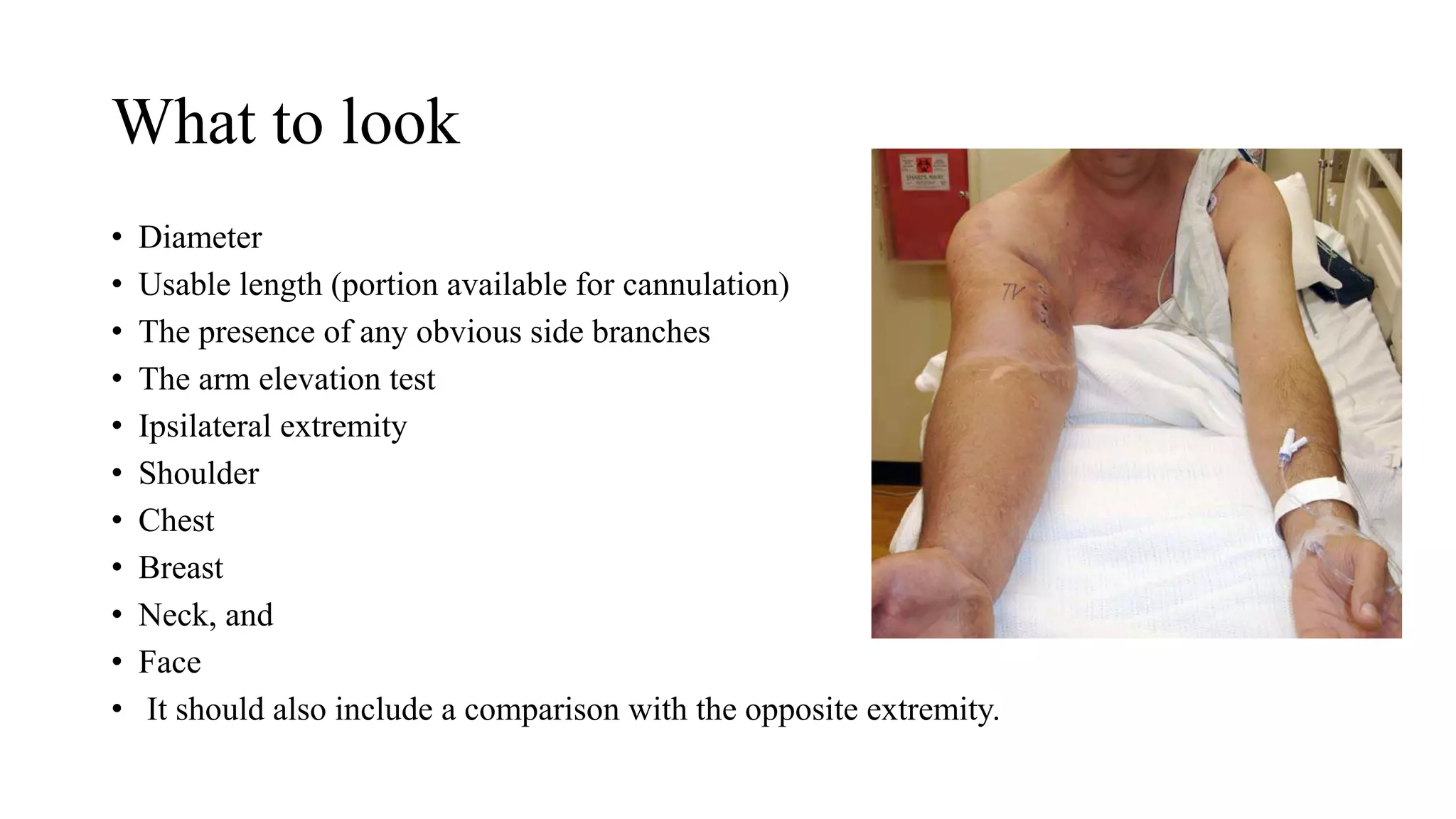
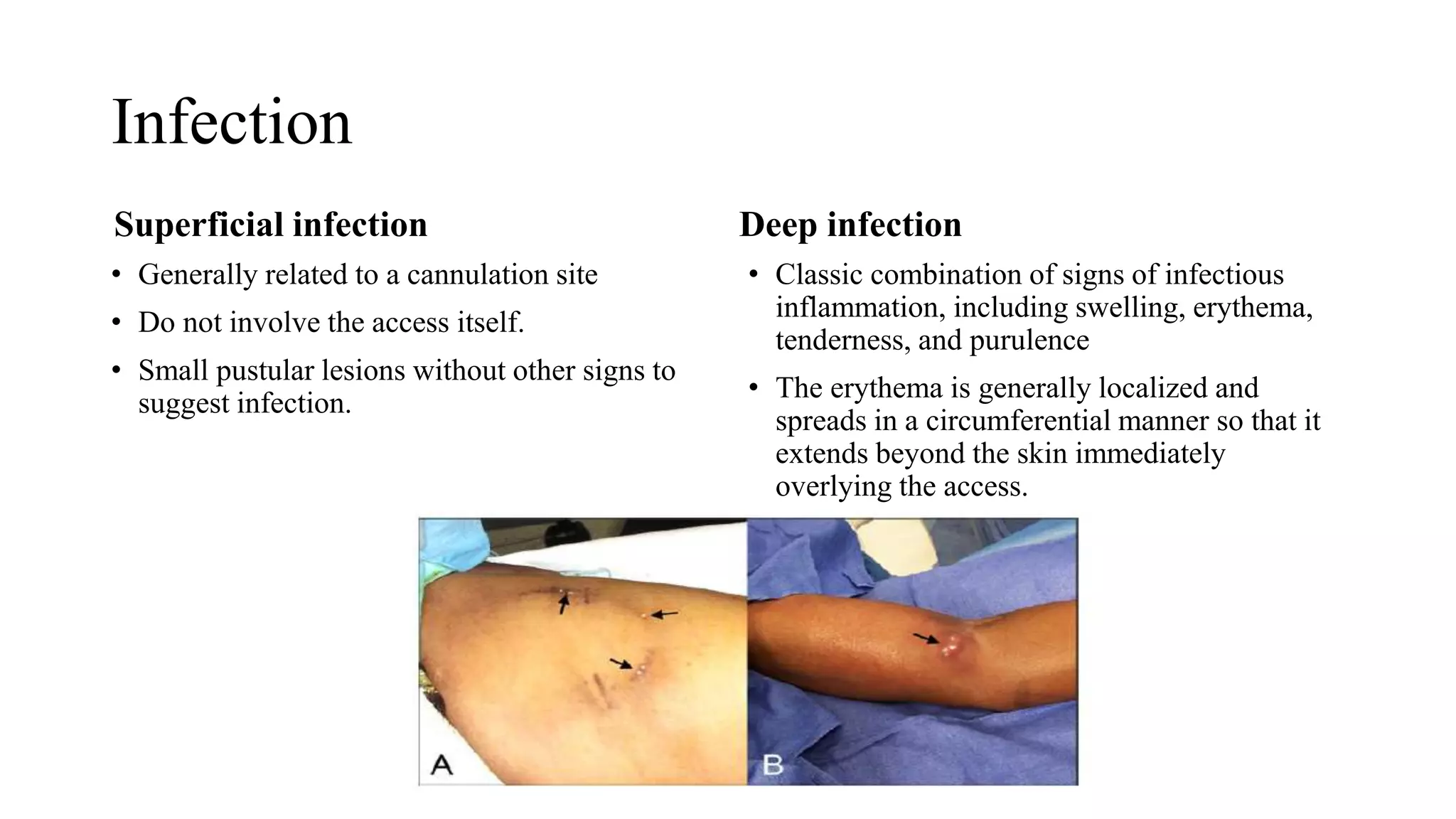
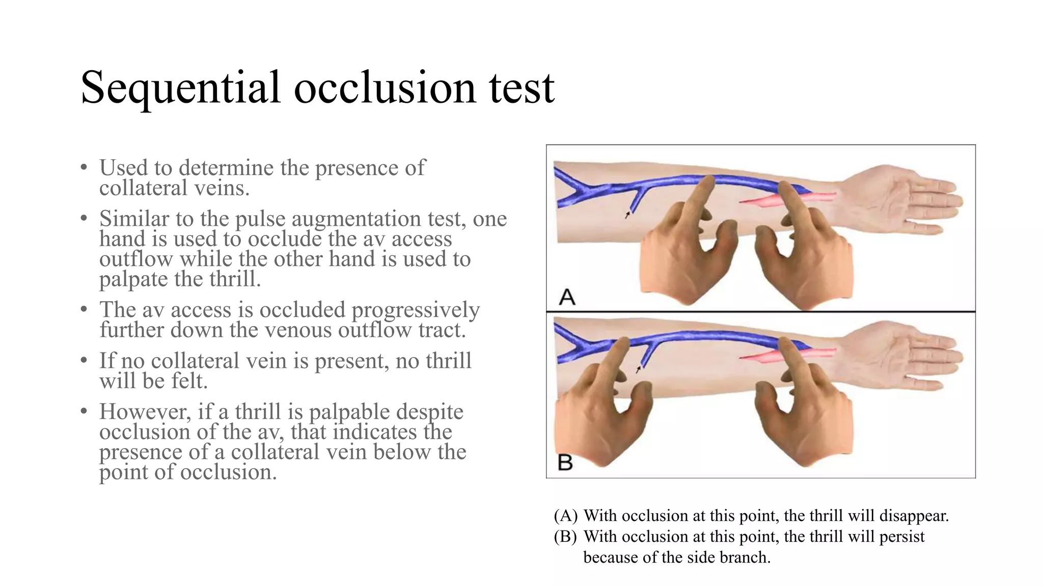
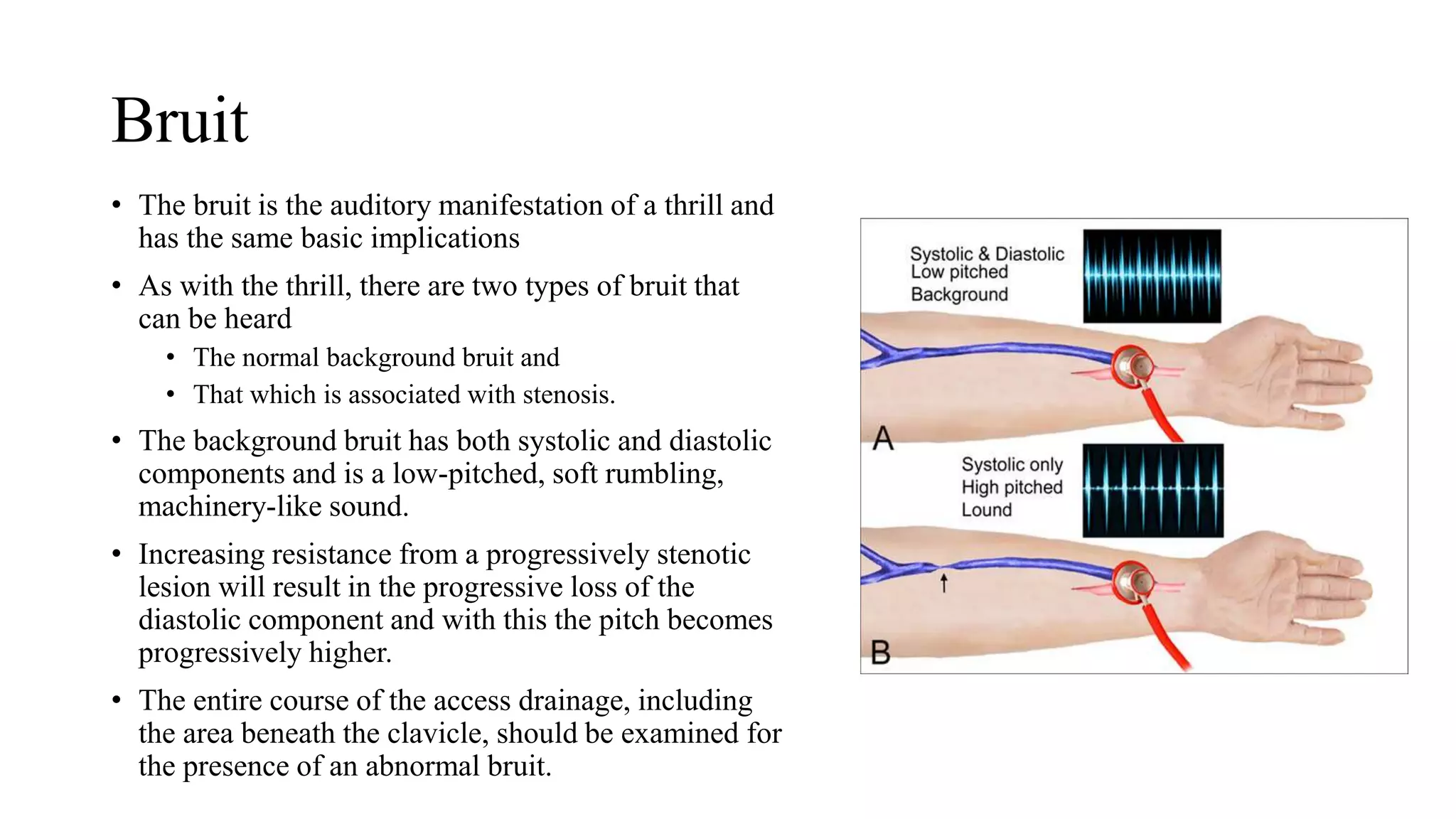
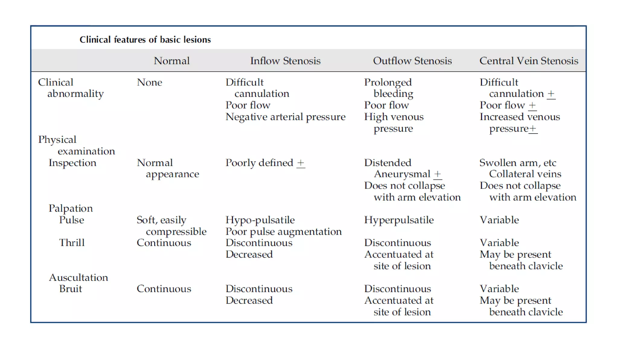
ARTERIO-VENOUS
ACCESS EXAMINATION
Department of Nephrology
MGM Medical College and Hospital
Aurangabad
The KDOQI guidelines recommend that Physical
Examination of the AV access be performed by a
qualified individual on regular basis (at least
monthly) when used for surveillance
Why examination??
• Detect vascular stenosis or other abnormalities that might lead to dysfunction.
• Simple to perform
• Does not require
• Additional machines,
• Regular calibrations,
• Additional cost, or
• Additional staff
• It is easily learned
Important terms
• Inflow refers to the feeding artery,
the arterial anastomosis, and
the juxta-anastomotic segment (the
structures within 2 cm of the arterial
anastomosis),
• Outflow segment refers to either the
entire draining vein until the
subclavian vein in the upper
extremity
• Central veins include the subclavian
vein, innominate vein, and superior
vena cava in the upper torso and the
iliac veins and inferior vena cava in
the lower torso.
Inspection (to look)
What to look
• Diameter
• Usable length (portion available for cannulation)
• The presence of any obvious side branches
• The arm elevation test
• Ipsilateral extremity
• Shoulder
• Chest
• Breast
• Neck, and
• Face
• It should also include a comparison with the opposite extremity.
Infection
Superficial infection
• Generally related to a cannulation site
• Do not involve the access itself.
• Small pustular lesions without other signs to
suggest infection.
Deep infection
• Classic combination of signs of infectious
inflammation, including swelling, erythema,
tenderness, and purulence
• The erythema is generally localized and
spreads in a circumferential manner so that it
extends beyond the skin immediately
overlying the access.
Edema
Scars on chest and neck
• Might indicate the sites of
previous temporary or tunneled
hemodialysis catheters
• Presence of a cardiovascular
implantable electronic device on
the chest wall. These devices are
frequently associated with
central vein stenosis.
• The presence of collateral veins
in the arm or chest should be
noted because these are
frequently indicative of
downstream stenosis.
Aneurysm and pseudoaneurysm
• The overlying skin should be
examined for evidence of
• Marked thinning,
• Depigmentation,
• Tightening of the overlying skin,
• Ulceration, or
• Spontaneous bleeding.
• If the skin is tissue-paper thin and
cannot be pinched between the
index finger and thumb, it has
advanced to a dangerous degree.
(A) Arteriovenous fistula with aneurysms with ulceration
(arrows; depigmentation with early skin break-down).
(B) Arteriovenous graft with large pseudoaneurysms (fusiform pseudoaneurysm)
Vascular steal syndrome
• leads to hand ischemia
• In the mildest cases, the affected hand is
pale or cyanotic in appearance compared
with the opposite side.
• In more severe cases, evidence of
ischemic changes in the skin, especially
at the fingertips, may be present
(A) Early chronic ischemia of hand without tissue loss
(hand was cold).
(B) Tissue loss at the fingertip is evident (dry gangrene).
Arm Elevation Test
• Unfortunately, this maneuver does not Work with
an AVG because of the higher level of pressure
• In the patient with an AVF, the access is generally
distended when the arm is dependent due to the
effects of gravity
• If the arm is then elevated to a level above that of
the heart, the AVF should collapse. Even if the
patient has a large dilated AVF, it should become
flaccid.
• However, if a venous stenosis is present, only the
portion of the AVF proximal to the lesion will
collapse while that portion distal to the site will
remain distended.
• One can conclude with a reasonable degree of
confidence that the outflow of the access is normal
if the patient’s AVF collapses when the arm is
elevated.
Palpation (to feel)
What to feel?
• Thrill
• Pulse
• 2 special maneuvers
• Pulse augmentation test
• Sequential occlusion test
Pulse
• Best appreciated using the fingertips
• Normally, the AV access is easily
compressible with very little pulse.
• In general, a pulsatile av access is an
adverse finding, and it is indicative of a
downstream lesion
• The degree of hyperpulsatility that is
present is proportional to the severity of
the stenosis.
• An unusually weak pulse (hypopulsatile
access) or “flat access” suggests the
presence of a stenotic lesion in the
inflow side of the access
Thrill
• Be best evaluated by using the palm of the hand
• A thrill is a palpable vibration (“buzz”). It is related to flow; when present, it indicates that there is
flow within the access.
• Two different types of thrill—a diffuse background thrill and a localized accentuated thrill.
• The presence of a soft, continuous (systolic and diastolic), diffuse background thrill palpable over
the course of the access, either an avf or an avg, is normal. This is most obvious at the venous
anastomosis.
• A stenotic lesion creates a localized area of turbulent flow within the vessel. As the lesion develops
with progressively increasing resistance to flow, the thrill becomes shortened and eventually loses
its diastolic component.
• The entire course of the access drainage should be examined for the presence of an abnormal thrill.
• With subclavian or cephalic arch stenosis, a thrill can often be detected below the clavicle.
• The absence of a thrill indicates a lack of flow. This finding, along with the absence of any pulse, is
characteristic of a thrombosed or “clotted” access.
Pulse Augmentation test
• Used to evaluate the inflow.
• The av access is completely occluded several centimeters above the arterial anastomosis
with one hand, while the other hand is used to assess the quality of the pulse.
• Increased pulse intensity (augmentation) with occlusion of the outflow vein is a normal
finding.
• Failure of the pulse to augment when the outflow vein is occluded suggests the presence
of inflow stenosis.
Sequential occlusion test
• Used to determine the presence of
collateral veins.
• Similar to the pulse augmentation test, one
hand is used to occlude the av access
outflow while the other hand is used to
palpate the thrill.
• The av access is occluded progressively
further down the venous outflow tract.
• If no collateral vein is present, no thrill
will be felt.
• However, if a thrill is palpable despite
occlusion of the av, that indicates the
presence of a collateral vein below the
point of occlusion.
(A) With occlusion at this point, the thrill will disappear.
(B) With occlusion at this point, the thrill will persist
because of the side branch.
Auscultation (to listen)
Bruit
• The bruit is the auditory manifestation of a thrill and
has the same basic implications
• As with the thrill, there are two types of bruit that
can be heard
• The normal background bruit and
• That which is associated with stenosis.
• The background bruit has both systolic and diastolic
components and is a low-pitched, soft rumbling,
machinery-like sound.
• Increasing resistance from a progressively stenotic
lesion will result in the progressive loss of the
diastolic component and with this the pitch becomes
progressively higher.
• The entire course of the access drainage, including
the area beneath the clavicle, should be examined for
the presence of an abnormal bruit.
Summary
Thank you

Arterio-Venous Access examination

  • 1.
    ARTERIO-VENOUS ACCESS EXAMINATION Department ofNephrology MGM Medical College and Hospital Aurangabad
  • 2.
    The KDOQI guidelinesrecommend that Physical Examination of the AV access be performed by a qualified individual on regular basis (at least monthly) when used for surveillance
  • 3.
    Why examination?? • Detectvascular stenosis or other abnormalities that might lead to dysfunction. • Simple to perform • Does not require • Additional machines, • Regular calibrations, • Additional cost, or • Additional staff • It is easily learned
  • 4.
    Important terms • Inflowrefers to the feeding artery, the arterial anastomosis, and the juxta-anastomotic segment (the structures within 2 cm of the arterial anastomosis), • Outflow segment refers to either the entire draining vein until the subclavian vein in the upper extremity • Central veins include the subclavian vein, innominate vein, and superior vena cava in the upper torso and the iliac veins and inferior vena cava in the lower torso.
  • 5.
  • 6.
    What to look •Diameter • Usable length (portion available for cannulation) • The presence of any obvious side branches • The arm elevation test • Ipsilateral extremity • Shoulder • Chest • Breast • Neck, and • Face • It should also include a comparison with the opposite extremity.
  • 7.
    Infection Superficial infection • Generallyrelated to a cannulation site • Do not involve the access itself. • Small pustular lesions without other signs to suggest infection. Deep infection • Classic combination of signs of infectious inflammation, including swelling, erythema, tenderness, and purulence • The erythema is generally localized and spreads in a circumferential manner so that it extends beyond the skin immediately overlying the access.
  • 8.
  • 9.
    Scars on chestand neck • Might indicate the sites of previous temporary or tunneled hemodialysis catheters • Presence of a cardiovascular implantable electronic device on the chest wall. These devices are frequently associated with central vein stenosis. • The presence of collateral veins in the arm or chest should be noted because these are frequently indicative of downstream stenosis.
  • 10.
    Aneurysm and pseudoaneurysm •The overlying skin should be examined for evidence of • Marked thinning, • Depigmentation, • Tightening of the overlying skin, • Ulceration, or • Spontaneous bleeding. • If the skin is tissue-paper thin and cannot be pinched between the index finger and thumb, it has advanced to a dangerous degree. (A) Arteriovenous fistula with aneurysms with ulceration (arrows; depigmentation with early skin break-down). (B) Arteriovenous graft with large pseudoaneurysms (fusiform pseudoaneurysm)
  • 11.
    Vascular steal syndrome •leads to hand ischemia • In the mildest cases, the affected hand is pale or cyanotic in appearance compared with the opposite side. • In more severe cases, evidence of ischemic changes in the skin, especially at the fingertips, may be present (A) Early chronic ischemia of hand without tissue loss (hand was cold). (B) Tissue loss at the fingertip is evident (dry gangrene).
  • 12.
    Arm Elevation Test •Unfortunately, this maneuver does not Work with an AVG because of the higher level of pressure • In the patient with an AVF, the access is generally distended when the arm is dependent due to the effects of gravity • If the arm is then elevated to a level above that of the heart, the AVF should collapse. Even if the patient has a large dilated AVF, it should become flaccid. • However, if a venous stenosis is present, only the portion of the AVF proximal to the lesion will collapse while that portion distal to the site will remain distended. • One can conclude with a reasonable degree of confidence that the outflow of the access is normal if the patient’s AVF collapses when the arm is elevated.
  • 13.
  • 14.
    What to feel? •Thrill • Pulse • 2 special maneuvers • Pulse augmentation test • Sequential occlusion test
  • 15.
    Pulse • Best appreciatedusing the fingertips • Normally, the AV access is easily compressible with very little pulse. • In general, a pulsatile av access is an adverse finding, and it is indicative of a downstream lesion • The degree of hyperpulsatility that is present is proportional to the severity of the stenosis. • An unusually weak pulse (hypopulsatile access) or “flat access” suggests the presence of a stenotic lesion in the inflow side of the access
  • 16.
    Thrill • Be bestevaluated by using the palm of the hand • A thrill is a palpable vibration (“buzz”). It is related to flow; when present, it indicates that there is flow within the access. • Two different types of thrill—a diffuse background thrill and a localized accentuated thrill. • The presence of a soft, continuous (systolic and diastolic), diffuse background thrill palpable over the course of the access, either an avf or an avg, is normal. This is most obvious at the venous anastomosis. • A stenotic lesion creates a localized area of turbulent flow within the vessel. As the lesion develops with progressively increasing resistance to flow, the thrill becomes shortened and eventually loses its diastolic component. • The entire course of the access drainage should be examined for the presence of an abnormal thrill. • With subclavian or cephalic arch stenosis, a thrill can often be detected below the clavicle. • The absence of a thrill indicates a lack of flow. This finding, along with the absence of any pulse, is characteristic of a thrombosed or “clotted” access.
  • 18.
    Pulse Augmentation test •Used to evaluate the inflow. • The av access is completely occluded several centimeters above the arterial anastomosis with one hand, while the other hand is used to assess the quality of the pulse. • Increased pulse intensity (augmentation) with occlusion of the outflow vein is a normal finding. • Failure of the pulse to augment when the outflow vein is occluded suggests the presence of inflow stenosis.
  • 19.
    Sequential occlusion test •Used to determine the presence of collateral veins. • Similar to the pulse augmentation test, one hand is used to occlude the av access outflow while the other hand is used to palpate the thrill. • The av access is occluded progressively further down the venous outflow tract. • If no collateral vein is present, no thrill will be felt. • However, if a thrill is palpable despite occlusion of the av, that indicates the presence of a collateral vein below the point of occlusion. (A) With occlusion at this point, the thrill will disappear. (B) With occlusion at this point, the thrill will persist because of the side branch.
  • 20.
  • 21.
    Bruit • The bruitis the auditory manifestation of a thrill and has the same basic implications • As with the thrill, there are two types of bruit that can be heard • The normal background bruit and • That which is associated with stenosis. • The background bruit has both systolic and diastolic components and is a low-pitched, soft rumbling, machinery-like sound. • Increasing resistance from a progressively stenotic lesion will result in the progressive loss of the diastolic component and with this the pitch becomes progressively higher. • The entire course of the access drainage, including the area beneath the clavicle, should be examined for the presence of an abnormal bruit.
  • 22.
  • 24.